The Optic Nerve In creatingrevising this slideset I


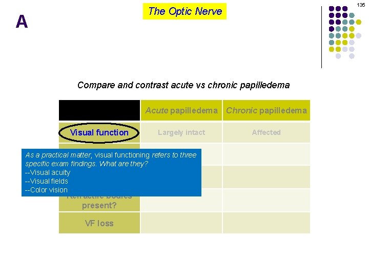
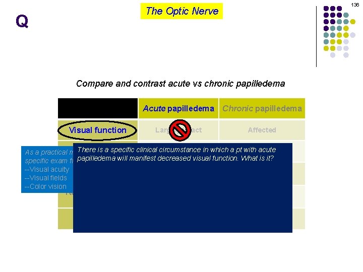
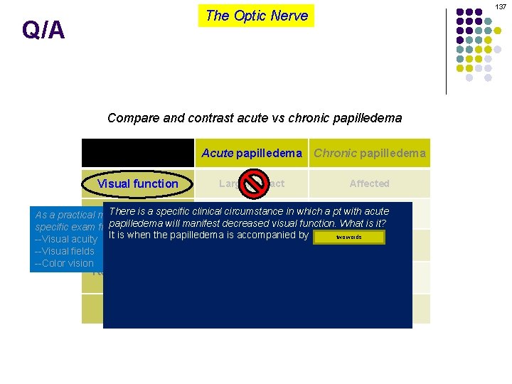
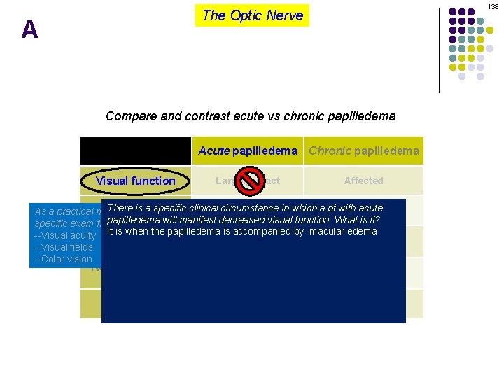
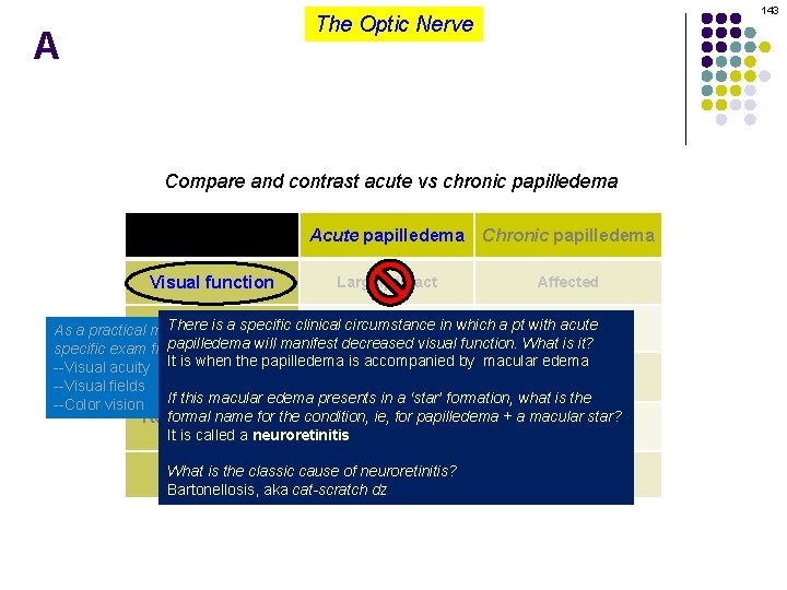
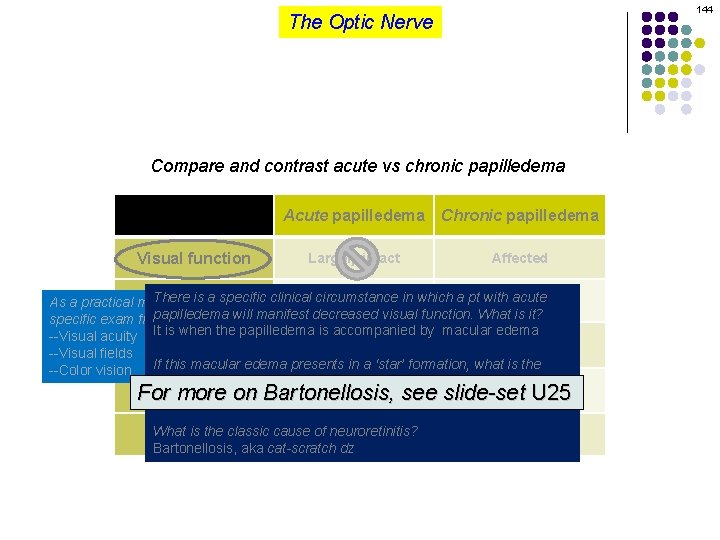
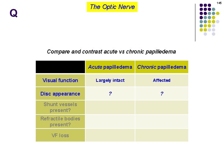
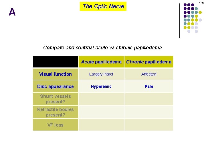
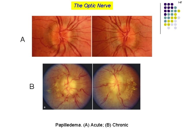
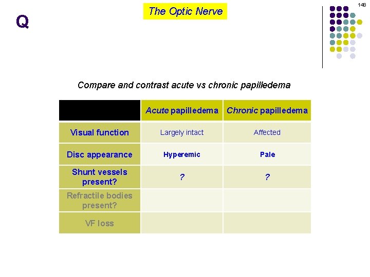
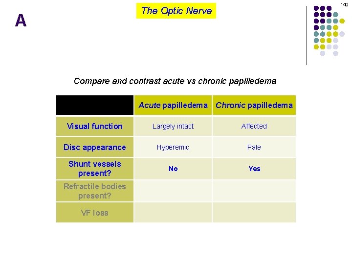
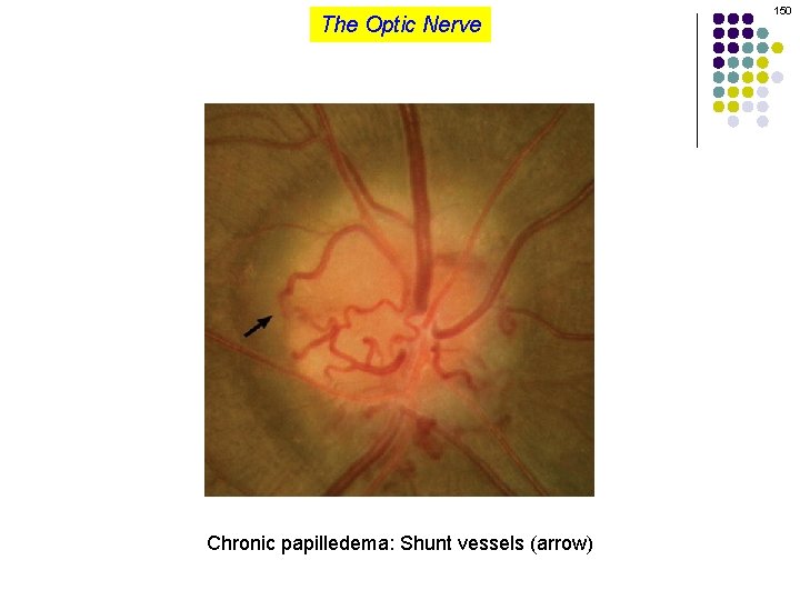
- Slides: 169

The Optic Nerve In creating/revising this slide-set, I consulted the four BCSC books that have a lot to say on the subject: Fundamentals, Neuro-Oph, Path and Glaucoma. Unfortunately, all four differed from one another regarding many aspects of optic nerve anatomy. Some of these differences were trivial; others not so much. As a comprehensive ophthalmologist, I have no familiarity with the primary literature concerning ophthalmic anatomy and histology. Thus, I am in no position to declare which book is correct regarding points on which they differ. The following slides represent my best attempt at compiling the disparate information in a manner that is reasonable and memorable. (As a matter of both interest and information, I have included some of the differing answers regarding certain aspects of the nerve. ) My main point: When answering questions regarding the optic nerve-whether such questions occur in a pimping session, on the OKAP or during the Boards--adopt and maintain a stance of flexibility. 1

The Optic Nerve In creating/revising this slide-set, I consulted the four BCSC books that have a lot to say on the subject: Fundamentals, Neuro-Oph, Path and Glaucoma. Unfortunately, all four differed from one another regarding many aspects of optic nerve anatomy. Some of these differences were trivial; others not so tl; dr: much. --When asked an optic-nerve question requiring a numeric response, phrase your answerophthalmologist, along these lines: ‘Well, bearing in mind the As a comprehensive I have no familiarity with the primary considerable anatomic variability that characterizes the optic literature concerning ophthalmic anatomy and histology. Thus, I amnerve, in no aposition reasonable estimate x. ’ regarding points on which they to declare whichwould book isbe correct --When asked a question about optic-nerve vasculature, begin differ. The following slides represent my best attempt at compiling theyour disparate information in ain manner that is reasonable and memorable. (As a response with ‘Bearing mind that there is not universal agreement matter of both information, I have included some of the differing regarding this, interest many and experts believe…’ answers regarding certain aspects of the nerve. ) My main point: When answering questions regarding the optic nerve-whether such questions occur in a pimping session, on the OKAP or during the Boards--adopt and maintain a stance of flexibility. 2

Q The Optic Nerve The optic nerves are composed of what? 3

A The Optic Nerve The optic nerves are composed of what? The axons of retinal ganglion cells 4

Q The Optic Nerve The optic nerves are composed of what? The axons of retinal ganglion cells How many fibers (axons) comprise an optic nerve? 5

A 6 The Optic Nerve The optic nerves are composed of what? The axons of retinal ganglion cells How many fibers (axons) comprise an optic nerve? Depends upon which book you ask, but the answer 1. 2 M works Glaucoma book: 1. 2 -1. 5 M Neuro: 1 -1. 2 M Fundamentals: “more than a million”

Q The Optic Nerve The optic nerves are composed of what? The axons of retinal ganglion cells Do they synapse in the region of the optic nerve head? 7

A The Optic Nerve The optic nerves are composed of what? The axons of retinal ganglion cells Do they synapse in the region of the optic nerve head? No 8

Q The Optic Nerve The optic nerves are composed of what? The axons of retinal ganglion cells Do they synapse in the region of the optic nerve head? No Where will they synapse? 9

A The Optic Nerve The optic nerves are composed of what? The axons of retinal ganglion cells Do they synapse in the region of the optic nerve head? No Where will they synapse? Most will synapse in the lateral geniculate nucleus (LGN) 10

Q The Optic Nerve The optic nerves are composed of what? The axons of retinal ganglion cells Do they synapse in the region of the optic nerve head? No Where will they synapse? Most will synapse in the lateral geniculate nucleus (LGN) Most? Where will the others synapse, and what are they responsible for? 11

A The Optic Nerve The optic nerves are composed of what? The axons of retinal ganglion cells Do they synapse in the region of the optic nerve head? No Where will they synapse? Most will synapse in the lateral geniculate nucleus (LGN) Most? Where will the others synapse, and what are they responsible for? Most of the others are involved in the pupillary light reflex; they peel off just prior to reaching the LGN, heading instead to the pretectum of the dorsal midbrain to synapse in the pretectal nuclei 12

Q The Optic Nerve The optic nerves are composed of what? The axons of retinal ganglion cells Do they synapse in the region of the optic nerve head? No Where will they synapse? Most will synapse in the lateral geniculate nucleus (LGN) Most? Where will the others synapse, and what are they responsible for? Most of the others are involved in the pupillary light reflex; they peel off just prior to reaching the LGN, heading instead to the pretectum of the dorsal midbrain to synapse in the pretectal nuclei ‘Most’? Where will the others synapse, and what are they responsible for? 13

A The Optic Nerve The optic nerves are composed of what? The axons of retinal ganglion cells Do they synapse in the region of the optic nerve head? No Where will they synapse? Most will synapse in the lateral geniculate nucleus (LGN) Most? Where will the others synapse, and what are they responsible for? Most of the others are involved in the pupillary light reflex; they peel off just prior to reaching the LGN, heading instead to the pretectum of the dorsal midbrain to synapse in the pretectal nuclei ‘Most’? Where will the others synapse, and what are they responsible for? The hypothalamus, where they are involved in modulating circadian responses 14

Q The Optic Nerve The optic nerves are composed of what? The axons of retinal ganglion cells Do they synapse in the region of the optic nerve head? No Where will they synapse? Most will synapse in the lateral geniculate nucleus (LGN) Most? Where will the others synapse, and what are they responsible for? Most of the others are involved in the pupillary light reflex; they peel off just prior to reaching the LGN, heading instead to the pretectum of the dorsal midbrain to synapse in the pretectal nuclei ‘Most’? Where will the others synapse, and what are they responsible for? The hypothalamus, where they are involved in modulating circadian There is an important clinical entity caused by damage to the pretectum. This entity has four classic responses findings, one of which involves the pupils. What is the eponymous name of this clinical entity? Parinaud syndrome What is the classic pupil finding in Parinaud syndrome? Light-near dissociation What are the two noneponymous names for Parinaud syndrome? 1) Dorsal midbrain syndrome 2) Pretectal syndrome 15

A The Optic Nerve The optic nerves are composed of what? The axons of retinal ganglion cells Do they synapse in the region of the optic nerve head? No Where will they synapse? Most will synapse in the lateral geniculate nucleus (LGN) Most? Where will the others synapse, and what are they responsible for? Most of the others are involved in the pupillary light reflex; they peel off just prior to reaching the LGN, heading instead to the pretectum of the dorsal midbrain to synapse in the pretectal nuclei ‘Most’? Where will the others synapse, and what are they responsible for? The hypothalamus, where they are involved in modulating circadian There is an important clinical entity caused by damage to the pretectum. This entity has four classic responses findings, one of which involves the pupils. What is the eponymous name of this clinical entity? Parinaud syndrome What is the classic pupil finding in Parinaud syndrome? Light-near dissociation What are the two noneponymous names for Parinaud syndrome? 1) Dorsal midbrain syndrome 2) Pretectal syndrome 16

Q The Optic Nerve The optic nerves are composed of what? The axons of retinal ganglion cells Do they synapse in the region of the optic nerve head? No Where will they synapse? Most will synapse in the lateral geniculate nucleus (LGN) Most? Where will the others synapse, and what are they responsible for? Most of the others are involved in the pupillary light reflex; they peel off just prior to reaching the LGN, heading instead to the pretectum of the dorsal midbrain to synapse in the pretectal nuclei ‘Most’? Where will the others synapse, and what are they responsible for? The hypothalamus, where they are involved in modulating circadian There is an important clinical entity caused by damage to the pretectum. This entity has four classic responses findings, one of which involves the pupils. What is the eponymous name of this clinical entity? Parinaud syndrome What is the classic pupil finding in Parinaud syndrome? Light-near dissociation What are the two noneponymous names for Parinaud syndrome? 1) Dorsal midbrain syndrome 2) Pretectal syndrome 17

A The Optic Nerve The optic nerves are composed of what? The axons of retinal ganglion cells Do they synapse in the region of the optic nerve head? No Where will they synapse? Most will synapse in the lateral geniculate nucleus (LGN) Most? Where will the others synapse, and what are they responsible for? Most of the others are involved in the pupillary light reflex; they peel off just prior to reaching the LGN, heading instead to the pretectum of the dorsal midbrain to synapse in the pretectal nuclei ‘Most’? Where will the others synapse, and what are they responsible for? The hypothalamus, where they are involved in modulating circadian There is an important clinical entity caused by damage to the pretectum. This entity has four classic responses findings, one of which involves the pupils. What is the eponymous name of this clinical entity? Parinaud syndrome What is the classic pupil finding in Parinaud syndrome? Light-near dissociation What are the two noneponymous names for Parinaud syndrome? 1) Dorsal midbrain syndrome 2) Pretectal syndrome 18

Q The Optic Nerve The optic nerves are composed of what? The axons of retinal ganglion cells Do they synapse in the region of the optic nerve head? No Where will they synapse? Most will synapse in the lateral geniculate nucleus (LGN) Most? Where will the others synapse, and what are they responsible for? Most of the others are involved in the pupillary light reflex; they peel off just prior to reaching the LGN, heading instead to the pretectum of the dorsal midbrain to synapse in the pretectal nuclei ‘Most’? Where will the others synapse, and what are they responsible for? The hypothalamus, where they are involved in modulating circadian responses There is an important clinical entity caused by damage to the pretectum. This entity has four classic findings, one of which involves the pupils. What is the eponymous name of this clinical entity? Parinaud syndrome What is the classic pupil finding in Parinaud syndrome? Light-near dissociation What is. What light-near are thedissociation? two noneponymous names for Parinaud syndrome? A phenomena which thesyndrome pupils miose less robustly in 1) Dorsalinmidbrain response to light than they do as part of the near response 2) Pretectal syndrome 19

A The Optic Nerve The optic nerves are composed of what? The axons of retinal ganglion cells Do they synapse in the region of the optic nerve head? No Where will they synapse? Most will synapse in the lateral geniculate nucleus (LGN) Most? Where will the others synapse, and what are they responsible for? Most of the others are involved in the pupillary light reflex; they peel off just prior to reaching the LGN, heading instead to the pretectum of the dorsal midbrain to synapse in the pretectal nuclei ‘Most’? Where will the others synapse, and what are they responsible for? The hypothalamus, where they are involved in modulating circadian There is an important clinical entity caused by damage to the pretectum. This entity has four classic responses findings, one of which involves the pupils. What is the eponymous name of this clinical entity? Parinaud syndrome What is the classic pupil finding in Parinaud syndrome? Light-near dissociation What is. What light-near are thedissociation? two noneponymous names for Parinaud syndrome? A phenomena which thesyndrome pupils miose less robustly in 1) Dorsalinmidbrain response to light than they do as part of the near response 2) Pretectal syndrome 20

Q The Optic Nerve The optic nerves are composed of what? The axons of retinal ganglion cells Do they synapse in the region of the optic nerve head? No Where will they synapse? Most will synapse in the lateral geniculate nucleus (LGN) Most? Where will the others synapse, and what are they responsible for? Most of the others are involved in the pupillary light reflex; they peel off just prior to reaching the LGN, heading instead to the pretectum of the dorsal midbrain to synapse in the pretectal nuclei ‘Most’? Where will the others synapse, and what are they responsible for? The hypothalamus, where they are involved in modulating circadian There is an important clinical entity caused by damage to the pretectum. This entity has four classic responses Theofnear response is often referred byeponymous what number-related findings, one which involves the pupils. What istothe name of thisname? clinical entity? The near triad Parinaud syndrome What is the classic pupil findingwhat in Parinaud Other than miosis, are the syndrome? other ocular responses of the near triad? Light-near--Miosis dissociation --Convergence What is. What light-near dissociation? are the two noneponymous names for Parinaud syndrome? --Accommodation A phenomena which thesyndrome pupils miose less robustly in 1) Dorsalinmidbrain response to light than they do as part of the near response 2) Pretectal syndrome 21

A The Optic Nerve The optic nerves are composed of what? The axons of retinal ganglion cells Do they synapse in the region of the optic nerve head? No Where will they synapse? Most will synapse in the lateral geniculate nucleus (LGN) Most? Where will the others synapse, and what are they responsible for? Most of the others are involved in the pupillary light reflex; they peel off just prior to reaching the LGN, heading instead to the pretectum of the dorsal midbrain to synapse in the pretectal nuclei ‘Most’? Where will the others synapse, and what are they responsible for? The hypothalamus, where they are involved in modulating circadian There is an important clinical entity caused by damage to the pretectum. This entity has four classic responses Theofnear response is often referred byeponymous what number-related findings, one which involves the pupils. What istothe name of thisname? clinical entity? The near triad Parinaud syndrome What is the classic pupil findingwhat in Parinaud Other than miosis, are the syndrome? other ocular responses of the near triad? Light-near--Miosis dissociation --Convergence What is. What light-near dissociation? are the two noneponymous names for Parinaud syndrome? --Accommodation A phenomena which thesyndrome pupils miose less robustly in 1) Dorsalinmidbrain response to light than they do as part of the near response 2) Pretectal syndrome 22

Q The Optic Nerve The optic nerves are composed of what? The axons of retinal ganglion cells Do they synapse in the region of the optic nerve head? No Where will they synapse? Most will synapse in the lateral geniculate nucleus (LGN) Most? Where will the others synapse, and what are they responsible for? Most of the others are involved in the pupillary light reflex; they peel off just prior to reaching the LGN, heading instead to the pretectum of the dorsal midbrain to synapse in the pretectal nuclei ‘Most’? Where will the others synapse, and what are they responsible for? The hypothalamus, where they are involved in modulating circadian There is an important clinical entity caused by damage to the pretectum. This entity has four classic responses Theofnear response is often referred byeponymous what number-related findings, one which involves the pupils. What istothe name of thisname? clinical entity? The near triad Parinaud syndrome What is the classic pupil findingwhat in Parinaud Other than miosis, are the syndrome? other ocular responses of the near triad? Light-near--Miosis dissociation -What is. What light-near are the two noneponymous names for Parinaud syndrome? -- dissociation? A phenomena which thesyndrome pupils miose less robustly in 1) Dorsalinmidbrain response to light than they do as part of the near response 2) Pretectal syndrome 23

A The Optic Nerve The optic nerves are composed of what? The axons of retinal ganglion cells Do they synapse in the region of the optic nerve head? No Where will they synapse? Most will synapse in the lateral geniculate nucleus (LGN) Most? Where will the others synapse, and what are they responsible for? Most of the others are involved in the pupillary light reflex; they peel off just prior to reaching the LGN, heading instead to the pretectum of the dorsal midbrain to synapse in the pretectal nuclei ‘Most’? Where will the others synapse, and what are they responsible for? The hypothalamus, where they are involved in modulating circadian There is an important clinical entity caused by damage to the pretectum. This entity has four classic responses Theofnear response is often referred byeponymous what number-related findings, one which involves the pupils. What istothe name of thisname? clinical entity? The near triad Parinaud syndrome What is the classic pupil findingwhat in Parinaud Other than miosis, are the syndrome? other ocular responses of the near triad? Light-near--Miosis dissociation --Convergence What is. What light-near dissociation? are the two noneponymous names for Parinaud syndrome? --Accommodation A phenomena which thesyndrome pupils miose less robustly in 1) Dorsalinmidbrain response to light than they do as part of the near response 2) Pretectal syndrome 24

Q The Optic Nerve The optic nerves are composed of what? The axons of retinal ganglion cells Do they synapse in the region of the optic nerve head? No Where will they synapse? Most will synapse in the lateral geniculate nucleus (LGN) Most? Where will the others synapse, and what are they responsible for? Most of the others are involved in the pupillary light reflex; they peel off just prior to reaching the LGN, heading instead to the pretectum of the dorsal midbrain to synapse in the pretectal nuclei ‘Most’? Where will the others synapse, and what are they responsible for? The hypothalamus, where they are involved in modulating circadian There is an important clinical entity caused by damage to the pretectum. This entity has four classic responses findings, one of which involves the pupils. What is the eponymous name of this clinical entity? Parinaud syndrome What is the classic pupil finding in Parinaud syndrome? Light-near dissociation What are the two noneponymous names for Parinaud syndrome? 1) Dorsal midbrain syndrome 2) Pretectal syndrome 25

A The Optic Nerve The optic nerves are composed of what? The axons of retinal ganglion cells Do they synapse in the region of the optic nerve head? No Where will they synapse? Most will synapse in the lateral geniculate nucleus (LGN) Most? Where will the others synapse, and what are they responsible for? Most of the others are involved in the pupillary light reflex; they peel off just prior to reaching the LGN, heading instead to the pretectum of the dorsal midbrain to synapse in the pretectal nuclei ‘Most’? Where will the others synapse, and what are they responsible for? The hypothalamus, where they are involved in modulating circadian There is an important clinical entity caused by damage to the pretectum. This entity has four classic responses findings, one of which involves the pupils. What is the eponymous name of this clinical entity? Parinaud syndrome What is the classic pupil finding in Parinaud syndrome? Light-near dissociation What are the two noneponymous names for Parinaud syndrome? 1) Dorsal midbrain syndrome 2) Pretectal syndrome 26

The Optic Nerve Q (anterior) 27 Portion Length (mm) ? 1 ? (posterior) Anatomically speaking, the optic nerve is considered to have four portions. What are they?

The Optic Nerve Q/A (anterior) 28 Portion Length (mm) Intraocular 1 ? ? (posterior) Anatomically speaking, the optic nerve is considered to have four portions. What are they?

The Optic Nerve Q/A (anterior) 29 Portion Length (mm) Intraocular 1 Orbital ? ? (posterior) Anatomically speaking, the optic nerve is considered to have four portions. What are they?

The Optic Nerve Q/A (anterior) 30 Portion Length (mm) Intraocular 1 Orbital ? Canalicular (posterior) ? Anatomically speaking, the optic nerve is considered to have four portions. What are they?

The Optic Nerve A (anterior) 31 Portion Length (mm) Intraocular 1 Orbital ? Canalicular (posterior) Intracranial Anatomically speaking, the optic nerve is considered to have four portions. What are they?

The Optic Nerve Optic nerve (don’t memorize the lengths) 32

33 The Optic Nerve Q Portion Length (mm) Intraocular ? Orbital Canalicular Intracranial Anatomically speaking, the optic nerve is considered to have four portions. What are they? How long is each?

34 The Optic Nerve Q/A Portion Length (mm) Intraocular 1 Orbital ? Canalicular Intracranial Anatomically speaking, the optic nerve is considered to have four portions. What are they? How long is each?

35 The Optic Nerve Q/A Portion Length (mm) Intraocular 1 Orbital 30 Canalicular ? Fundamentals: 25 Path: 25 -30 Neuro: 30 Intracranial Anatomically speaking, the optic nerve is considered to have four portions. What are they? How long is each?

36 The Optic Nerve Q/A Portion Length (mm) Intraocular 1 Orbital 30 Canalicular 10 Intracranial ? Fundamentals: 4 -10 Path: 4 -10 Neuro: 8 -10 Anatomically speaking, the optic nerve is considered to have four portions. What are they? How long is each?

37 The Optic Nerve A Portion Length (mm) Intraocular 1 Orbital 30 Canalicular 10 Intracranial 10 Fundamentals: 10 Path: 10 Neuro: 8 -12 Anatomically speaking, the optic nerve is considered to have four portions. What are they? How long is each?

38 The Optic Nerve Q Portion Length (mm) Intraocular 1 Orbital 30 Canalicular 10 How long is the distance between the back of the eye and the orbital apex? 10 About 18 mm Intracranial Anatomically speaking, the optic nerve is considered to have four portions. What are they?

39 The Optic Nerve A Portion Length (mm) Intraocular 1 Orbital 30 Canalicular 10 How long is the distance between the back of the eye and the orbital apex? 10 About 18 mm Intracranial Anatomically speaking, the optic nerve is considered to have four portions. What are they?

40 The Optic Nerve Q Portion Length (mm) Intraocular 1 Orbital 30 Canalicular 10 Intracranial 10 Anatomically speaking, the optic nerve is considered to have four portions. What are they? How long is each? The intraocular portion is also considered to have four portions. What are they? (innermost) Portion Blood supply ? Central retinal artery (CRA) Short posterior ciliary arteries Arterial circle of Zinn & Haller (outermost) Centripetal CRA branches, centrifugal pial branches

41 The Optic Nerve Q/A Portion Length (mm) Intraocular 1 Orbital 30 Canalicular 10 Intracranial 10 Anatomically speaking, the optic nerve is considered to have four portions. What are they? How long is each? The intraocular portion is also considered to have four portions. What are they? (innermost) Portion Blood supply NFL portion Central retinal artery (CRA) ? Short posterior ciliary arteries Arterial circle of Zinn & Haller (outermost) Centripetal CRA branches, centrifugal pial branches

42 The Optic Nerve Q/A Portion Length (mm) Intraocular 1 Orbital 30 Canalicular 10 Intracranial 10 Anatomically speaking, the optic nerve is considered to have four portions. What are they? How long is each? The intraocular portion is also considered to have four portions. What are they? (innermost) (outermost) Portion Blood supply NFL portion Central retinal artery (CRA) Pre-laminar Short posterior ciliary arteries ? Arterial circle of Zinn & Haller Centripetal CRA branches, centrifugal pial branches

43 The Optic Nerve Q/A Portion Length (mm) Intraocular 1 Orbital 30 Canalicular 10 Intracranial 10 Anatomically speaking, the optic nerve is considered to have four portions. What are they? How long is each? The intraocular portion is also considered to have four portions. What are they? (innermost) (outermost) Portion Blood supply NFL portion Central retinal artery (CRA) Pre-laminar Short posterior ciliary arteries Laminar Arterial circle of Zinn & Haller ? Centripetal CRA branches, centrifugal pial branches

44 The Optic Nerve A Portion Length (mm) Intraocular 1 Orbital 30 Canalicular 10 Intracranial 10 Anatomically speaking, the optic nerve is considered to have four portions. What are they? How long is each? The intraocular portion is also considered to have four portions. What are they? (innermost) (outermost) Portion Blood supply NFL portion Central retinal artery (CRA) Pre-laminar Short posterior ciliary arteries Laminar Arterial circle of Zinn & Haller Retrolaminar Centripetal CRA branches, centrifugal pial branches

45 The Optic Nerve NFL Prelaminar Laminar Retrolaminar Optic nerve: Intraocular portion

46 The Optic Nerve Q Portion Length (mm) Intraocular 1 Orbital 30 Canalicular 10 Intracranial 10 Anatomically speaking, optic nerve is considered to have four portions. What are they? To what lamina the is this referring? How long is each? The lamina cribrosa The intraocular portion is also considered to have four portions. What are they? What is What the blood supply forcribrosa? each? is the lamina The fenestrated hole in the posterior sclera through which the optic nerve exits Portion Blood supply Does the lamina extend the entire thickness of the eye wall? Centralsclera retinal artery (CRA) NFL it is about 1/3 theportion thickness of the adjacent (innermost) No, (outermost) Pre-laminar Short posterior ciliary arteries Laminar Arterial circle of Zinn & Haller Retrolaminar Centripetal CRA branches, centrifugal pial branches

47 The Optic Nerve A Portion Length (mm) Intraocular 1 Orbital 30 Canalicular 10 Intracranial 10 Anatomically speaking, optic nerve is considered to have four portions. What are they? To what lamina the is this referring? How long is each? The lamina cribrosa The intraocular portion is also considered to have four portions. What are they? What is What the blood supply forcribrosa? each? is the lamina The fenestrated hole in the posterior sclera through which the optic nerve exits Portion Blood supply Does the lamina extend the entire thickness of the eye wall? Centralsclera retinal artery (CRA) NFL it is about 1/3 theportion thickness of the adjacent (innermost) No, (outermost) Pre-laminar Short posterior ciliary arteries Laminar Arterial circle of Zinn & Haller Retrolaminar Centripetal CRA branches, centrifugal pial branches

48 The Optic Nerve Q Portion Length (mm) Intraocular 1 Orbital 30 Canalicular 10 Intracranial 10 Anatomically speaking, optic nerve is considered to have four portions. What are they? To what lamina the is this referring? How long is each? The lamina cribrosa The intraocular portion is also considered to have four portions. What are they? Lamina cribrosa? Ifor thought that was the super-thin part of the medial orbital wall. What is What the blood supply each? is the lamina cribrosa? You’re thinking of the lamina papyracea The fenestrated hole in the posterior sclera through which the optic nerve exits Portion Blood supply Does the lamina extend the entire thickness of the eye wall? Centralsclera retinal artery (CRA) NFL it is about 1/3 theportion thickness of the adjacent (innermost) No, (outermost) Pre-laminar Short posterior ciliary arteries Laminar Arterial circle of Zinn & Haller Retrolaminar Centripetal CRA branches, centrifugal pial branches

49 The Optic Nerve A Portion Length (mm) Intraocular 1 Orbital 30 Canalicular 10 Intracranial 10 Anatomically speaking, optic nerve is considered to have four portions. What are they? To what lamina the is this referring? How long is each? The lamina cribrosa The intraocular portion is also considered to have four portions. What are they? Lamina cribrosa? Ifor thought that was the super-thin part of the medial orbital wall. What is What the blood supply each? is the lamina cribrosa? You’re thinking of the lamina papyracea The fenestrated hole in the posterior sclera through which the optic nerve exits Portion Blood supply Does the lamina extend the entire thickness of the eye wall? Centralsclera retinal artery (CRA) NFL it is about 1/3 theportion thickness of the adjacent (innermost) No, (outermost) Pre-laminar Short posterior ciliary arteries Laminar Arterial circle of Zinn & Haller Retrolaminar Centripetal CRA branches, centrifugal pial branches

50 The Optic Nerve Q Portion Length (mm) Intraocular 1 Orbital 30 Canalicular 10 Intracranial 10 Anatomically speaking, optic nerve is considered to have four portions. What are they? To what lamina the is this referring? How long is each? The lamina cribrosa The intraocular portion is also considered to have four portions. What are they? What is What the blood supply forcribrosa? each? is the lamina The fenestrated hole in the posterior sclera through which the optic nerve exits Portion Blood supply Does the lamina extend the entire thickness of the eye wall? Centralsclera retinal artery (CRA) NFL it is about 1/3 theportion thickness of the adjacent (innermost) No, (outermost) Pre-laminar Short posterior ciliary arteries Laminar Arterial circle of Zinn & Haller Retrolaminar Centripetal CRA branches, centrifugal pial branches

51 The Optic Nerve A Portion Length (mm) Intraocular 1 Orbital 30 Canalicular 10 Intracranial 10 Anatomically speaking, optic nerve is considered to have four portions. What are they? To what lamina the is this referring? How long is each? The lamina cribrosa The intraocular portion is also considered to have four portions. What are they? What is What the blood supply forcribrosa? each? is the lamina The fenestrated hole in the posterior sclera through which the optic nerve exits Portion Blood supply Does the lamina extend the entire thickness of the eye wall? Centralsclera retinal artery (CRA) NFL it is about 1/3 theportion thickness of the adjacent (innermost) No, (outermost) Pre-laminar Short posterior ciliary arteries Laminar Arterial circle of Zinn & Haller Retrolaminar Centripetal CRA branches, centrifugal pial branches

52 The Optic Nerve Q Portion Length (mm) Intraocular 1 Orbital 30 Canalicular 10 Intracranial 10 Anatomically speaking, optic nerve is considered to have four portions. What are they? To what lamina the is this referring? How long is each? The lamina cribrosa The intraocular portion is also considered to have four portions. What are they? What is What the blood supply forcribrosa? each? is the lamina The fenestrated hole in the posterior sclera through which the optic nerve exits Portion How many fenestrations are there? Blood supply Does the lamina extend the entire thickness of the eye wall? 200 -300 (innermost) Centralsclera retinal artery (CRA) NFL No, it is about 1/3 theportion thickness of the adjacent (outermost) Pre-laminar Short posterior ciliary arteries Laminar Arterial circle of Zinn & Haller Retrolaminar Centripetal CRA branches, centrifugal pial branches

53 The Optic Nerve A Portion Length (mm) Intraocular 1 Orbital 30 Canalicular 10 Intracranial 10 Anatomically speaking, optic nerve is considered to have four portions. What are they? To what lamina the is this referring? How long is each? The lamina cribrosa The intraocular portion is also considered to have four portions. What are they? What is What the blood supply forcribrosa? each? is the lamina The fenestrated hole in the posterior sclera through which the optic nerve exits Portion How many fenestrations are there? Blood supply Does the lamina extend the entire thickness of the eye wall? 200 -300 (innermost) Centralsclera retinal artery (CRA) NFL No, it is about 1/3 theportion thickness of the adjacent (outermost) Pre-laminar Short posterior ciliary arteries Laminar Arterial circle of Zinn & Haller Retrolaminar Centripetal CRA branches, centrifugal pial branches

54 The Optic Nerve Q Portion Length (mm) Intraocular 1 Orbital 30 Canalicular 10 Intracranial 10 Anatomically speaking, optic nerve is considered to have four portions. What are they? To what lamina the is this referring? How long is each? The lamina cribrosa The intraocular portion is also considered to have four portions. What are they? What is What the blood supply forcribrosa? each? is the lamina The fenestrated hole in the posterior sclera through which the optic nerve exits Portion Blood supply How many fenestrations Two fenestrations are there? are much larger than the Does the lamina extend the entire thickness of the eye wall? 200 -300 others. What passes through the larger retinal ones? artery (CRA) (innermost) Central NFL portion No, it is about. The 1/3 the thickness of the adjacent sclera central retinal artery and vein (outermost) Pre-laminar Short posterior ciliary arteries Laminar Arterial circle of Zinn & Haller Retrolaminar Centripetal CRA branches, centrifugal pial branches

55 The Optic Nerve A Portion Length (mm) Intraocular 1 Orbital 30 Canalicular 10 Intracranial 10 Anatomically speaking, optic nerve is considered to have four portions. What are they? To what lamina the is this referring? How long is each? The lamina cribrosa The intraocular portion is also considered to have four portions. What are they? What is What the blood supply forcribrosa? each? is the lamina The fenestrated hole in the posterior sclera through which the optic nerve exits Portion Blood supply How many fenestrations Two fenestrations are there? are much larger than the Does the lamina extend the entire thickness of the eye wall? 200 -300 others. What passes through the larger retinal ones? artery (CRA) (innermost) Central NFL portion No, it is about. The 1/3 the thickness of the adjacent sclera central retinal artery and vein (outermost) Pre-laminar Short posterior ciliary arteries Laminar Arterial circle of Zinn & Haller Retrolaminar Centripetal CRA branches, centrifugal pial branches

The Optic Nerve Lamina cribrosa 56

57 The Optic Nerve Q Portion Length (mm) Intraocular 1 Orbital 30 Canalicular 10 Intracranial 10 Anatomically speaking, optic nerve is considered to have four portions. What are they? To what lamina the is this referring? How long is each? The lamina cribrosa The intraocular portion is also considered to have four portions. What are they? What is What the blood supply forcribrosa? each? is the lamina The fenestrated hole in the posterior sclera through which the optic nerve exits Portion Blood supply Does the lamina extend the entire thickness of the eye wall? Centralsclera retinal artery (CRA) NFL it is about 1/3 theportion thickness of the adjacent (innermost) No, (outermost) Pre-laminar Short posterior ciliary arteries Laminar Arterial circle of Zinn & Haller Retrolaminar Centripetal CRA branches, centrifugal pial branches

58 The Optic Nerve A Portion Length (mm) Intraocular 1 Orbital 30 Canalicular 10 Intracranial 10 Anatomically speaking, optic nerve is considered to have four portions. What are they? To what lamina the is this referring? How long is each? The lamina cribrosa The intraocular portion is also considered to have four portions. What are they? What is What the blood supply forcribrosa? each? is the lamina The fenestrated hole in the posterior sclera through which the optic nerve exits Portion Blood supply Does the lamina extend the entire thickness of the eye wall? Centralsclera retinal artery (CRA) NFL it is about 1/3 theportion thickness of the adjacent (innermost) No, (outermost) Pre-laminar Short posterior ciliary arteries Laminar Arterial circle of Zinn & Haller Retrolaminar Centripetal CRA branches, centrifugal pial branches

59 The Optic Nerve Q Portion Length (mm) Intraocular 1 Orbital 30 Canalicular 10 Intracranial 10 Anatomically speaking, optic nerve is considered to have four portions. What are they? To what lamina the is this referring? How long is each? The lamina cribrosa The intraocular portion is also considered to have four portions. What are they? What is What the blood supply forcribrosa? each? is the lamina Withhole whichinportion of the eye wall isthrough the lamina aligned; The fenestrated the posterior sclera which the optic nerve exits supply ie, is it. Portion the inner third, the middle third Blood or the outer third? The inner third Does the lamina extend the entire thickness of the eye wall? (innermost) No, (outermost) Central retinal artery (CRA) NFL it is about 1/3 theportion thickness of the adjacent sclera Pre-laminar Short posterior ciliary arteries Laminar Arterial circle of Zinn & Haller Retrolaminar Centripetal CRA branches, centrifugal pial branches

60 The Optic Nerve A Portion Length (mm) Intraocular 1 Orbital 30 Canalicular 10 Intracranial 10 Anatomically speaking, optic nerve is considered to have four portions. What are they? To what lamina the is this referring? How long is each? The lamina cribrosa The intraocular portion is also considered to have four portions. What are they? What is What the blood supply forcribrosa? each? is the lamina Withhole whichinportion of the eye wall isthrough the lamina aligned; The fenestrated the posterior sclera which the optic nerve exits supply ie, is it. Portion the inner third, the middle third Blood or the outer third? The inner third Does the lamina extend the entire thickness of the eye wall? (innermost) No, (outermost) Central retinal artery (CRA) NFL it is about 1/3 theportion thickness of the adjacent sclera Pre-laminar Short posterior ciliary arteries Laminar Arterial circle of Zinn & Haller Retrolaminar Centripetal CRA branches, centrifugal pial branches

61 The Optic Nerve Q Portion Length (mm) Intraocular 1 Orbital 30 Canalicular 10 Intracranial 10 Anatomically speaking, the optic nerve is considered to have four portions. What are they? How long is each? The intraocular portion is also considered to have four portions. What are they? What is the blood supply for each? (innermost) Portion Blood supply NFL portion ? Pre-laminar Laminar (outermost) Retrolaminar

62 The Optic Nerve Q/A Portion Length (mm) Intraocular 1 Orbital 30 Canalicular 10 Intracranial 10 Anatomically speaking, the optic nerve is considered to have four portions. What are they? How long is each? The intraocular portion is also considered to have four portions. What are they? What is the blood supply for each? (innermost) Portion Blood supply NFL portion Central retinal artery (CRA) Pre-laminar ? Laminar (outermost) Retrolaminar

63 The Optic Nerve Q/A Portion Length (mm) Intraocular 1 Orbital 30 Canalicular 10 Intracranial 10 Anatomically speaking, the optic nerve is considered to have four portions. What are they? How long is each? The intraocular portion is also considered to have four portions. What are they? What is the blood supply for each? (innermost) (outermost) Portion Blood supply NFL portion Central retinal artery (CRA) Pre-laminar Short posterior ciliary arteries Laminar ? Retrolaminar

64 The Optic Nerve Q/A Portion Length (mm) Intraocular 1 Orbital 30 Canalicular 10 Intracranial 10 Anatomically speaking, the optic nerve is considered to have four portions. What are they? How long is each? The intraocular portion is also considered to have four portions. What are they? What is the blood supply for each? (innermost) (outermost) Portion Blood supply NFL portion Central retinal artery (CRA) Pre-laminar Short posterior ciliary arteries Laminar Arterial circle of Zinn & Haller Retrolaminar ?

65 The Optic Nerve A Portion Length (mm) Intraocular 1 Orbital 30 Canalicular 10 Intracranial 10 Anatomically speaking, the optic nerve is considered to have four portions. What are they? How long is each? The intraocular portion is also considered to have four portions. What are they? What is the blood supply for each? (innermost) (outermost) Portion Blood supply NFL portion Central retinal artery (CRA) Pre-laminar Short posterior ciliary arteries Laminar Arterial circle of Zinn & Haller Retrolaminar Centripetal CRA branches, centrifugal pial branches

The Optic Nerve Intraocular optic nerve: Blood supply 66

67 The Optic Nerve Q Portion Length (mm) Intraocular 1 Orbital 30 Canalicular 10 Intracranial 10 Anatomically speaking, the optic nerve is considered to have four portions. What are they? How long is each? To which portion(s) ofconsidered the intraocular nerve does the term optic apply? The intraocular portion is also to have four portions. What aredisc they? Theblood portion visible ophthalmoscopy, ie, the NFL portion What is the supply foron each? (innermost) (outermost) Portion Blood supply NFL portion? Central retinal artery (CRA) Pre-laminar? Short posterior ciliary arteries Laminar? Arterial circle of Zinn & Haller Retrolaminar? Centripetal CRA branches, centrifugal pial branches

68 The Optic Nerve A Portion Length (mm) Intraocular 1 Orbital 30 Canalicular 10 Intracranial 10 Anatomically speaking, the optic nerve is considered to have four portions. What are they? How long is each? To which portion(s) ofconsidered the intraocular nerve does the term optic apply? The intraocular portion is also to have four portions. What aredisc they? Theblood portion visible ophthalmoscopy, ie, the NFL What is the supply foron each? (innermost) (outermost) Portion Blood supply NFL portion Central retinal artery (CRA) Pre-laminar Short posterior ciliary arteries Laminar Arterial circle of Zinn & Haller Retrolaminar Centripetal CRA branches, centrifugal pial branches

69 The Optic Nerve Q Portion Length (mm) Intraocular 1 Orbital 30 Canalicular 10 Intracranial 10 Anatomically speaking, the optic nerve is considered to have four portions. What are they? How long is each? To which portion(s) ofconsidered the intraocular nerve does the term optic apply? The intraocular portion is also to have four portions. What aredisc they? Theblood portion visible ophthalmoscopy, ie, the NFL What is the supply foron each? Portion (innermost) (outermost) NFL portion What is the diameter of the optic disc? Well, bearing. Blood in mind the considerable anatomic variability supply that characterizes the optic nerve, a reasonable estimate would be 1. 6 mm, with the vertical diameter usually a little Central retinal artery (CRA) larger than the horizontal Pre-laminar Short posterior ciliary arteries Laminar Arterial circle of Zinn & Haller Retrolaminar Centripetal CRA branches, centrifugal pial branches

70 The Optic Nerve A Portion Length (mm) Intraocular 1 Orbital 30 Canalicular 10 Intracranial 10 Anatomically speaking, the optic nerve is considered to have four portions. What are they? How long is each? To which portion(s) ofconsidered the intraocular nerve does the term optic apply? The intraocular portion is also to have four portions. What aredisc they? Theblood portion visible ophthalmoscopy, ie, the NFL What is the supply foron each? Portion (innermost) (outermost) NFL portion What is the diameter of the optic disc? Well, bearing. Blood in mind the considerable anatomic variability supply that characterizes the optic nerve, a reasonable estimate would be 1. 6 mm, with the vertical diameter usually a little Central retinal artery (CRA) larger than the horizontal Fundamentals: 1. 76 x 1 92 Glaucoma: 1. 5 -1. 7 Neuro: 1. 5 Pre-laminar Short posterior ciliary arteries Laminar Arterial circle of Zinn & Haller Retrolaminar Centripetal CRA branches, centrifugal pial branches

71 The Optic Nerve Q Portion Length (mm) Intraocular 1 Orbital 30 Canalicular 10 Intracranial 10 Anatomically speaking, the optic nerve is considered to have four portions. What are they? How long is each? To which portion(s) ofconsidered the intraocular nerve does the term optic apply? The intraocular portion is also to have four portions. What aredisc they? Theblood portion visible ophthalmoscopy, ie, the NFL What is the supply foron each? What is the diameter of the optic disc? Well, bearing. Blood in mind the considerable anatomic variability Portion supply that characterizes the optic nerve, a reasonable estimate would be 1. 6 mm, with the vertical diameter usually a little (innermost) Central retinal artery (CRA) NFL portion larger than the horizontal What is the diameter of the nerve after it passes through the lamina cribrosa? It doubles to 3 -4 mm or so Pre-laminar Short posterior ciliary arteries Why does it double in size? Laminar Arterial circle of Zinn & Haller Because it as at this point the fibers become myelinated (outermost) Retrolaminar Centripetal CRA branches, centrifugal pial branches

72 The Optic Nerve A Portion Length (mm) Intraocular 1 Orbital 30 Canalicular 10 Intracranial 10 Anatomically speaking, the optic nerve is considered to have four portions. What are they? How long is each? To which portion(s) ofconsidered the intraocular nerve does the term optic apply? The intraocular portion is also to have four portions. What aredisc they? Theblood portion visible ophthalmoscopy, ie, the NFL What is the supply foron each? What is the diameter of the optic disc? Well, bearing. Blood in mind the considerable anatomic variability Portion supply that characterizes the optic nerve, a reasonable estimate would be 1. 6 mm, with the vertical diameter usually a little (innermost) Central retinal artery (CRA) NFL portion larger than the horizontal What is the diameter of the nerve after it passes through the lamina cribrosa? It doubles to 3 -4 mm or so Pre-laminar Short posterior ciliary arteries Why does it double in size? Laminar Arterial circle of Zinn & Haller Because it as at this point the fibers become myelinated (outermost) Retrolaminar Centripetal CRA branches, centrifugal pial branches

73 The Optic Nerve Q Portion Length (mm) Intraocular 1 Orbital 30 Canalicular 10 Intracranial 10 Anatomically speaking, the optic nerve is considered to have four portions. What are they? How long is each? To which portion(s) ofconsidered the intraocular nerve does the term optic apply? The intraocular portion is also to have four portions. What aredisc they? Theblood portion visible ophthalmoscopy, ie, the NFL What is the supply foron each? What is the diameter of the optic disc? Well, bearing. Blood in mind the considerable anatomic variability Portion supply that characterizes the optic nerve, a reasonable estimate would be 1. 6 mm, with the vertical diameter usually a little (innermost) Central retinal artery (CRA) NFL portion larger than the horizontal What is the diameter of the nerve after it passes through the lamina cribrosa? It doubles to 3 -4 mm or so Pre-laminar Short posterior ciliary arteries Why does it double in size? Laminar Arterial circle of Zinn & Haller Because it as at this point the fibers become myelinated (outermost) Retrolaminar Centripetal CRA branches, centrifugal pial branches

74 The Optic Nerve Q/A Portion Length (mm) Intraocular 1 Orbital 30 Canalicular 10 Intracranial 10 Anatomically speaking, the optic nerve is considered to have four portions. What are they? How long is each? To which portion(s) ofconsidered the intraocular nerve does the term optic apply? The intraocular portion is also to have four portions. What aredisc they? Theblood portion visible ophthalmoscopy, ie, the NFL What is the supply foron each? What is the diameter of the optic disc? Well, bearing. Blood in mind the considerable anatomic variability Portion supply that characterizes the optic nerve, a reasonable estimate would be 1. 6 mm, with the vertical diameter usually a little (innermost) Central retinal artery (CRA) NFL portion larger than the horizontal What is the diameter of the nerve after it passes through the lamina cribrosa? It doubles to 3 -4 mm or so Pre-laminar Short posterior ciliary arteries Why does it double in size? Laminar Arterial circle of Zinn & Haller Because it as at this point the fibers become myelinated (outermost) Retrolaminar Centripetal CRA branches, centrifugal pial branches

75 The Optic Nerve A Portion Length (mm) Intraocular 1 Orbital 30 Canalicular 10 Intracranial 10 Anatomically speaking, the optic nerve is considered to have four portions. What are they? How long is each? To which portion(s) ofconsidered the intraocular nerve does the term optic apply? The intraocular portion is also to have four portions. What aredisc they? Theblood portion visible ophthalmoscopy, ie, the NFL What is the supply foron each? What is the diameter of the optic disc? Well, bearing. Blood in mind the considerable anatomic variability Portion supply that characterizes the optic nerve, a reasonable estimate would be 1. 6 mm, with the vertical diameter usually a little (innermost) Central retinal artery (CRA) NFL portion larger than the horizontal What is the diameter of the nerve after it passes through the lamina cribrosa? It doubles to 3 -4 mm or so Pre-laminar Short posterior ciliary arteries Why does it double in size? Laminar Arterial circle of Zinn & Haller Because it as at this point the fibers become myelinated (outermost) Retrolaminar Centripetal CRA branches, centrifugal pial branches

76 The Optic Nerve Q Portion Length (mm) Intraocular 1 Orbital 30 Canalicular 10 Intracranial 10 Can myelin appear prior to this point? Yes Anatomically speaking, the optic nerve is considered to have four portions. What are they? When myelinated retinal nerve fibers are present, what are they called? How long is each? They are called ‘myelinated retinal nerve fibers’ To which portion(s) ofconsidered the intraocular nerve does the term optic apply? The intraocular portion is also to have four portions. What aredisc they? The portion visible ophthalmoscopy, ie, thenerve NFLfibers? What blood supply foron each? What is is thethe ophthalmoscopic appearance of myelinated retinal What is the diameter of the optic disc? They appear as white patches usually near the optic disc Well, bearing. Blood in mind the considerable anatomic variability Portion supply that characterizes the optic nerve, a reasonable estimate How large are the patches? It varies widely--they can be very big, or very smallwould be 1. 6 mm, with the vertical diameter usually a little (innermost) Central retinal artery (CRA) NFL portion larger than the horizontal What is the diameter of the nerve after it passes through the lamina cribrosa? Can multiple patches in the same eye? It doubles to 3 -4 mm or be so present Short posterior ciliary arteries Pre-laminar Yes Why does it double in size? Arterial Laminar Because it as at this point the fibers become myelinated (outermost) Retrolaminar circle of Zinn & Haller Centripetal CRA branches, centrifugal pial branches

77 The Optic Nerve A Portion Length (mm) Intraocular 1 Orbital 30 Canalicular 10 Intracranial 10 Can myelin appear prior to this point? Yes Anatomically speaking, the optic nerve is considered to have four portions. What are they? When myelinated retinal nerve fibers are present, what are they called? How long is each? They are called ‘myelinated retinal nerve fibers’ To which portion(s) ofconsidered the intraocular nerve does the term optic apply? The intraocular portion is also to have four portions. What aredisc they? The portion visible ophthalmoscopy, ie, thenerve NFLfibers? What blood supply foron each? What is is thethe ophthalmoscopic appearance of myelinated retinal What is the diameter of the optic disc? They appear as white patches usually near the optic disc Well, bearing. Blood in mind the considerable anatomic variability Portion supply that characterizes the optic nerve, a reasonable estimate How large are the patches? It varies widely--they can be very big, or very smallwould be 1. 6 mm, with the vertical diameter usually a little (innermost) Central retinal artery (CRA) NFL portion larger than the horizontal What is the diameter of the nerve after it passes through the lamina cribrosa? Can multiple patches in the same eye? It doubles to 3 -4 mm or be so present Short posterior ciliary arteries Pre-laminar Yes Why does it double in size? Arterial Laminar Because it as at this point the fibers become myelinated (outermost) Retrolaminar circle of Zinn & Haller Centripetal CRA branches, centrifugal pial branches

78 The Optic Nerve Q Portion Length (mm) Intraocular 1 Orbital 30 Canalicular 10 Intracranial 10 Can myelin appear prior to this point? Yes Anatomically speaking, the optic nerve is considered to have four portions. What are they? When myelinated retinal nerve fibers are present, what are they called? How long is each? They are called ‘myelinated retinal nerve fibers’ To which portion(s) ofconsidered the intraocular nerve does the term optic apply? The intraocular portion is also to have four portions. What aredisc they? The portion visible ophthalmoscopy, ie, thenerve NFLfibers? What blood supply foron each? What is is thethe ophthalmoscopic appearance of myelinated retinal What is the diameter of the optic disc? They appear as white patches usually near the optic disc Well, bearing. Blood in mind the considerable anatomic variability Portion supply that characterizes the optic nerve, a reasonable estimate How large are the patches? It varies widely--they can be very big, or very smallwould be 1. 6 mm, with the vertical diameter usually a little (innermost) Central retinal artery (CRA) NFL portion larger than the horizontal What is the diameter of the nerve after it passes through the lamina cribrosa? Can multiple patches in the same eye? It doubles to 3 -4 mm or be so present Short posterior ciliary arteries Pre-laminar Yes Why does it double in size? Arterial Laminar Because it as at this point the fibers become myelinated (outermost) Retrolaminar circle of Zinn & Haller Centripetal CRA branches, centrifugal pial branches

79 The Optic Nerve A Portion Length (mm) Intraocular 1 Orbital 30 Canalicular 10 Intracranial 10 Can myelin appear prior to this point? Yes Anatomically speaking, the optic nerve is considered to have four portions. What are they? When myelinated retinal nerve fibers are present, what are they called? How long is each? They are called ‘myelinated retinal nerve fibers’ To which portion(s) ofconsidered the intraocular nerve does the term optic apply? The intraocular portion is also to have four portions. What aredisc they? The portion visible ophthalmoscopy, ie, thenerve NFLfibers? What blood supply foron each? What is is thethe ophthalmoscopic appearance of myelinated retinal What is the diameter of the optic disc? They appear as white patches usually near the optic disc Well, bearing. Blood in mind the considerable anatomic variability Portion supply that characterizes the optic nerve, a reasonable estimate How large are the patches? It varies widely--they can be very big, or very smallwould be 1. 6 mm, with the vertical diameter usually a little (innermost) Central retinal artery (CRA) NFL portion larger than the horizontal What is the diameter of the nerve after it passes through the lamina cribrosa? Can multiple patches in the same eye? It doubles to 3 -4 mm or be so present Short posterior ciliary arteries Pre-laminar Yes Why does it double in size? Arterial Laminar Because it as at this point the fibers become myelinated (outermost) Retrolaminar circle of Zinn & Haller Centripetal CRA branches, centrifugal pial branches

80 The Optic Nerve Q Portion Length (mm) Intraocular 1 Orbital 30 Canalicular 10 Intracranial 10 Can myelin appear prior to this point? Yes Anatomically speaking, the optic nerve is considered to have four portions. What are they? When myelinated retinal nerve ? fibers are present, what are they called? How long is each? They are called ‘myelinated retinal nerve fibers’ To which portion(s) ofconsidered the intraocular nerve does the term optic apply? The intraocular portion to have four portions. What aredisc they? ^is also The portion visible ophthalmoscopy, ie, thenerve NFLfibers? What blood supply foron each? What is is thethe ophthalmoscopic appearance of myelinated retinal What word is sometimes used instead of myelinated? What is the diameter of the optic disc? They appear as white patches usually near the optic disc Medullated retinal nerve fibers Well, bearing. Blood in mind the considerable anatomic variability Portion supply that characterizes the optic nerve, a reasonable estimate How large are the patches? It varies widely--they can be very big, or very smallwould be 1. 6 mm, with the vertical diameter usually a little (innermost) Central retinal artery (CRA) NFL portion larger than the horizontal What is the diameter of the nerve after it passes through the lamina cribrosa? Can multiple patches in the same eye? It doubles to 3 -4 mm or be so present Short posterior ciliary arteries Pre-laminar Yes Why does it double in size? Arterial Laminar Because it as at this point the fibers become myelinated (outermost) Retrolaminar circle of Zinn & Haller Centripetal CRA branches, centrifugal pial branches

81 The Optic Nerve A Portion Length (mm) Intraocular 1 Orbital 30 Canalicular 10 Intracranial 10 Can myelin appear prior to this point? Yes Anatomically speaking, the optic nerve is considered to have four portions. What are they? When myelinated retinal nerve fibers are present, what are they called? medullated How long is each? They are called ‘myelinated retinal nerve fibers’ To which portion(s) ofconsidered the intraocular nerve does the term optic apply? The intraocular portion to have four portions. What aredisc they? ^is also The portion visible ophthalmoscopy, ie, thenerve NFLfibers? What blood supply foron each? What is is thethe ophthalmoscopic appearance of myelinated retinal What word is sometimes used instead of myelinated? What is the diameter of the optic disc? They appear as white patches usually near the optic disc Medullated retinal nerve fibers Well, bearing. Blood in mind the considerable anatomic variability Portion supply that characterizes the optic nerve, a reasonable estimate How large are the patches? It varies widely--they can be very big, or very smallwould be 1. 6 mm, with the vertical diameter usually a little (innermost) Central retinal artery (CRA) NFL portion larger than the horizontal What is the diameter of the nerve after it passes through the lamina cribrosa? Can multiple patches in the same eye? It doubles to 3 -4 mm or be so present Short posterior ciliary arteries Pre-laminar Yes Why does it double in size? Arterial Laminar Because it as at this point the fibers become myelinated (outermost) Retrolaminar circle of Zinn & Haller Centripetal CRA branches, centrifugal pial branches

82 The Optic Nerve Q Portion Length (mm) Intraocular 1 Orbital 30 Canalicular 10 Intracranial 10 Can myelin appear prior to this point? Yes Anatomically speaking, the optic nerve is considered to have four portions. What are they? When myelinated retinal nerve fibers are present, what are they called? How long is each? They are called ‘myelinated retinal nerve fibers’ To which portion(s) ofconsidered the intraocular nerve does the term optic apply? The intraocular portion is also to have four portions. What aredisc they? The portion visible ophthalmoscopy, ie, thenerve NFLfibers? What blood supply foron each? What is is thethe ophthalmoscopic appearance of myelinated retinal What is the diameter of the optic disc? They appear as white patches usually near the optic disc Well, bearing. Blood in mind the considerable anatomic variability Portion supply that characterizes the optic nerve, a reasonable estimate How large are the patches? It varies widely--they can be very big, or very smallwould be 1. 6 mm, with the vertical diameter usually a little (innermost) Central retinal artery (CRA) NFL portion larger than the horizontal What is the diameter of the nerve after it passes through the lamina cribrosa? Can multiple patches in the same eye? It doubles to 3 -4 mm or be so present Short posterior ciliary arteries Pre-laminar Yes Why does it double in size? Arterial Laminar Because it as at this point the fibers become myelinated (outermost) Retrolaminar circle of Zinn & Haller Centripetal CRA branches, centrifugal pial branches

83 The Optic Nerve A Portion Length (mm) Intraocular 1 Orbital 30 Canalicular 10 Intracranial 10 Can myelin appear prior to this point? Yes Anatomically speaking, the optic nerve is considered to have four portions. What are they? When myelinated retinal nerve fibers are present, what are they called? How long is each? They are called ‘myelinated retinal nerve fibers’ To which portion(s) ofconsidered the intraocular nerve does the term optic apply? The intraocular portion is also to have four portions. What aredisc they? The portion visible ophthalmoscopy, ie, thenerve NFLfibers? What blood supply foron each? What is is thethe ophthalmoscopic appearance of myelinated retinal What is the diameter of the optic disc? They appear as white patches usually near the optic disc Well, bearing. Blood in mind the considerable anatomic variability Portion supply that characterizes the optic nerve, a reasonable estimate How large are the patches? It varies widely--they can be very big, or very smallwould be 1. 6 mm, with the vertical diameter usually a little (innermost) Central retinal artery (CRA) NFL portion larger than the horizontal What is the diameter of the nerve after it passes through the lamina cribrosa? Can multiple patches in the same eye? It doubles to 3 -4 mm or be so present Short posterior ciliary arteries Pre-laminar Yes Why does it double in size? Arterial Laminar Because it as at this point the fibers become myelinated (outermost) Retrolaminar circle of Zinn & Haller Centripetal CRA branches, centrifugal pial branches

The Optic Nerve Myelinated retinal nerve fiber layer 84

85 The Optic Nerve Q Portion Length (mm) Intraocular 1 Orbital 30 Canalicular 10 Intracranial 10 Can myelin appear prior to this point? Yes Anatomically speaking, the optic nerve is considered to have four portions. What are they? When myelinated retinal nerve fibers are present, what are they called? How long is each? They are called ‘myelinated retinal nerve fibers’ To which portion(s) ofconsidered the intraocular nerve does the term optic apply? The intraocular portion is also to have four portions. What aredisc they? The portion visible ophthalmoscopy, ie, thenerve NFLfibers? What blood supply foron each? What is is thethe ophthalmoscopic appearance of myelinated retinal What is the diameter of the optic disc? They appear as white patches usually near the optic disc Well, bearing. Blood in mind the considerable anatomic variability Portion supply that characterizes the optic nerve, a reasonable estimate How large are the patches? It varies widely--they can be very big, or very smallwould be 1. 6 mm, with the vertical diameter usually a little (innermost) Central retinal artery (CRA) NFL portion larger than the horizontal What is the diameter of the nerve after it passes through the lamina cribrosa? Can multiple patches in the same eye? It doubles to 3 -4 mm or be so present Short posterior ciliary arteries Pre-laminar Yes Why does it double in size? Arterial Laminar Because it as at this point the fibers become myelinated (outermost) Retrolaminar circle of Zinn & Haller Centripetal CRA branches, centrifugal pial branches

86 The Optic Nerve A Portion Length (mm) Intraocular 1 Orbital 30 Canalicular 10 Intracranial 10 Can myelin appear prior to this point? Yes Anatomically speaking, the optic nerve is considered to have four portions. What are they? When myelinated retinal nerve fibers are present, what are they called? How long is each? They are called ‘myelinated retinal nerve fibers’ To which portion(s) ofconsidered the intraocular nerve does the term optic apply? The intraocular portion is also to have four portions. What aredisc they? The portion visible ophthalmoscopy, ie, thenerve NFLfibers? What blood supply foron each? What is is thethe ophthalmoscopic appearance of myelinated retinal What is the diameter of the optic disc? They appear as white patches usually near the optic disc Well, bearing. Blood in mind the considerable anatomic variability Portion supply that characterizes the optic nerve, a reasonable estimate How large are the patches? It varies widely--they can be very big, or very smallwould be 1. 6 mm, with the vertical diameter usually a little (innermost) Central retinal artery (CRA) NFL portion larger than the horizontal What is the diameter of the nerve after it passes through the lamina cribrosa? Can multiple patches in the same eye? It doubles to 3 -4 mm or be so present Short posterior ciliary arteries Pre-laminar Yes Why does it double in size? Arterial Laminar Because it as at this point the fibers become myelinated (outermost) Retrolaminar circle of Zinn & Haller Centripetal CRA branches, centrifugal pial branches

The Optic Nerve Myelinated retinal nerve fiber layer: Very big, and very small 87

88 The Optic Nerve Q Portion Length (mm) Intraocular 1 Orbital 30 Canalicular 10 Intracranial 10 Can myelin appear prior to this point? Yes Anatomically speaking, the optic nerve is considered to have four portions. What are they? When myelinated retinal nerve fibers are present, what are they called? How long is each? They are called ‘myelinated retinal nerve fibers’ To which portion(s) ofconsidered the intraocular nerve does the term optic apply? The intraocular portion is also to have four portions. What aredisc they? The portion visible ophthalmoscopy, ie, thenerve NFLfibers? What blood supply foron each? What is is thethe ophthalmoscopic appearance of myelinated retinal What is the diameter of the optic disc? They appear as white patches usually near the optic disc Well, bearing. Blood in mind the considerable anatomic variability Portion supply that characterizes the optic nerve, a reasonable estimate How large are the patches? It varies widely--they can be very big, or very smallwould be 1. 6 mm, with the vertical diameter usually a little (innermost) Central retinal artery (CRA) NFL portion larger than the horizontal What is the diameter of the nerve after it passes through the lamina cribrosa? Can multiple patches in the same eye? It doubles to 3 -4 mm or be so present Short posterior ciliary arteries Pre-laminar Yes Why does it double in size? Arterial Laminar Because it as at this point the fibers become myelinated (outermost) Retrolaminar circle of Zinn & Haller Centripetal CRA branches, centrifugal pial branches

89 The Optic Nerve A Portion Length (mm) Intraocular 1 Orbital 30 Canalicular 10 Intracranial 10 Can myelin appear prior to this point? Yes Anatomically speaking, the optic nerve is considered to have four portions. What are they? When myelinated retinal nerve fibers are present, what are they called? How long is each? They are called ‘myelinated retinal nerve fibers’ To which portion(s) ofconsidered the intraocular nerve does the term optic apply? The intraocular portion is also to have four portions. What aredisc they? The portion visible ophthalmoscopy, ie, thenerve NFLfibers? What blood supply foron each? What is is thethe ophthalmoscopic appearance of myelinated retinal What is the diameter of the optic disc? They appear as white patches usually near the optic disc Well, bearing. Blood in mind the considerable anatomic variability Portion supply that characterizes the optic nerve, a reasonable estimate How large are the patches? It varies widely--they can be very big, or very smallwould be 1. 6 mm, with the vertical diameter usually a little (innermost) Central retinal artery (CRA) NFL portion larger than the horizontal What is the diameter of the nerve after it passes through the lamina cribrosa? Can multiple patches in the same eye? It doubles to 3 -4 mm or be so present Short posterior ciliary arteries Pre-laminar Yes Why does it double in size? Arterial Laminar Because it as at this point the fibers become myelinated (outermost) Retrolaminar circle of Zinn & Haller Centripetal CRA branches, centrifugal pial branches

The Optic Nerve Myelinated retinal nerve fiber layer: Multiple 90

91 The Optic Nerve Q Portion Length (mm) Intraocular 1 In addition to myelin, the retrolaminar optic nerve acquires something else 30 of significance. What? Orbital Its meningeal sheaths Canalicular 10 Does it pick up all three meningeal layers? Yes 10 Intracranial Can myelin appear prior to this point? Yes Does it have a subarachnoid space, and if so, is this space filled with CSF? Yes and yes Anatomically speaking, the optic nerve is considered to have four portions. What are they? When myelinated retinal nerve fibers are present, what are they called? How long is each? They called ‘myelinated retinal fibers’ Is theare CSF-filled subarachnoid space of the retrolaminar optic nerve continuous with the CSF-filled To which portion(s) ofnerve the intraocular nerve does the term optic apply? The intraocular portion is also considered to have four portions. What aredisc they? subarachnoid space of the rest of the CNS? The portion visible ophthalmoscopy, ie, thenerve NFLfibers? What blood supply foron each? What is is thethe ophthalmoscopic appearance of myelinated retinal Yes What is the diameter of the optic disc? They appear as white patches usually near the optic disc Well, space bearingof. Blood inthe mind the considerable anatomic variability How does the pressure in the CSF-filled retrolaminar optic nerve Portionsubarachnoid supply that characterizes the. CNS optic(ie, nerve, a reasonable estimate How largetoare theofpatches? compare that the CSF-filled subarachnoid space of the rest of the compared to Itintracranial varies widely--they be very big, or very smallwould be 1. 6 mm, with the vertical diameter usually a little pressure, can ICP)? (innermost) Central retinal artery (CRA) NFL portion larger than the horizontal They are exactly the same What is the diameter of the nerve after it passes through the lamina cribrosa? Can multiple patches in the same eye? It doubles to 3 -4 mm or be so present Short posterior ciliary arteries Pre-laminar Yes Why does it double in size? Arterial Laminar Because it as at this point the fibers become myelinated (outermost) Retrolaminar circle of Zinn & Haller Centripetal CRA branches, centrifugal pial branches

92 The Optic Nerve A Portion Length (mm) Intraocular 1 In addition to myelin, the retrolaminar optic nerve acquires something else 30 of significance. What? Orbital Its meningeal sheaths Canalicular 10 Does it pick up all three meningeal layers? Yes 10 Intracranial Can myelin appear prior to this point? Yes Does it have a subarachnoid space, and if so, is this space filled with CSF? Yes and yes Anatomically speaking, the optic nerve is considered to have four portions. What are they? When myelinated retinal nerve fibers are present, what are they called? How long is each? They called ‘myelinated retinal fibers’ Is theare CSF-filled subarachnoid space of the retrolaminar optic nerve continuous with the CSF-filled To which portion(s) ofnerve the intraocular nerve does the term optic apply? The intraocular portion is also considered to have four portions. What aredisc they? subarachnoid space of the rest of the CNS? The portion visible ophthalmoscopy, ie, thenerve NFLfibers? What blood supply foron each? What is is thethe ophthalmoscopic appearance of myelinated retinal Yes What is the diameter of the optic disc? They appear as white patches usually near the optic disc Well, space bearingof. Blood inthe mind the considerable anatomic variability How does the pressure in the CSF-filled retrolaminar optic nerve Portionsubarachnoid supply that characterizes the. CNS optic(ie, nerve, a reasonable estimate How largetoare theofpatches? compare that the CSF-filled subarachnoid space of the rest of the compared to Itintracranial varies widely--they be very big, or very smallwould be 1. 6 mm, with the vertical diameter usually a little pressure, can ICP)? (innermost) Central retinal artery (CRA) NFL portion larger than the horizontal They are exactly the same What is the diameter of the nerve after it passes through the lamina cribrosa? Can multiple patches in the same eye? It doubles to 3 -4 mm or be so present Short posterior ciliary arteries Pre-laminar Yes Why does it double in size? Arterial Laminar Because it as at this point the fibers become myelinated (outermost) Retrolaminar circle of Zinn & Haller Centripetal CRA branches, centrifugal pial branches

93 The Optic Nerve Q Portion Length (mm) Intraocular 1 In addition to myelin, the retrolaminar optic nerve acquires something else 30 of significance. What? Orbital Its meningeal sheaths Canalicular 10 Does it pick up all three meningeal layers? Yes 10 Intracranial Can myelin appear prior to this point? Yes Does it have a subarachnoid space, and if so, is this space filled with CSF? Yes and yes Anatomically speaking, the optic nerve is considered to have four portions. What are they? When myelinated retinal nerve fibers are present, what are they called? How long is each? They called ‘myelinated retinal fibers’ Is theare CSF-filled subarachnoid space of the retrolaminar optic nerve continuous with the CSF-filled To which portion(s) ofnerve the intraocular nerve does the term optic apply? The intraocular portion is also considered to have four portions. What aredisc they? subarachnoid space of the rest of the CNS? The portion visible ophthalmoscopy, ie, thenerve NFLfibers? What blood supply foron each? What is is thethe ophthalmoscopic appearance of myelinated retinal Yes What is the diameter of the optic disc? They appear as white patches usually near the optic disc Well, space bearingof. Blood inthe mind the considerable anatomic variability How does the pressure in the CSF-filled retrolaminar optic nerve Portionsubarachnoid supply that characterizes the. CNS optic(ie, nerve, a reasonable estimate How largetoare theofpatches? compare that the CSF-filled subarachnoid space of the rest of the compared to Itintracranial varies widely--they be very big, or very smallwould be 1. 6 mm, with the vertical diameter usually a little pressure, can ICP)? (innermost) Central retinal artery (CRA) NFL portion larger than the horizontal They are exactly the same What is the diameter of the nerve after it passes through the lamina cribrosa? Can multiple patches in the same eye? It doubles to 3 -4 mm or be so present Short posterior ciliary arteries Pre-laminar Yes Why does it double in size? Arterial Laminar Because it as at this point the fibers become myelinated (outermost) Retrolaminar circle of Zinn & Haller Centripetal CRA branches, centrifugal pial branches

94 The Optic Nerve A Portion Length (mm) Intraocular 1 In addition to myelin, the retrolaminar optic nerve acquires something else 30 of significance. What? Orbital Its meningeal sheaths Canalicular 10 Does it pick up all three meningeal layers? Yes 10 Intracranial Can myelin appear prior to this point? Yes Does it have a subarachnoid space, and if so, is this space filled with CSF? Yes and yes Anatomically speaking, the optic nerve is considered to have four portions. What are they? When myelinated retinal nerve fibers are present, what are they called? How long is each? They called ‘myelinated retinal fibers’ Is theare CSF-filled subarachnoid space of the retrolaminar optic nerve continuous with the CSF-filled To which portion(s) ofnerve the intraocular nerve does the term optic apply? The intraocular portion is also considered to have four portions. What aredisc they? subarachnoid space of the rest of the CNS? The portion visible ophthalmoscopy, ie, thenerve NFLfibers? What blood supply foron each? What is is thethe ophthalmoscopic appearance of myelinated retinal Yes What is the diameter of the optic disc? They appear as white patches usually near the optic disc Well, space bearingof. Blood inthe mind the considerable anatomic variability How does the pressure in the CSF-filled retrolaminar optic nerve Portionsubarachnoid supply that characterizes the. CNS optic(ie, nerve, a reasonable estimate How largetoare theofpatches? compare that the CSF-filled subarachnoid space of the rest of the compared to Itintracranial varies widely--they be very big, or very smallwould be 1. 6 mm, with the vertical diameter usually a little pressure, can ICP)? (innermost) Central retinal artery (CRA) NFL portion larger than the horizontal They are exactly the same What is the diameter of the nerve after it passes through the lamina cribrosa? Can multiple patches in the same eye? It doubles to 3 -4 mm or be so present Short posterior ciliary arteries Pre-laminar Yes Why does it double in size? Arterial Laminar Because it as at this point the fibers become myelinated (outermost) Retrolaminar circle of Zinn & Haller Centripetal CRA branches, centrifugal pial branches

The Optic Nerve Retrolaminar optic nerve: Meninges 95

96 The Optic Nerve Q Portion Length (mm) Intraocular 1 In addition to myelin, the retrolaminar optic nerve acquires something else 30 of significance. What? Orbital Its meningeal sheaths Canalicular 10 Does it pick up all three meningeal layers? Yes 10 Intracranial Can myelin appear prior to this point? Yes Does it have a subarachnoid space, and if so, is this space filled with CSF? Yes and yes Anatomically speaking, the optic nerve is considered to have four portions. What are they? When myelinated retinal nerve fibers are present, what are they called? How long is each? They called ‘myelinated retinal fibers’ Is theare CSF-filled subarachnoid space of the retrolaminar optic nerve continuous with the CSF-filled To which portion(s) ofnerve the intraocular nerve does the term optic apply? The intraocular portion is also considered to have four portions. What aredisc they? subarachnoid space of the rest of the CNS? The portion visible ophthalmoscopy, ie, thenerve NFLfibers? What blood supply foron each? What is is thethe ophthalmoscopic appearance of myelinated retinal Yes What is the diameter of the optic disc? They appear as white patches usually near the optic disc Well, space bearingof. Blood inthe mind the considerable anatomic variability How does the pressure in the CSF-filled retrolaminar optic nerve Portionsubarachnoid supply that characterizes the. CNS optic(ie, nerve, a reasonable estimate How largetoare theofpatches? compare that the CSF-filled subarachnoid space of the rest of the compared to Itintracranial varies widely--they be very big, or very smallwould be 1. 6 mm, with the vertical diameter usually a little pressure, can ICP)? (innermost) Central retinal artery (CRA) NFL portion larger than the horizontal They are exactly the same What is the diameter of the nerve after it passes through the lamina cribrosa? Can multiple patches in the same eye? It doubles to 3 -4 mm or be so present Short posterior ciliary arteries Pre-laminar Yes Why does it double in size? Arterial Laminar Because it as at this point the fibers become myelinated (outermost) Retrolaminar circle of Zinn & Haller Centripetal CRA branches, centrifugal pial branches

97 The Optic Nerve A Portion Length (mm) Intraocular 1 In addition to myelin, the retrolaminar optic nerve acquires something else 30 of significance. What? Orbital Its meningeal sheaths Canalicular 10 Does it pick up all three meningeal layers? Yes 10 Intracranial Can myelin appear prior to this point? Yes Does it have a subarachnoid space, and if so, is this space filled with CSF? Yes and yes Anatomically speaking, the optic nerve is considered to have four portions. What are they? When myelinated retinal nerve fibers are present, what are they called? How long is each? They called ‘myelinated retinal fibers’ Is theare CSF-filled subarachnoid space of the retrolaminar optic nerve continuous with the CSF-filled To which portion(s) ofnerve the intraocular nerve does the term optic apply? The intraocular portion is also considered to have four portions. What aredisc they? subarachnoid space of the rest of the CNS? The portion visible ophthalmoscopy, ie, thenerve NFLfibers? What blood supply foron each? What is is thethe ophthalmoscopic appearance of myelinated retinal Yes What is the diameter of the optic disc? They appear as white patches usually near the optic disc Well, space bearingof. Blood inthe mind the considerable anatomic variability How does the pressure in the CSF-filled retrolaminar optic nerve Portionsubarachnoid supply that characterizes the. CNS optic(ie, nerve, a reasonable estimate How largetoare theofpatches? compare that the CSF-filled subarachnoid space of the rest of the compared to Itintracranial varies widely--they be very big, or very smallwould be 1. 6 mm, with the vertical diameter usually a little pressure, can ICP)? (innermost) Central retinal artery (CRA) NFL portion larger than the horizontal They are exactly the same What is the diameter of the nerve after it passes through the lamina cribrosa? Can multiple patches in the same eye? It doubles to 3 -4 mm or be so present Short posterior ciliary arteries Pre-laminar Yes Why does it double in size? Arterial Laminar Because it as at this point the fibers become myelinated (outermost) Retrolaminar circle of Zinn & Haller Centripetal CRA branches, centrifugal pial branches

98 The Optic Nerve Q Portion Length (mm) Intraocular 1 In addition to myelin, the retrolaminar optic nerve acquires something else 30 of significance. What? Orbital Its meningeal sheaths Canalicular 10 Does it pick up all three meningeal layers? Yes 10 Intracranial Can myelin appear prior to this point? Yes Does it have a subarachnoid space, and if so, is this space filled with CSF? Yes and yes Anatomically speaking, the optic nerve is considered to have four portions. What are they? When myelinated retinal nerve fibers are present, what are they called? How long is each? They called ‘myelinated retinal fibers’ Is theare CSF-filled subarachnoid space of the retrolaminar optic nerve continuous with the CSF-filled To which portion(s) ofnerve the intraocular nerve does the term optic apply? The intraocular portion is also considered to have four portions. What aredisc they? subarachnoid space of the rest of the CNS? The portion visible ophthalmoscopy, ie, thenerve NFLfibers? What blood supply foron each? What is is thethe ophthalmoscopic appearance of myelinated retinal Yes What is the diameter of the optic disc? They appear as white patches usually near the optic disc Well, space bearingof. Blood inthe mind the considerable anatomic variability How does the pressure in the CSF-filled retrolaminar optic nerve Portionsubarachnoid supply that characterizes the. CNS optic(ie, nerve, a reasonable estimate How largetoare theofpatches? compare that the CSF-filled subarachnoid space of the rest of the compared to Itintracranial varies widely--they be very big, or very smallwould be 1. 6 mm, with the vertical diameter usually a little pressure, can ICP)? (innermost) Central retinal artery (CRA) NFL portion larger than the horizontal They are exactly the same What is the diameter of the nerve after it passes through the lamina cribrosa? Can multiple patches in the same eye? It doubles to 3 -4 mm or be so present Short posterior ciliary arteries Pre-laminar Yes Why does it double in size? Arterial Laminar Because it as at this point the fibers become myelinated (outermost) Retrolaminar circle of Zinn & Haller Centripetal CRA branches, centrifugal pial branches

99 The Optic Nerve A Portion Length (mm) Intraocular 1 In addition to myelin, the retrolaminar optic nerve acquires something else 30 of significance. What? Orbital Its meningeal sheaths Canalicular 10 Does it pick up all three meningeal layers? Yes 10 Intracranial Can myelin appear prior to this point? Yes Does it have a subarachnoid space, and if so, is this space filled with CSF? Yes and yes Anatomically speaking, the optic nerve is considered to have four portions. What are they? When myelinated retinal nerve fibers are present, what are they called? How long is each? They called ‘myelinated retinal fibers’ Is theare CSF-filled subarachnoid space of the retrolaminar optic nerve continuous with the CSF-filled To which portion(s) ofnerve the intraocular nerve does the term optic apply? The intraocular portion is also considered to have four portions. What aredisc they? subarachnoid space of the rest of the CNS? The portion visible ophthalmoscopy, ie, thenerve NFLfibers? What blood supply foron each? What is is thethe ophthalmoscopic appearance of myelinated retinal Yes What is the diameter of the optic disc? They appear as white patches usually near the optic disc Well, space bearingof. Blood inthe mind the considerable anatomic variability How does the pressure in the CSF-filled retrolaminar optic nerve Portionsubarachnoid supply that characterizes the. CNS optic(ie, nerve, a reasonable estimate How largetoare theofpatches? compare that the CSF-filled subarachnoid space of the rest of the compared to Itintracranial varies widely--they be very big, or very smallwould be 1. 6 mm, with the vertical diameter usually a little pressure, can ICP)? (innermost) Central retinal artery (CRA) NFL portion larger than the horizontal They are exactly the same What is the diameter of the nerve after it passes through the lamina cribrosa? Can multiple patches in the same eye? It doubles to 3 -4 mm or be so present Short posterior ciliary arteries Pre-laminar Yes Why does it double in size? Arterial Laminar Because it as at this point the fibers become myelinated (outermost) Retrolaminar circle of Zinn & Haller Centripetal CRA branches, centrifugal pial branches

100 The Optic Nerve Q Portion Length (mm) Intraocular 1 In addition to myelin, the retrolaminar optic nerve acquires something else 30 of significance. What? Orbital Its meningeal sheaths Canalicular 10 Does it pick up all three meningeal layers? Yes 10 Intracranial Can myelin appear prior to this point? Yes Does it have a subarachnoid space, and if so, is this space filled with CSF? Yes and yes Anatomically speaking, the optic nerve is considered to have four portions. What are they? When myelinated retinal nerve fibers are present, what are they called? How long is each? They called ‘myelinated retinal fibers’ Is theare CSF-filled subarachnoid space of the retrolaminar optic nerve continuous with the CSF-filled To which portion(s) ofnerve the intraocular nerve does the term optic apply? The intraocular portion is also considered to have four portions. What aredisc they? subarachnoid space of the rest of the CNS? The portion visible ophthalmoscopy, ie, thenerve NFLfibers? What blood supply foron each? What is is thethe ophthalmoscopic appearance of myelinated retinal Yes What is the diameter of the optic disc? They appear as white patches usually near the optic disc Well, space bearingof. Blood inthe mind the considerable anatomic variability How does the pressure in the CSF-filled retrolaminar optic nerve Portionsubarachnoid supply that characterizes the. CNS optic(ie, nerve, a reasonable estimate How largetoare theofpatches? compare that the CSF-filled subarachnoid space of the rest of the compared to Itintracranial varies widely--they be very big, or very smallwould be 1. 6 mm, with the vertical diameter usually a little pressure, can ICP)? (innermost) Central retinal artery (CRA) NFL portion larger than the horizontal They are exactly the same What is the diameter of the nerve after it passes through the lamina cribrosa? Can multiple patches in the same eye? It doubles to 3 -4 mm or be so present Short posterior ciliary arteries Pre-laminar Yes Why does it double in size? Arterial Laminar Because it as at this point the fibers become myelinated (outermost) Retrolaminar circle of Zinn & Haller Centripetal CRA branches, centrifugal pial branches

101 The Optic Nerve A Portion Length (mm) Intraocular 1 In addition to myelin, the retrolaminar optic nerve acquires something else 30 of significance. What? Orbital Its meningeal sheaths Canalicular 10 Does it pick up all three meningeal layers? Yes 10 Intracranial Can myelin appear prior to this point? Yes Does it have a subarachnoid space, and if so, is this space filled with CSF? Yes and yes Anatomically speaking, the optic nerve is considered to have four portions. What are they? When myelinated retinal nerve fibers are present, what are they called? How long is each? They called ‘myelinated retinal fibers’ Is theare CSF-filled subarachnoid space of the retrolaminar optic nerve continuous with the CSF-filled To which portion(s) ofnerve the intraocular nerve does the term optic apply? The intraocular portion is also considered to have four portions. What aredisc they? subarachnoid space of the rest of the CNS? The portion visible ophthalmoscopy, ie, thenerve NFLfibers? What blood supply foron each? What is is thethe ophthalmoscopic appearance of myelinated retinal Yes What is the diameter of the optic disc? They appear as white patches usually near the optic disc Well, space bearingof. Blood inthe mind the considerable anatomic variability How does the pressure in the CSF-filled retrolaminar optic nerve Portionsubarachnoid supply that characterizes the. CNS optic(ie, nerve, a reasonable estimate How largetoare theofpatches? compare that the CSF-filled subarachnoid space of the rest of the compared to Itintracranial varies widely--they be very big, or very smallwould be 1. 6 mm, with the vertical diameter usually a little pressure, can ICP)? (innermost) Central retinal artery (CRA) NFL portion larger than the horizontal They are exactly the same What is the diameter of the nerve after it passes through the lamina cribrosa? Can multiple patches in the same eye? It doubles to 3 -4 mm or be so present Short posterior ciliary arteries Pre-laminar Yes Why does it double in size? Arterial Laminar Because it as at this point the fibers become myelinated (outermost) Retrolaminar circle of Zinn & Haller Centripetal CRA branches, centrifugal pial branches

102 The Optic Nerve Q Portion Length (mm) Intraocular 1 In addition to myelin, the retrolaminar optic nerve acquires something else 30 of significance. What? Orbital Its meningeal sheaths Canalicular 10 Does it pick up all three meningeal layers? Yes 10 Intracranial Can myelin appear prior to this point? Yes Does it have a subarachnoid space, and if so, is this space filled with CSF? Yes and yes How far forward in the optic nerve doesnerve the CSF-filled space extend, ie, four whatportions. structure provides thethey? Anatomically speaking, the optic is considered to have What are When myelinated retinal nerve fibers are present, what are they called? anterior limit is to each? the space? How long They are called ‘myelinated retinal nerve fibers’ Is the CSF-filled subarachnoid space of the retrolaminar optic nerve continuous withretrolaminar the CSF-filled The lamina cribrosa (which also isof the structure delimiting the anterior of the To which portion(s) the intraocular nerve does theextent term optic disc apply? The intraocular portion is also considered to have four portions. What are they? subarachnoid space of the rest of the CNS? space) The portion visible ophthalmoscopy, ie, thenerve NFLfibers? What blood supply foron each? What is is thethe ophthalmoscopic appearance of myelinated retinal Yes What is the diameter of the optic disc? They appear as white patches usually near the optic disc Well, space bearingof. Blood inthe mind the considerable anatomic variability How does the pressure in the CSF-filled retrolaminar optic nerve Portionsubarachnoid supply that characterizes the. CNS optic(ie, nerve, a reasonable estimate How largetoare theofpatches? compare that the CSF-filled subarachnoid space of the rest of the compared to Itintracranial varies widely--they be very big, or very smallwould be 1. 6 mm, with the vertical diameter usually a little pressure, can ICP)? (innermost) Central retinal artery (CRA) NFL portion larger than the horizontal They are exactly the same What is the diameter of the nerve after it passes through the lamina cribrosa? Can multiple patches in the same eye? It doubles to 3 -4 mm or be so present Short posterior ciliary arteries Pre-laminar Yes Why does it double in size? Arterial Laminar Because it as at this point the fibers become myelinated (outermost) Retrolaminar circle of Zinn & Haller Centripetal CRA branches, centrifugal pial branches

103 The Optic Nerve A Portion Length (mm) Intraocular 1 In addition to myelin, the retrolaminar optic nerve acquires something else 30 of significance. What? Orbital Its meningeal sheaths Canalicular 10 Does it pick up all three meningeal layers? Yes 10 Intracranial Can myelin appear prior to this point? Yes Does it have a subarachnoid space, and if so, is this space filled with CSF? Yes and yes How far forward in the optic nerve doesnerve the CSF-filled space extend, ie, four whatportions. structure provides thethey? Anatomically speaking, the optic is considered to have What are When myelinated retinal nerve fibers are present, what are they called? anterior limit is to each? the space? How long They are called ‘myelinated retinal nerve fibers’ Is the CSF-filled subarachnoid space of the retrolaminar optic nerve continuous withretrolaminar the CSF-filled The lamina cribrosa (which also isof the structure delimiting the anterior of the To which portion(s) the intraocular nerve does theextent term optic disc apply? The intraocular portion is also considered to have four portions. What are they? subarachnoid space of the rest of the CNS? space) The portion visible ophthalmoscopy, ie, thenerve NFLfibers? What blood supply foron each? What is is thethe ophthalmoscopic appearance of myelinated retinal Yes What is the diameter of the optic disc? They appear as white patches usually near the optic disc Well, space bearingof. Blood inthe mind the considerable anatomic variability How does the pressure in the CSF-filled retrolaminar optic nerve Portionsubarachnoid supply that characterizes the. CNS optic(ie, nerve, a reasonable estimate How largetoare theofpatches? compare that the CSF-filled subarachnoid space of the rest of the compared to Itintracranial varies widely--they be very big, or very smallwould be 1. 6 mm, with the vertical diameter usually a little pressure, can ICP)? (innermost) Central retinal artery (CRA) NFL portion larger than the horizontal They are exactly the same What is the diameter of the nerve after it passes through the lamina cribrosa? Can multiple patches in the same eye? It doubles to 3 -4 mm or be so present Short posterior ciliary arteries Pre-laminar Yes Why does it double in size? Arterial Laminar Because it as at this point the fibers become myelinated (outermost) Retrolaminar circle of Zinn & Haller Centripetal CRA branches, centrifugal pial branches

104 The Optic Nerve Q Portion Length (mm) Intraocular 1 Orbital 30 Canalicular 10 Intracranial 10 To which portion(s) of the intraocular nerve does the term optic nerve head apply? Anatomically optic nerve considered to have four What are they? This onespeaking, is tougherthe to answer. Theis. Glaucoma book treats theportions. terms optic nerve How head long is each? and optic disc as synonyms. The Fundamentals book initially does as well… The intraocular portion is also considered to have fourisportions. Whatwith are they? but three pages later states that the nerve head synonymous Whatintraocular is the blood supplyoffor portion theeach? nerve. (innermost) (outermost) Portion Blood supply NFL portion? Central retinal artery (CRA) Pre-laminar? Short posterior ciliary arteries Laminar? Arterial circle of Zinn & Haller Retrolaminar? Centripetal CRA branches, centrifugal pial branches

105 The Optic Nerve A Portion Length (mm) Intraocular 1 Orbital 30 Canalicular 10 Intracranial 10 To which portion(s) of the intraocular nerve does the term optic nerve head apply? Anatomically optic nerve considered to have four What are they? This onespeaking, is tougherthe to answer. Theis. Glaucoma book treats theportions. terms optic nerve How head long is each? and optic disc as synonyms. The Fundamentals book initially does as well… The intraocular portion is also considered to have fourisportions. Whatwith are they? but three pages later states that the nerve head synonymous Whatintraocular is the blood supplyoffor portion theeach? nerve. Portion (innermost) (outermost) NFL portion Blood supply Glaucoma Fundamentals Central retinal artery (CRA) Pre-laminar Short posterior ciliary arteries Laminar Arterial circle of Zinn & Haller Retrolaminar Centripetal CRA branches, centrifugal pial branches

106 The Optic Nerve A Portion Intraocular Length (mm) Fundamentals 1 Orbital 30 Canalicular 10 Intracranial 10 To which portion(s) of the intraocular nerve does the term optic nerve head apply? Anatomically optic nerve considered to have four What are they? This onespeaking, is tougherthe to answer. Theis. Glaucoma book treats theportions. terms optic nerve How head long is each? and optic disc as synonyms. The Fundamentals book initially does as well… The intraocular portion is also considered to have fourisportions. Whatwith are they? but three pages later states that the nerve head synonymous entire Whatintraocular is the blood supply for each? portion of the nerve. (innermost) Portion Blood supply NFL portion Central retinal artery (CRA) Pre-laminar Short posterior ciliary arteries Fundamentals (outermost) Laminar Arterial circle of Zinn & Haller Retrolaminar Centripetal CRA branches, centrifugal pial branches

107 The Optic Nerve Q Portion Length (mm) Intraocular 1 Orbital 30 Canalicular 10 Intracranial 10 Anatomically speaking, the optic nerve is considered to have four portions. What are they? way to think about the layers of the intraocular portion of the optic nerve How. One longuseful is each? to relate them to the tissue surrounding them. Obviously, laminar layer is Theisintraocular portion is also considered to have four portions. the What are they? surrounded bysupply the lamina cribrosa. What are the others surrounded by? What is the blood for each? (innermost) Portion Surrounded by… NFL portion ? Pre-laminar Laminar (outermost) Retrolaminar Lamina cribrosa

108 The Optic Nerve Q/A Portion Length (mm) Intraocular 1 Orbital 30 Canalicular 10 Intracranial 10 Anatomically speaking, the optic nerve is considered to have four portions. What are they? way to think about the layers of the intraocular portion of the optic nerve How. One longuseful is each? to relate them to the tissue surrounding them. Obviously, laminar layer is Theisintraocular portion is also considered to have four portions. the What are they? surrounded bysupply the lamina cribrosa. What are the others surrounded by? What is the blood for each? (innermost) (outermost) Portion Surrounded by… NFL portion Retina Pre-laminar ? Laminar Lamina cribrosa Retrolaminar

109 The Optic Nerve Q/A Portion Length (mm) Intraocular 1 Orbital 30 Canalicular 10 Intracranial 10 Anatomically speaking, the optic nerve is considered to have four portions. What are they? way to think about the layers of the intraocular portion of the optic nerve How. One longuseful is each? to relate them to the tissue surrounding them. Obviously, laminar layer is Theisintraocular portion is also considered to have four portions. the What are they? surrounded bysupply the lamina cribrosa. What are the others surrounded by? What is the blood for each? (innermost) (outermost) Portion Surrounded by… NFL portion Retina Pre-laminar Choroid Laminar Lamina cribrosa Retrolaminar ?

110 The Optic Nerve A Portion Length (mm) Intraocular 1 Orbital 30 Canalicular 10 Intracranial 10 Anatomically speaking, the optic nerve is considered to have four portions. What are they? way to think about the layers of the intraocular portion of the optic nerve How. One longuseful is each? to relate them to the tissue surrounding them. Obviously, laminar layer is Theisintraocular portion is also considered to have four portions. the What are they? surrounded bysupply the lamina cribrosa. What are the others surrounded by? What is the blood for each? (innermost) (outermost) Portion Surrounded by… NFL portion Retina Pre-laminar Choroid Laminar Lamina cribrosa Retrolaminar Sclera

Q Define papilledema. The Optic Nerve 111

A The Optic Nerve Define papilledema. Disc edema secondary to increased ICP 112

Q The Optic Nerve Define papilledema. Disc edema secondary to increased ICP Where along the course of the optic nerve does ICP exert its nefarious influence? 113

A The Optic Nerve Define papilledema. Disc edema secondary to increased ICP Where along the course of the optic nerve does ICP exert its nefarious influence? As anterior as it can go--the posterior aspect of the lamina cribrosa 114

Q The Optic Nerve Define papilledema. Disc edema secondary to increased ICP Where along the course of the optic nerve does ICP exert its nefarious influence? As anterior as it can go--the posterior aspect of the lamina cribrosa How does increased pressure at the lamina lead to edema of the optic disc? 115

A The Optic Nerve Define papilledema. Disc edema secondary to increased ICP Where along the course of the optic nerve does ICP exert its nefarious influence? As anterior as it can go--the posterior aspect of the lamina cribrosa How does increased pressure at the lamina lead to edema of the optic disc? By interfering with anterograde axoplasmic flow. (Remember, the optic nerve fibers are simply the axons of retinal ganglion cells. ) Axoplasmic stasis at the lamina cribrosa leads to swelling of the fibers in the pre-laminar and NFL portion of the nerve, which in the aggregate manifests as disc edema. Fiber swelling may also compromise blood flow to the pre-laminar/NFL portions of the nerve, which could lead to further axon compromise (and therefore further swelling) as well as fluid accumulation (ditto). 116

Q The Optic Nerve Define papilledema. Disc edema secondary to increased ICP Where along the course of the optic nerve does ICP exert its nefarious influence? As anterior as it can go--the posterior aspect of the lamina cribrosa How does increased pressure at the lamina lead to edema of the optic disc? By interfering with anterograde axoplasmic flow. (Remember, the optic nerve fibers are simply the axons of retinal ganglion cells. ) Axoplasmic stasis at the lamina cribrosa leads to swelling of the fibers in the pre-laminar and NFL portion of the nerve, which in the aggregate manifests as disc edema. Fiber swelling may also compromise blood flow to the pre-laminar/NFL portions of the nerve, which could lead to further axon compromise (and therefore further swelling) as well as fluid accumulation (ditto). Is papilledema a unilateral, or bilateral condition? 117

A The Optic Nerve Define papilledema. Disc edema secondary to increased ICP Where along the course of the optic nerve does ICP exert its nefarious influence? As anterior as it can go--the posterior aspect of the lamina cribrosa How does increased pressure at the lamina lead to edema of the optic disc? By interfering with anterograde axoplasmic flow. (Remember, the optic nerve fibers are simply the axons of retinal ganglion cells. ) Axoplasmic stasis at the lamina cribrosa leads to swelling of the fibers in the pre-laminar and NFL portion of the nerve, which in the aggregate manifests as disc edema. Fiber swelling may also compromise blood flow to the pre-laminar/NFL portions of the nerve, which could lead to further axon compromise (and therefore further swelling) as well as fluid accumulation (ditto). Is papilledema a unilateral, or bilateral condition? Absent pre-existing damage to one nerve, it is almost always bilateral 118

The Optic Nerve Papilledema 119

Q The Optic Nerve Define papilledema. Disc edema secondary to increased ICP Where along the course of the optic nerve does ICP exert its nefarious influence? As anterior as it can go--the posterior aspect of the lamina cribrosa How does increased pressure at the lamina lead to edema of the optic disc? By interfering with anterograde axoplasmic flow. (Remember, the optic nerve fibers are simply the axons of retinal ganglion cells. ) Axoplasmic stasis at the lamina cribrosa leads to swelling of the fibers in the pre-laminar and NFL portion of the nerve, which in the aggregate manifests as disc edema. Fiber swelling may also compromise blood flow to the pre-laminar/NFL portions of the nerve, which could lead to further axon compromise (and therefore further swelling) as well as fluid accumulation (ditto). Is papilledema a unilateral, or bilateral condition? Absent pre-existing damage to one nerve, it is almost always bilateral There is a classic syndrome which presents with unilateral papilledema--what is it? Foster Kennedy syndrome (FKS) What is the pathophysiology of FKS? An intracranial mass is located such as to compress one optic nerve, thereby causing it to atrophy. By dint of its space-occupying capacity, the mass increases ICP enough to induce papilledema in the other, non-atrophied optic nerve. Ergo, unilateral papilledema. 120

A The Optic Nerve Define papilledema. Disc edema secondary to increased ICP Where along the course of the optic nerve does ICP exert its nefarious influence? As anterior as it can go--the posterior aspect of the lamina cribrosa How does increased pressure at the lamina lead to edema of the optic disc? By interfering with anterograde axoplasmic flow. (Remember, the optic nerve fibers are simply the axons of retinal ganglion cells. ) Axoplasmic stasis at the lamina cribrosa leads to swelling of the fibers in the pre-laminar and NFL portion of the nerve, which in the aggregate manifests as disc edema. Fiber swelling may also compromise blood flow to the pre-laminar/NFL portions of the nerve, which could lead to further axon compromise (and therefore further swelling) as well as fluid accumulation (ditto). Is papilledema a unilateral, or bilateral condition? Absent pre-existing damage to one nerve, it is almost always bilateral There is a classic syndrome which presents with unilateral papilledema--what is it? Foster Kennedy syndrome (FKS) What is the pathophysiology of FKS? An intracranial mass is located such as to compress one optic nerve, thereby causing it to atrophy. By dint of its space-occupying capacity, the mass increases ICP enough to induce papilledema in the other, non-atrophied optic nerve. Ergo, unilateral papilledema. 121

Q The Optic Nerve Define papilledema. Disc edema secondary to increased ICP Where along the course of the optic nerve does ICP exert its nefarious influence? As anterior as it can go--the posterior aspect of the lamina cribrosa How does increased pressure at the lamina lead to edema of the optic disc? By interfering with anterograde axoplasmic flow. (Remember, the optic nerve fibers are simply the axons of retinal ganglion cells. ) Axoplasmic stasis at the lamina cribrosa leads to swelling of the fibers in the pre-laminar and NFL portion of the nerve, which in the aggregate manifests as disc edema. Fiber swelling may also compromise blood flow to the pre-laminar/NFL portions of the nerve, which could lead to further axon compromise (and therefore further swelling) as well as fluid accumulation (ditto). Is papilledema a unilateral, or bilateral condition? Absent pre-existing damage to one nerve, it is almost always bilateral There is a classic syndrome which presents with unilateral papilledema--what is it? Foster Kennedy syndrome (FKS) What is the pathophysiology of FKS? An intracranial mass is located such as to compress one optic nerve, thereby causing it to atrophy. By dint of its space-occupying capacity, the mass increases ICP enough to induce papilledema in the other, non-atrophied optic nerve. Ergo, unilateral papilledema. 122

A The Optic Nerve Define papilledema. Disc edema secondary to increased ICP Where along the course of the optic nerve does ICP exert its nefarious influence? As anterior as it can go--the posterior aspect of the lamina cribrosa How does increased pressure at the lamina lead to edema of the optic disc? By interfering with anterograde axoplasmic flow. (Remember, the optic nerve fibers are simply the axons of retinal ganglion cells. ) Axoplasmic stasis at the lamina cribrosa leads to swelling of the fibers in the pre-laminar and NFL portion of the nerve, which in the aggregate manifests as disc edema. Fiber swelling may also compromise blood flow to the pre-laminar/NFL portions of the nerve, which could lead to further axon compromise (and therefore further swelling) as well as fluid accumulation (ditto). Is papilledema a unilateral, or bilateral condition? Absent pre-existing damage to one nerve, it is almost always bilateral There is a classic syndrome which presents with unilateral papilledema--what is it? Foster Kennedy syndrome (FKS) What is the pathophysiology of FKS? An intracranial mass is located such as to compress one optic nerve, thereby causing it to atrophy. By dint of its space-occupying capacity, the mass increases ICP enough to induce papilledema in the other, non-atrophied optic nerve. Ergo, unilateral papilledema. 123

The Optic Nerve Foster Kennedy Syndrome in a 52 -year-old woman (A) Fundus picture of the right eye showing optic disc pallor. (B) Fundus picture of the left eye showing disc edema with tortuosity of the peripapillary vessels. (C & D) T 2 weighted MRI images in axial and sagittal view demonstrating a extra-axial, well circumscribed, homogenous, isointense mass lesion in the fronto-parietal cortex with broad based dural attachment and tenting with surrounding hyperintense cerebral oedema suggestive of meningioma. 124

Q The Optic Nerve Define papilledema. Disc edema secondary to increased ICP Where along the course of the optic nerve does ICP exert its nefarious influence? As anterior as it can go--the posterior aspect of the lamina cribrosa How does increased pressure at the lamina lead to edema of the optic disc? By interfering with anterograde axoplasmic flow. (Remember, the optic nerve fibers are simply the axons of retinal ganglion cells. ) Axoplasmic stasis at the lamina cribrosa leads to swelling of the fibers in the pre-laminar and NFL portion of the nerve, which in the aggregate manifests as disc edema. Fiber swelling may also compromise blood flow to the pre-laminar/NFL portions of the nerve, which could lead to further axon compromise (and therefore further swelling) as well as fluid accumulation (ditto). Is papilledema a unilateral, or bilateral condition? Absent pre-existing damage to one nerve, it is almost always bilateral There is a classic syndrome which presents with unilateral papilledema--what is it? Foster Kennedy syndrome (FKS) What is tumor is the classic cause of FKS? An olfactory-groove meningioma What is the pathophysiology of FKS? An intracranial mass is located such as to compress one optic nerve, thereby causing it to atrophy. By dint of its space-occupying capacity, the mass increases ICP enough to induce papilledema in the other, non-atrophied optic nerve. Ergo, unilateral papilledema. 125

A The Optic Nerve Define papilledema. Disc edema secondary to increased ICP Where along the course of the optic nerve does ICP exert its nefarious influence? As anterior as it can go--the posterior aspect of the lamina cribrosa How does increased pressure at the lamina lead to edema of the optic disc? By interfering with anterograde axoplasmic flow. (Remember, the optic nerve fibers are simply the axons of retinal ganglion cells. ) Axoplasmic stasis at the lamina cribrosa leads to swelling of the fibers in the pre-laminar and NFL portion of the nerve, which in the aggregate manifests as disc edema. Fiber swelling may also compromise blood flow to the pre-laminar/NFL portions of the nerve, which could lead to further axon compromise (and therefore further swelling) as well as fluid accumulation (ditto). Is papilledema a unilateral, or bilateral condition? Absent pre-existing damage to one nerve, it is almost always bilateral There is a classic syndrome which presents with unilateral papilledema--what is it? Foster Kennedy syndrome (FKS) What is tumor is the classic cause of FKS? An olfactory-groove meningioma What is the pathophysiology of FKS? An intracranial mass is located such as to compress one optic nerve, thereby causing it to atrophy. By dint of its space-occupying capacity, the mass increases ICP enough to induce papilledema in the other, non-atrophied optic nerve. Ergo, unilateral papilledema. 126

Q The Optic Nerve 127 Define papilledema. Disc edema secondary to increased ICP Where along the course of the optic nerve does ICP exert its nefarious influence? As anterior as it can go--the posterior aspect of the lamina cribrosa How does increased pressure at the lamina lead to edema of the optic disc? By interfering with anterograde axoplasmic flow. (Remember, the optic nerve fibers are simply the axons of retinal ganglion cells. ) Axoplasmic stasis at the lamina cribrosa leads to swelling of the fibers in the pre-laminar and NFL portion of the nerve, which in the aggregate manifests Mark Twain once said a classic [book] one that everyone talks about reads. Likewise, all as disc edema. Fiber swelling mayisalso compromise blood flowbut tonobody the pre-laminar/NFL ophthalmologists are aware of FKS, butlead no one (I know) axon has ever seen it. However, pseudo-Foster portions of the nerve, which could to further compromise (and therefore further Kennedy syndrome--disc edema in one eye + a pale atrophic nerve in the fellow eye--is a relatively common swelling) as well as fluid accumulation (ditto). entity (at least a hundred times more common than FKS). What is the classic cause of pseudo-FKS? (No question yet—keep going) A pt with a remote history of NAION in one eye (so that nerve is now pale and atrophic) with a recent Is papilledema unilateral, or bilateral condition? NAION in the eyeawith disc edema Absent pre-existing damage to one nerve, it is almost always bilateral There is a classic syndrome which presents with unilateral papilledema--what is it? Foster Kennedy syndrome (FKS) What is the pathophysiology of FKS? An intracranial mass is located such as to compress one optic nerve, thereby causing it to atrophy. By dint of its space-occupying capacity, the mass increases ICP enough to induce papilledema in the other, non-atrophied optic nerve. Ergo, unilateral papilledema.

Q The Optic Nerve 128 Define papilledema. Disc edema secondary to increased ICP Where along the course of the optic nerve does ICP exert its nefarious influence? As anterior as it can go--the posterior aspect of the lamina cribrosa How does increased pressure at the lamina lead to edema of the optic disc? By interfering with anterograde axoplasmic flow. (Remember, the optic nerve fibers are simply the axons of retinal ganglion cells. ) Axoplasmic stasis at the lamina cribrosa leads to swelling of the fibers in the pre-laminar and NFL portion of the nerve, which in the aggregate manifests Mark Twain once said a classic [book] one that everyone talks about reads. Likewise, all as disc edema. Fiber swelling mayisalso compromise blood flowbut tonobody the pre-laminar/NFL ophthalmologists are aware of FKS, butlead no one (I know) axon has ever seen it. However, pseudo-Foster portions of the nerve, which could to further compromise (and therefore further Kennedy syndrome--disc edema in one eye + a pale atrophic nerve in the fellow eye--is a relatively common swelling) as well as fluid accumulation (ditto). entity (at least a hundred times more common than FKS). What is the classic cause of pseudo-FKS? A pt with a remote history of NAION in one eye (so that nerve is now pale and atrophic) with a recent Is papilledema unilateral, or bilateral condition? NAION in the eyeawith disc edema Absent pre-existing damage to one nerve, it is almost always bilateral There is a classic syndrome which presents with unilateral papilledema--what is it? Foster Kennedy syndrome (FKS) What is the pathophysiology of FKS? An intracranial mass is located such as to compress one optic nerve, thereby causing it to atrophy. By dint of its space-occupying capacity, the mass increases ICP enough to induce papilledema in the other, non-atrophied optic nerve. Ergo, unilateral papilledema.

A The Optic Nerve 129 Define papilledema. Disc edema secondary to increased ICP Where along the course of the optic nerve does ICP exert its nefarious influence? As anterior as it can go--the posterior aspect of the lamina cribrosa How does increased pressure at the lamina lead to edema of the optic disc? By interfering with anterograde axoplasmic flow. (Remember, the optic nerve fibers are simply the axons of retinal ganglion cells. ) Axoplasmic stasis at the lamina cribrosa leads to swelling of the fibers in the pre-laminar and NFL portion of the nerve, which in the aggregate manifests Mark Twain once said a classic [book] one that everyone talks about reads. Likewise, all as disc edema. Fiber swelling mayisalso compromise blood flowbut tonobody the pre-laminar/NFL ophthalmologists are aware of FKS, butlead no one (I know) axon has ever seen it. However, pseudo-Foster portions of the nerve, which could to further compromise (and therefore further Kennedy syndrome--disc edema in one eye + a pale atrophic nerve in the fellow eye--is a relatively common swelling) as well as fluid accumulation (ditto). entity (at least a hundred times more common than FKS). What is the classic cause of pseudo-FKS? A pt with a remote history of NAION in one eye (so that nerve is now pale and atrophic) with a recent Is papilledema unilateral, or bilateral condition? NAION in the eyeawith disc edema Absent pre-existing damage to one nerve, it is almost always bilateral There is a classic syndrome which presents with unilateral papilledema--what is it? Foster Kennedy syndrome (FKS) What is the pathophysiology of FKS? An intracranial mass is located such as to compress one optic nerve, thereby causing it to atrophy. By dint of its space-occupying capacity, the mass increases ICP enough to induce papilledema in the other, non-atrophied optic nerve. Ergo, unilateral papilledema.

The Optic Nerve 66 -year-old vasculopathy with bilateral, sequential, acute painless vision loss from pseudo-Foster Kennedy syndrome. (A) He initially presented with acute vision loss OS and was noted to have disc edema with peripapillary hemorrhages OS from (NAION). 130

The Optic Nerve 66 -year-old vasculopathy with bilateral, sequential, acute painless vision loss from pseudo-Foster Kennedy syndrome. (A) He initially presented with acute vision loss OS and was noted to have disc edema with peripapillary hemorrhages OS from (NAION). (B) Three months later, he developed acute painless vision loss OD. Dilated fundus examination at that time showed diffuse pallor OS and hyperemic sectoral disc edema OD. 131

132 The Optic Nerve Q Compare and contrast acute vs chronic papilledema Visual function Disc appearance Shunt vessels present? Refractile bodies present? VF loss Acute papilledema Chronic papilledema ? ?

133 The Optic Nerve A Compare and contrast acute vs chronic papilledema Visual function Disc appearance Shunt vessels present? Refractile bodies present? VF loss Acute papilledema Chronic papilledema Largely intact Affected

134 The Optic Nerve Q Compare and contrast acute vs chronic papilledema Visual function Acute papilledema Chronic papilledema Largely intact Affected appearance As a practical. Disc matter, visual functioning refers to three specific exam findings. What are they? -Shunt vessels -present? -Refractile bodies present? VF loss

135 The Optic Nerve A Compare and contrast acute vs chronic papilledema Visual function Acute papilledema Chronic papilledema Largely intact Affected appearance As a practical. Disc matter, visual functioning refers to three specific exam findings. What are they? --Visual acuity Shunt vessels --Visual fields present? --Color vision Refractile bodies present? VF loss

136 The Optic Nerve Q Compare and contrast acute vs chronic papilledema Visual function Acute papilledema Chronic papilledema Largely intact Affected There is a specific clinical circumstance appearance As a practical. Disc matter, visual functioning refers to three in which a pt with acute papilledema will they? manifest decreased visual function. What is it? specific exam findings. What are It is when the papilledema is accompanied by macular edema --Visual acuity Shunt vessels --Visual fields present? If this macular edema presents in a ‘star’ formation, what is the --Color vision formal name for the condition, ie, for papilledema + a macular star? Refractile bodies Itpresent? is called a neuroretinitis What the classic cause of neuroretinitis? VF is loss Bartonellosis, aka cat-scratch dz

137 The Optic Nerve Q/A Compare and contrast acute vs chronic papilledema Visual function Acute papilledema Chronic papilledema Largely intact Affected There is a specific clinical circumstance appearance As a practical. Disc matter, visual functioning refers to three in which a pt with acute papilledema will they? manifest decreased visual function. What is it? specific exam findings. What are It is when the papilledema is accompanied by macular edema two words --Visual acuity Shunt vessels --Visual fields present? If this macular edema presents in a ‘star’ formation, what is the --Color vision formal name for the condition, ie, for papilledema + a macular star? Refractile bodies Itpresent? is called a neuroretinitis What the classic cause of neuroretinitis? VF is loss Bartonellosis, aka cat-scratch dz

138 The Optic Nerve A Compare and contrast acute vs chronic papilledema Visual function Acute papilledema Chronic papilledema Largely intact Affected There is a specific clinical circumstance appearance As a practical. Disc matter, visual functioning refers to three in which a pt with acute papilledema will they? manifest decreased visual function. What is it? specific exam findings. What are It is when the papilledema is accompanied by macular edema --Visual acuity Shunt vessels --Visual fields present? If this macular edema presents in a ‘star’ formation, what is the --Color vision formal name for the condition, ie, for papilledema + a macular star? Refractile bodies Itpresent? is called a neuroretinitis What the classic cause of neuroretinitis? VF is loss Bartonellosis, aka cat-scratch dz

139 The Optic Nerve Q Compare and contrast acute vs chronic papilledema Visual function Acute papilledema Chronic papilledema Largely intact Affected There is a specific clinical circumstance appearance As a practical. Disc matter, visual functioning refers to three in which a pt with acute papilledema will they? manifest decreased visual function. What is it? specific exam findings. What are It is when the papilledema is accompanied by macular edema --Visual acuity Shunt vessels --Visual fields present? If this macular edema presents in a ‘star’ formation, what is the --Color vision formal name for the condition, ie, for papilledema + a macular star? Refractile bodies Itpresent? is called a neuroretinitis What the classic cause of neuroretinitis? VF is loss Bartonellosis, aka cat-scratch dz

140 The Optic Nerve A Compare and contrast acute vs chronic papilledema Visual function Acute papilledema Chronic papilledema Largely intact Affected There is a specific clinical circumstance appearance As a practical. Disc matter, visual functioning refers to three in which a pt with acute papilledema will they? manifest decreased visual function. What is it? specific exam findings. What are It is when the papilledema is accompanied by macular edema --Visual acuity Shunt vessels --Visual fields present? If this macular edema presents in a ‘star’ formation, what is the --Color vision formal name for the condition, ie, for papilledema + a macular star? Refractile bodies Itpresent? is called a neuroretinitis What the classic cause of neuroretinitis? VF is loss Bartonellosis, aka cat-scratch dz

The Optic Nerve Neuroretinitis 141

142 The Optic Nerve Q Compare and contrast acute vs chronic papilledema Visual function Acute papilledema Chronic papilledema Largely intact Affected There is a specific clinical circumstance appearance As a practical. Disc matter, visual functioning refers to three in which a pt with acute papilledema will they? manifest decreased visual function. What is it? specific exam findings. What are It is when the papilledema is accompanied by macular edema --Visual acuity Shunt vessels --Visual fields present? If this macular edema presents in a ‘star’ formation, what is the --Color vision formal name for the condition, ie, for papilledema + a macular star? Refractile bodies Itpresent? is called a neuroretinitis What the classic cause of neuroretinitis? VF is loss Bartonellosis, aka cat-scratch dz

143 The Optic Nerve A Compare and contrast acute vs chronic papilledema Visual function Acute papilledema Chronic papilledema Largely intact Affected There is a specific clinical circumstance appearance As a practical. Disc matter, visual functioning refers to three in which a pt with acute papilledema will they? manifest decreased visual function. What is it? specific exam findings. What are It is when the papilledema is accompanied by macular edema --Visual acuity Shunt vessels --Visual fields present? If this macular edema presents in a ‘star’ formation, what is the --Color vision formal name for the condition, ie, for papilledema + a macular star? Refractile bodies Itpresent? is called a neuroretinitis What the classic cause of neuroretinitis? VF is loss Bartonellosis, aka cat-scratch dz

144 The Optic Nerve Compare and contrast acute vs chronic papilledema Visual function Acute papilledema Chronic papilledema Largely intact Affected There is a specific clinical circumstance appearance As a practical. Disc matter, visual functioning refers to three in which a pt with acute papilledema will they? manifest decreased visual function. What is it? specific exam findings. What are It is when the papilledema is accompanied by macular edema --Visual acuity Shunt vessels --Visual fields present? If this macular edema presents in a ‘star’ formation, what is the --Color vision formal name for the condition, ie, for papilledema + a macular star? Refractile bodies For on Bartonellosis, see slide-set U 25 Itpresent? is more called a neuroretinitis What the classic cause of neuroretinitis? VF is loss Bartonellosis, aka cat-scratch dz

145 The Optic Nerve Q Compare and contrast acute vs chronic papilledema Acute papilledema Chronic papilledema Visual function Largely intact Affected Disc appearance ? ? Shunt vessels present? Refractile bodies present? VF loss

146 The Optic Nerve A Compare and contrast acute vs chronic papilledema Acute papilledema Chronic papilledema Visual function Largely intact Affected Disc appearance Hyperemic Pale Shunt vessels present? Refractile bodies present? VF loss

The Optic Nerve A B Papilledema. (A) Acute; (B) Chronic 147

148 The Optic Nerve Q Compare and contrast acute vs chronic papilledema Acute papilledema Chronic papilledema Visual function Largely intact Affected Disc appearance Hyperemic Pale Shunt vessels present? ? ? Refractile bodies present? VF loss

149 The Optic Nerve A Compare and contrast acute vs chronic papilledema Acute papilledema Chronic papilledema Visual function Largely intact Affected Disc appearance Hyperemic Pale Shunt vessels present? No Yes Refractile bodies present? VF loss

The Optic Nerve Chronic papilledema: Shunt vessels (arrow) 150

151 The Optic Nerve Q Compare and contrast acute vs chronic papilledema Acute papilledema Chronic papilledema Visual function Largely intact Affected Disc appearance Hyperemic Pale Shunt vessels present? No Yes Are shunt vessels ‘new, ’ ie, do they represent neovascularization? Refractile bodies No present? What are they, then? VF loss They are pre-existing venules that, over time, have dilated in response to chronically elevated blood flow through them Why are these venules subject to chronic elevations in the amount of blood they must transmit? Because the normal pathway of egress from the retina, ie, the central retinal vein, is partially obstructed in these eyes, and thus blood is forced to find alternate routes out of the eye

152 The Optic Nerve A Compare and contrast acute vs chronic papilledema Acute papilledema Chronic papilledema Visual function Largely intact Affected Disc appearance Hyperemic Pale Shunt vessels present? No Yes Are shunt vessels ‘new, ’ ie, do they represent neovascularization? Refractile bodies No present? What are they, then? VF loss They are pre-existing venules that, over time, have dilated in response to chronically elevated blood flow through them Why are these venules subject to chronic elevations in the amount of blood they must transmit? Because the normal pathway of egress from the retina, ie, the central retinal vein, is partially obstructed in these eyes, and thus blood is forced to find alternate routes out of the eye

153 The Optic Nerve Q Compare and contrast acute vs chronic papilledema Acute papilledema Chronic papilledema Visual function Largely intact Affected Disc appearance Hyperemic Pale Shunt vessels present? No Yes Are shunt vessels ‘new, ’ ie, do they represent neovascularization? Refractile bodies No present? What are they, then? VF loss They are pre-existing venules that, over time, have dilated in response to chronically elevated blood flow through them Why are these venules subject to chronic elevations in the amount of blood they must transmit? Because the normal pathway of egress from the retina, ie, the central retinal vein, is partially obstructed in these eyes, and thus blood is forced to find alternate routes out of the eye

154 The Optic Nerve A Compare and contrast acute vs chronic papilledema Acute papilledema Chronic papilledema Visual function Largely intact Affected Disc appearance Hyperemic Pale Shunt vessels present? No Yes Are shunt vessels ‘new, ’ ie, do they represent neovascularization? Refractile bodies No present? What are they, then? VF loss They are pre-existing venules that, over time, have dilated in response to chronically elevated blood flow through them Why are these venules subject to chronic elevations in the amount of blood they must transmit? Because the normal pathway of egress from the retina, ie, the central retinal vein, is partially obstructed in these eyes, and thus blood is forced to find alternate routes out of the eye

155 The Optic Nerve Q Compare and contrast acute vs chronic papilledema Acute papilledema Chronic papilledema Visual function Largely intact Affected Disc appearance Hyperemic Pale Shunt vessels present? No Yes Are shunt vessels ‘new, ’ ie, do they represent neovascularization? Refractile bodies No present? What are they, then? VF loss They are pre-existing venules that, over time, have dilated in response to chronically elevated blood flow through them Why are these venules subject to chronic elevations in the amount of blood they must transmit? Because the normal pathway of egress from the retina, ie, the central retinal vein, is partially obstructed in these eyes, and thus blood is forced to find alternate routes out of the eye

156 The Optic Nerve A Compare and contrast acute vs chronic papilledema Acute papilledema Chronic papilledema Visual function Largely intact Affected Disc appearance Hyperemic Pale Shunt vessels present? No Yes Are shunt vessels ‘new, ’ ie, do they represent neovascularization? Refractile bodies No present? What are they, then? VF loss They are pre-existing venules that, over time, have dilated in response to chronically elevated blood flow through them Why are these venules subject to chronic elevations in the amount of blood they must transmit? Because the normal pathway of egress from the retina, ie, the central retinal vein, is partially obstructed in these eyes, and thus blood is forced to find alternate routes out of the eye

157 The Optic Nerve Q Compare and contrast acute vs chronic papilledema Acute papilledema Chronic papilledema Visual function Largely intact Affected Disc appearance Hyperemic Pale Shunt vessels present? No Yes Refractile bodies present? ? ? VF loss

158 The Optic Nerve A Compare and contrast acute vs chronic papilledema Acute papilledema Chronic papilledema Visual function Largely intact Affected Disc appearance Hyperemic Pale Shunt vessels present? No Yes Refractile bodies present? No Yes VF loss

159 The Optic Nerve Q Compare contrast acute chronic ‘Refractile bodies’? and Do you mean optic nervevs drusen? No, this is a completely different entity Acute papilledema Chronic papilledema OK, what are refractile bodies as seen in chronic papilledema? They are minute aggregations of lipid that leached into the optic disc interstitium Largely intact Affected Visual function Where on (in? ) the optic disc are they found? On the surface, often near the margin Hyperemic Pale Disc appearance Do they resolve along with resolution of the papilledema? Yes Shunt vessels No present? Refractile bodies present? VF loss No Yes

160 The Optic Nerve A Compare contrast acute chronic ‘Refractile bodies’? and Do you mean optic nervevs drusen? No, this is a completely different entity Acute papilledema Chronic papilledema OK, what are refractile bodies as seen in chronic papilledema? They are minute aggregations of lipid that leached into the optic disc interstitium Largely intact Affected Visual function Where on (in? ) the optic disc are they found? On the surface, often near the margin Hyperemic Pale Disc appearance Do they resolve along with resolution of the papilledema? Yes Shunt vessels No present? Refractile bodies present? VF loss No Yes

161 The Optic Nerve Q Compare contrast acute chronic ‘Refractile bodies’? and Do you mean optic nervevs drusen? No, this is a completely different entity Acute papilledema Chronic papilledema OK, what are refractile bodies as seen in chronic papilledema? They are minute aggregations of lipid that leached into the optic disc interstitium Largely intact Affected Visual function Where on (in? ) the optic disc are they found? On the surface, often near the margin Hyperemic Pale Disc appearance Do they resolve along with resolution of the papilledema? Yes Shunt vessels No present? Refractile bodies present? VF loss No Yes

162 The Optic Nerve A Compare contrast acute chronic ‘Refractile bodies’? and Do you mean optic nervevs drusen? No, this is a completely different entity Acute papilledema Chronic papilledema OK, what are refractile bodies as seen in chronic papilledema? They are minute aggregations of lipid that leached into the optic disc interstitium Largely intact Affected Visual function Where on (in? ) the optic disc are they found? On the surface, often near the margin Hyperemic Pale Disc appearance Do they resolve along with resolution of the papilledema? Yes Shunt vessels No present? Refractile bodies present? VF loss No Yes

163 The Optic Nerve Q Compare contrast acute chronic ‘Refractile bodies’? and Do you mean optic nervevs drusen? No, this is a completely different entity Acute papilledema Chronic papilledema OK, what are refractile bodies as seen in chronic papilledema? They are minute aggregations of lipid that leached into the optic disc interstitium Largely intact Affected Visual function Where on (in? ) the optic disc are they found? On the surface, often near the margin Hyperemic Pale Disc appearance Do they resolve along with resolution of the papilledema? Yes Shunt vessels No present? Refractile bodies present? VF loss No Yes

164 The Optic Nerve A Compare contrast acute chronic ‘Refractile bodies’? and Do you mean optic nervevs drusen? No, this is a completely different entity Acute papilledema Chronic papilledema OK, what are refractile bodies as seen in chronic papilledema? They are minute aggregations of lipid that leached into the optic disc interstitium Largely intact Affected Visual function Where on (in? ) the optic disc are they found? On the surface, often near the margin Hyperemic Pale Disc appearance Do they resolve along with resolution of the papilledema? Yes Shunt vessels No present? Refractile bodies present? VF loss No Yes

The Optic Nerve Chronic papilledema: Refractile bodies (arrow) 165

166 The Optic Nerve Q Compare contrast acute chronic ‘Refractile bodies’? and Do you mean optic nervevs drusen? No, this is a completely different entity Acute papilledema Chronic papilledema OK, what are refractile bodies as seen in chronic papilledema? They are minute aggregations of lipid that leached into the optic disc interstitium Largely intact Affected Visual function Where on (in? ) the optic disc are they found? On the surface, often near the margin Hyperemic Pale Disc appearance Do they resolve along with resolution of the papilledema? Yes Shunt vessels No present? Refractile bodies present? VF loss No Yes

167 The Optic Nerve A Compare contrast acute chronic ‘Refractile bodies’? and Do you mean optic nervevs drusen? No, this is a completely different entity Acute papilledema Chronic papilledema OK, what are refractile bodies as seen in chronic papilledema? They are minute aggregations of lipid that leached into the optic disc interstitium Largely intact Affected Visual function Where on (in? ) the optic disc are they found? On the surface, often near the margin Hyperemic Pale Disc appearance Do they resolve along with resolution of the papilledema? Yes Shunt vessels No present? Refractile bodies present? VF loss No Yes

168 The Optic Nerve Q Compare and contrast acute vs chronic papilledema Acute papilledema Chronic papilledema Visual function Largely intact Affected Disc appearance Hyperemic Pale Shunt vessels present? No Yes Refractile bodies present? No Yes VF loss ? ?

169 The Optic Nerve A Compare and contrast acute vs chronic papilledema Acute papilledema Chronic papilledema Visual function Largely intact Affected Disc appearance Hyperemic Pale Shunt vessels present? No Yes Refractile bodies present? No Yes VF loss None, or enlarged blind spot Varies, but often extensive