Eye and Ear AP I Lab Eye Outer

Eye and Ear AP I Lab

Eye • Outer most layer of eye: –Sclera–white part or external eye, supports the eye –Cornea–most anterior, clear part of eye, protective • Middle layer of eye: –Choroid–darkly pigmented, highly vascular layer that nourishes eye and prevents light scattering –Ciliary body–composed of: • Ora serrata –outermost ring of ciliary muscles • Ciliary process –secrete aqueous humor –Iris–pigmented smooth muscle that dilates and constricts to control amount of light entering eye –Pupil–hole in iris that allows light to enter eye

: –Retina–double-layered membrane that contains rods")
Eye • Inner most layer of eye (sensory layer): –Retina–double-layered membrane that contains rods and cones, which transduce light energy into nerve impulses, this is how you see –Macula lutea –Yellowish area of high cone density (color vision) on retina • Fovea centralis –a small depression in the macula lutea, produce sharpest image –Optic disc –spot where optic nerve leaves eye (blind spot)
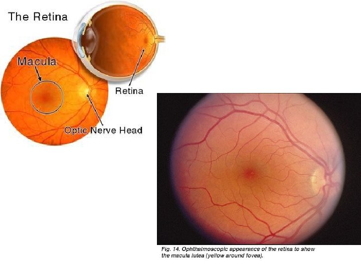

Eye • Lens–clear, flexible structure that focuses light on retina. Action of ciliary muscles in ciliary body changes shape of lens to refocus light • Anterior segment: –Anterior chamber –between cornea and iris –Posterior chamber –between iris and lens –Aqueous humor –liquid in anterior segment, keeps eye lubricated • Posterior segment–from lens to back of eye –Vitreous humor –gel-like fluid filling posterior chamber, supports shape of eye

Posterior segment

Extrinsic Eye Muscles • Superior rectus • Inferior rectus • Medial rectus • Lateral rectus • Superior oblique • Inferior oblique Superior rectus Lateral rectus Medial rectus Inferior oblique Inferior rectus
 Pupil (opening in iris)")
Iris Sclera Cornea (clear covering) Pupil (opening in iris)

Sclera Choroid Optic nerve
 Ciliary body Ciliary process Ora Serrata Cornea Lens Canal of")
Ciliary muscle (Gray dots) Ciliary body Ciliary process Ora Serrata Cornea Lens Canal of Schlemm (black dot) Sclera Choroid Retina
 Central Fovea Blind spot Posterior segment (Vitreous) Anterior")
Macula Lutea (area around central fovea) Central Fovea Blind spot Posterior segment (Vitreous) Anterior segment Sclera Choroid Retina
 –the")
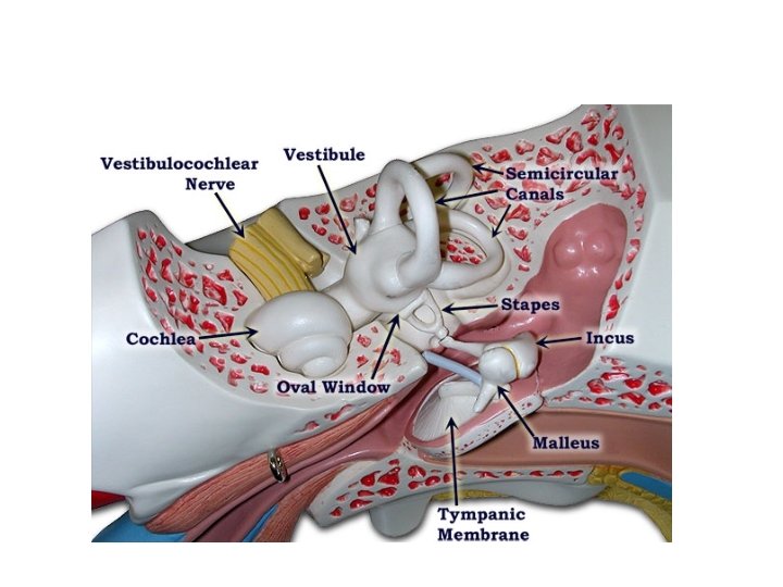
Ear • Pinna/Auricle –outer most part of ear • External auditory meatus (EAM) –the tube • Ceruminous glands –secrete cerumen (ear wax) • Tympanic membrane –separates external ear from middle ear; aka ear drum • Middle ear –chamber between tympanic membrane and inner ear; contains ossicles • Eustachian tube –connects middle ear to nasopharynx
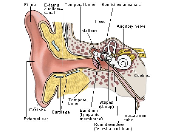

Ear • Ossicles: –Malleus –Incus –Stapes • Oval window –Stapes articulates here to send vibrations into inner ear • Round window –just inferior and medial to oval window

Ear • Inner ear –composed of a membrane-filled bony labyrinth • Vestibular apparatus –part of inner ear associated w/ balance: –Utricle –Saccule –Semicircular canals • Ampula–receptor at the base of each semicircular canal
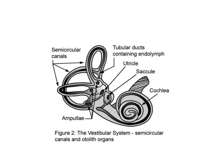

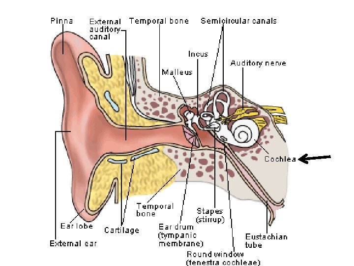
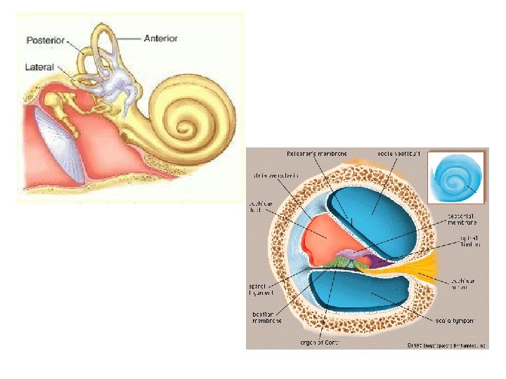
Ear • Cochlea–snail-shaped bony cavity that contains the mechanism of hearing • Cochlear duct –spiral membrane that fills cochlea –Has two levels: 1. Scala vestibuli –upper part of cochlear duct 2. Scala tympani –lower part of cochlear duct • Spiral Organ of Corti –contiguous w/ cochlear duct contains receptors for hearing (CN VIII) • Tectorial membrane –part of SOC
 Auditory ossicles EAM Tympanic membrane Auditory tube Ceruminous gland")
Pinna (Auricle) Auditory ossicles EAM Tympanic membrane Auditory tube Ceruminous gland

Inner Ear Labyrinth

Inner ear Labyrinth Round window

Cross Section of the Cochlea Scala vestibuli Cochlear duct Tectorial membrane Organ of corti Scala tympani

Cross Section of the Cochlea Enlarged Organ of Corti Tectorial membrane Organ of Corti
- Slides: 26