Part Sensory Organs The Sensory Organs Sensory organs


 Fibrous tunic 纤维膜 (outer layer) Cornea 角膜: p anterior 1/6;")
 Fibrous tunic 纤维 膜 Sclera巩膜: p posterior p 5/6, consists")

 Vascular tunic 血管膜 (middle layer): n iris 虹膜 lies")
 ciliary ring")

 Internal tunic of eyeball内膜— retina 视网膜 n division: 以锯状缘ora serrata为界")
 structure: The retina consists of two layers: n pigment")

, located medial to posterior")

 Aqueous humor房水 1) Chamber of eye 眼房: lies")
 Aqueous humor房水 • A clear watery fluid that fills chamber")

 Lens 晶状体 n position: lis behind the iris, anterior to")

 Vitreous body 玻璃体 Consists of colorless, transparent jelly-like substance胶")
 Refractive media: 屈光系统 include Cornea 角膜、 aqueous humor 房水、")


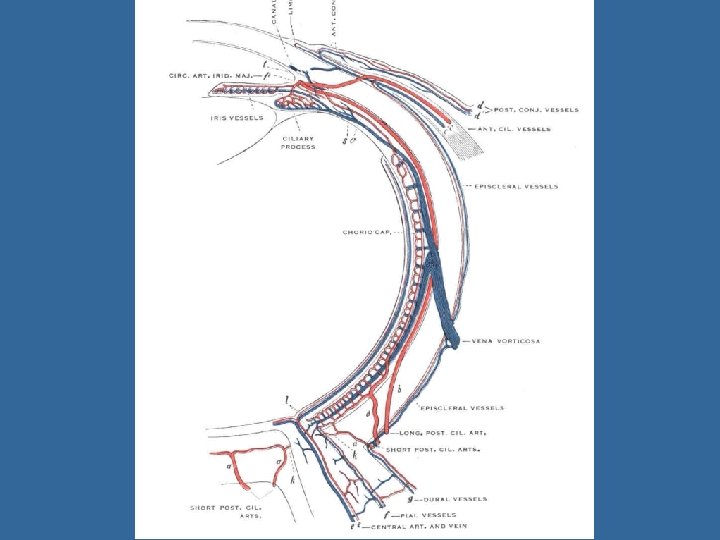
 central a. of retina 视网膜中 央动脉 branches:")
 short posterior ciliary a. 睫状后短动脉: 又称脉络膜动脉Choroidal artery")
 central v. of retina")


- Slides: 66
Part Ⅳ Sensory Organs 感 觉 器
The Sensory Organs Sensory organs include • receptors感受器 • accessory organs辅助装置. The receptors may be divided into three kinds: • exteroceptors 外感受器: 分布在皮肤、鼻腔和口腔粘膜、视器 和听器等处, receive stimuli such as touch, temperature, pain, light and sound from the external environment • interoceptors 内感受器: 分布于内脏和心血管等处pick up information about internal environment • proprioceptors 本体感受器: 分布在肌肉、肌腱、关节、韧带 和内耳平衡器等处receive stimuli from muscles, tendons, joints and ligaments
chapter 1 The Visual Organ 视器 The Visual Organ consist of n eyeball 眼球 n accessory organs of eyeball 眼附属器
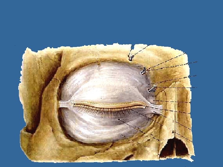
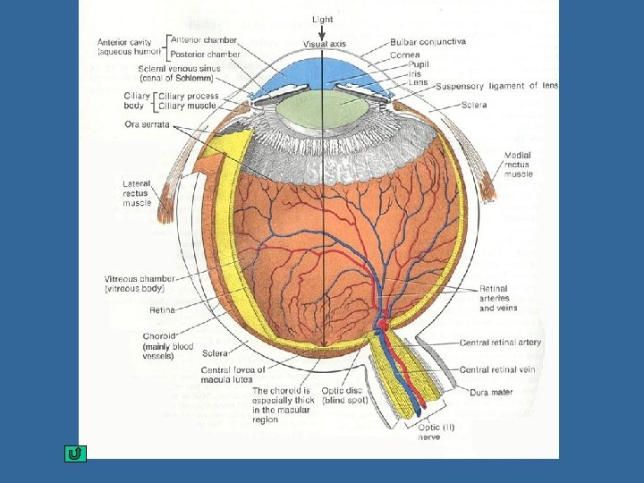
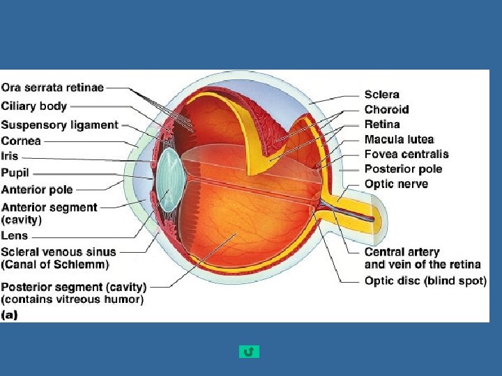
Section 1 Eyeball Ⅰ. Shape of eyeball anterior and posterior poles anterior pole 前、后极 Axis of eyeball 眼轴: a line joining the two poles Optic axis 视轴: a line joining the center of the pupil to the fovea centralis黄斑中央凹 Equator 赤道: an imaginary line encircling the eyeball, midway between anterior and posterior poles posterior pole
Ⅱ. Structure of eyeball 1. Walls of eyeball Fibrous tunic of eyeball Vascular tunic of eyeball Retina 视网膜 Cornea 角膜 Sclera 巩膜 Iris 虹膜 Cilliary body 睫状体 Choroid 脉络膜 Pars iridica retinae Pars caeca retinae盲部 虹膜部 Pars ciliaris retinae 睫状体部 Pars optica retinae 视部
Wall of eyeball (1) Fibrous tunic 纤维膜 (outer layer) Cornea 角膜: p anterior 1/6; p a nonvascular, transparent portion; p Ⅱ supplied richly by nerves; because it is curved, the cornea helps focus light. 感觉神经丰富,故感觉敏锐。有屈光 作用
Walls of eyeball (1) Fibrous tunic 纤维 膜 Sclera巩膜: p posterior p 5/6, consists of fibrous connective tissue having protection and surpporting for eyeball, p posteriorly it contineus with the sheath of optic n.
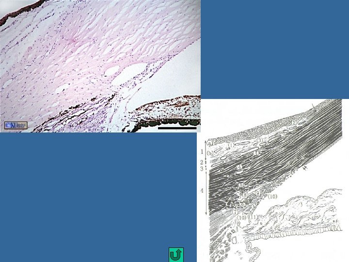
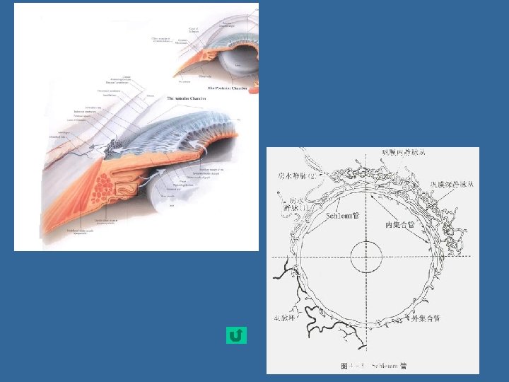
nsinus venous sclerae 巩膜静脉窦: lies beneath the junction of cornea and sclera 角膜与巩膜交界处, and is irregular circular canal. cribriform plate of sclera 巩膜筛板 cornea Sinus venous sclerae sclera
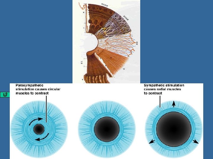
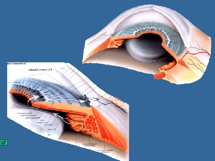
Wall of eyeball (2) Vascular tunic 血管膜 (middle layer): n iris 虹膜 lies the anterior part of the vascular tunic, and is a thin contractile membrane with a central opening, the pupil 瞳孔 iridocorneal angle 虹膜角膜角 sphincter pupillae 瞳孔括约肌 dilator pupillae 瞳孔开大肌
eyeball n Ciliary body 睫状体: Behind the iris,may be divided into a) ciliary ring 睫状环 b) ciliary processes 睫状突: 60~80条, producing aqueous humor房水 ciliary muscle睫状肌 ciliary zonules 睫状小带 d) 作用: 调节晶状体的曲度 secrete the aqueous humor房水 ciliary zonules
eyeball n choroid 脉络膜 p Thin, highly vascular in posterior 2/3 of eye p Contains brown pigmented cells and dense capillary plexus function: Nutrition 营养作用 Absorb the disperse light 吸收分散光作用。
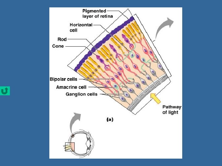
Wall of eyeball (3) Internal tunic of eyeball内膜— retina 视网膜 n division: 以锯状缘ora serrata为界 a. Pars caeca retinae盲部 : pars iridica retinae虹膜部 pars ciliaris retinae睫状体部 无感光细胞,仅有色素上皮层 b. Pars optica retinae视部 : 脉络膜部: 有感光作用
Wall of eyeball 2) structure: The retina consists of two layers: n pigment epithelial layer色素上皮层 n nervus layer神经层 :consist of three layers of cells p Photoreceptor cells:感光细胞 Cone cell 视锥细胞 Rod cell 视杆细胞 Rod cell Cone cell Pigment cell layer
Wall of eyeball p bipolar cell 双极细胞 p ganglion cell 节细胞, whose axons form the optic n. fibers Ganglion cell Bipolar neuron Rod cells Cone cells Pigment cell layer
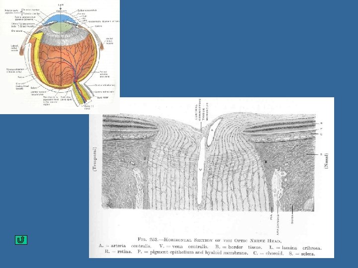
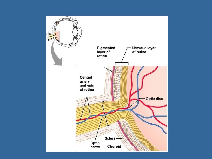
Wall of eyeball n Optic disc 视神经盘 (blind spot盲点), located medial to posterior pole of eye, and consists of optic nerve fibers and at where there are central a. and v. of retina n Macula lutea 黄斑 – Lies lateral about 3. 5 mm to optic disc, a shallow depression, and is yellowish in color
Wall of eyeball – Fovea centralis 中央凹, is an aera of greatest visual acuity and is completely free of blood vessels (concentration of cone cells). n The pigment epithelial layer色素上皮层 absorbs light that enter the eyeball preventing backscatter (blurring of vision)
2. Contents of eyeball 眼球内容物 (1) Aqueous humor房水 1) Chamber of eye 眼房: lies between cornea and lens, and divided by iris into: anterior chamber 眼前房 posterior chamber 眼后房
Contents of eyeball 2) Aqueous humor房水 • A clear watery fluid that fills chamber of eye,secreted by ciliary body. Functions • Helps focus light • Helps maintain constant pressure in eyeball • Helps nourish the lens and cornea
Production and circulation of aqueous humor房水: secreted by the ciliary body pupil posterior chamber anterior chamber iridocorneal angle anterior ciliary vein睫前静脉 sinus venosus sclera ophthalmic vein
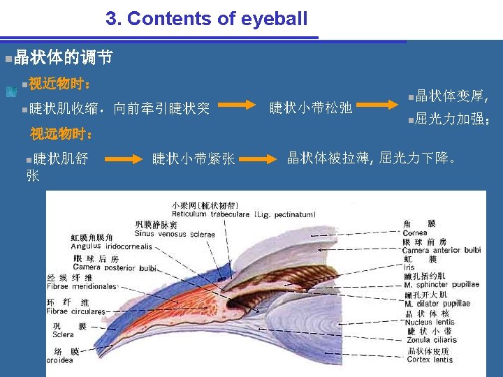
Contents of eyeball (2) Lens 晶状体 n position: lis behind the iris, anterior to the vitreous body 玻璃 体. n shape: Transparent biconvex structure, covered by an elastic transparent capsule which is connected by the ciliary zonules睫状小带 (suspensory lig. 晶状体悬韧带 )to the ciliary process
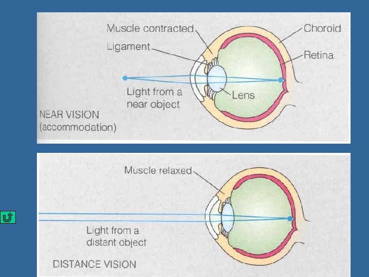
Contents of eyeball n Structure: lens capsule 晶状体囊 cortex of lens 晶状体皮质 lens nucleus 晶状体核 n Its shape is changed by the ciliary muscle: n for near vision, the ciliary muscle contracts and the lens rounds up, n while for distant vision the lens flattens out, so that the eye may be focused on distant objects
Contents of eyeball (3) Vitreous body 玻璃体 Consists of colorless, transparent jelly-like substance胶 状 物 质 in which there is a meshwork of fine fibrils, occupies the space between lens and retina n. Helps maintain the shape of eyeball and supports the retina
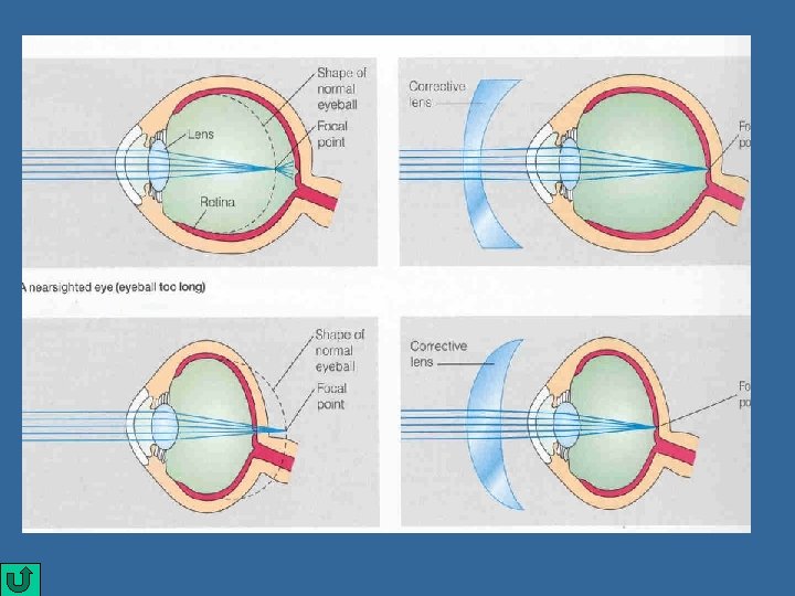
Contents of eyeball (4) Refractive media: 屈光系统 include Cornea 角膜、 aqueous humor 房水、 lens 晶状体 vitreous body 玻璃体 Bend entering light waves and focus them on the retina
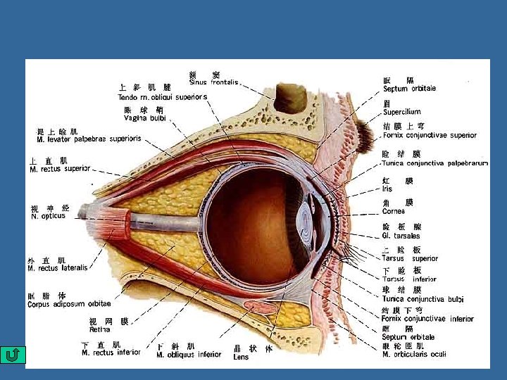
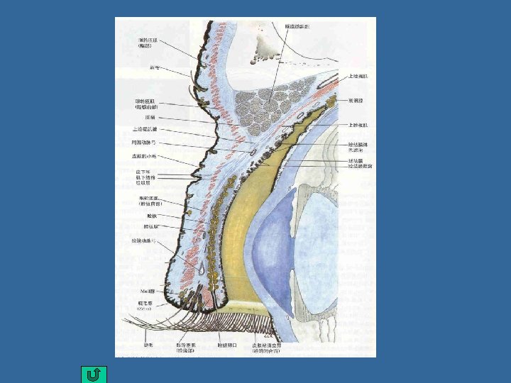
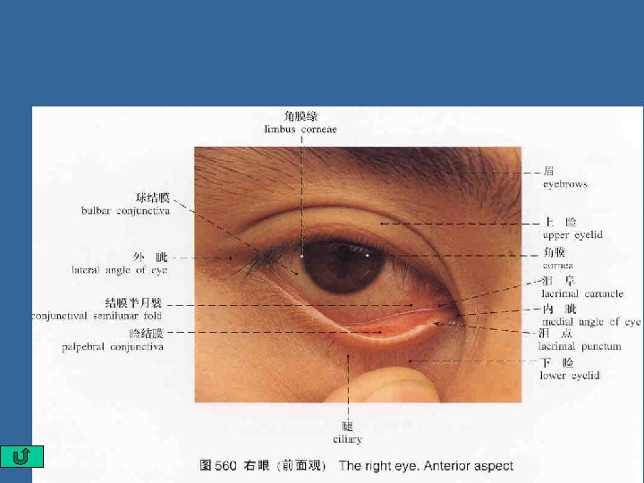
Section 2. Accessory organs Ⅰ. Eyelids眼睑: upper and lower ,consist of 5 layers, ①Skin 皮肤 ②subcutaneous adipose tissue 皮下组织, ③musclar layer 肌层: orbicularis oculi ④tarsus睑板: formed by dense connective tissue (tarsal glands) ⑤ palpebral conjunctiva 睑结膜
Accessory organs of eye Ⅱ. Conjunctiva 结膜: thin mucous membrane n 3 parts: • Palpebral conjunctiva 睑结膜: lining inner surface of eyelids; • Bulbar conjunctiva 球结膜: lining anterior part of sclera; • Conjunctival fornix 结膜穹隆 (superior and inferior): the reflected part of the conjunctiva from the superior and inferior eyelids onto the eyeball. Conjunctival sac 结膜囊
Accessory organs of eye Ⅲ. Lacrimal apparatus 泪器 1. Lacrimal gland 泪腺: 2. Lacrimal passages 泪道: p lacrimal punctum泪点: on each eyelid margin near medial angle p lacrimal ductules 泪小管: in each lid, pass medially, join and enter lacrimal sac p Lacrimal sac 泪囊: in fossa for lacrimal sac, opening into nasolacrimal duct p Nasolacrimal duct 鼻泪管: opening into inferior nasal meatus
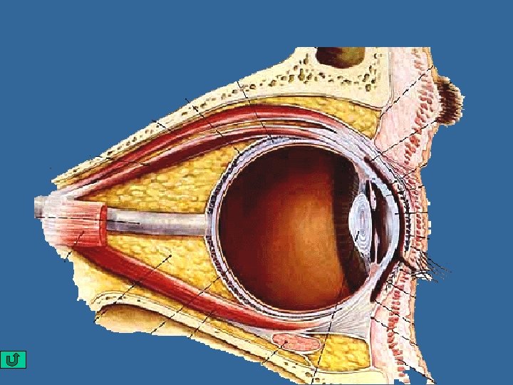
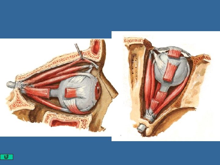
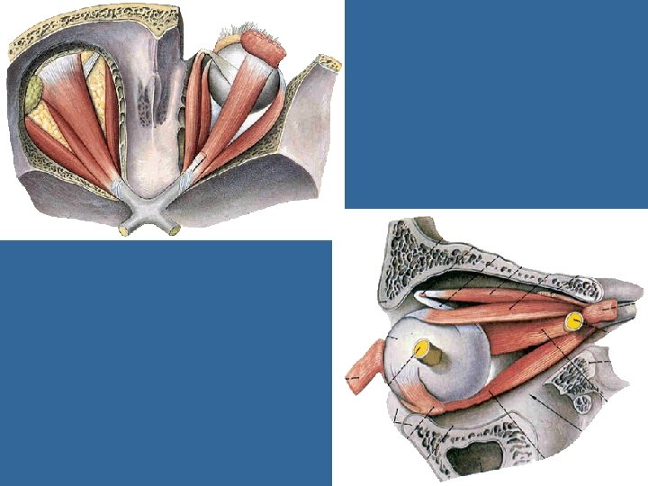
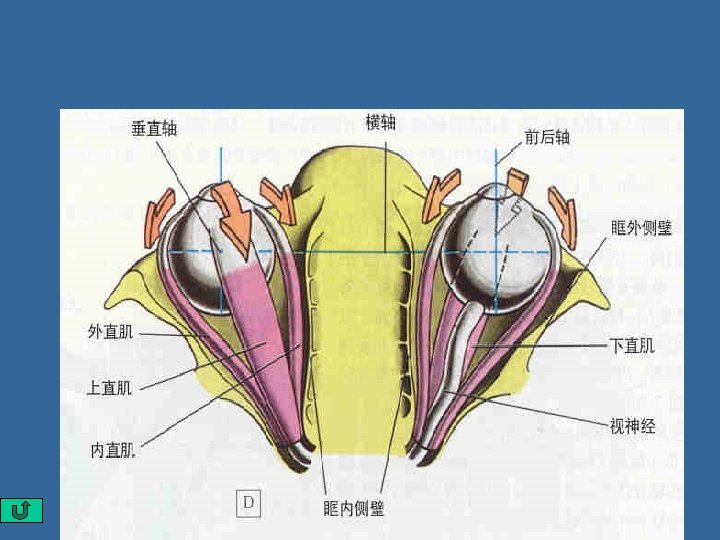
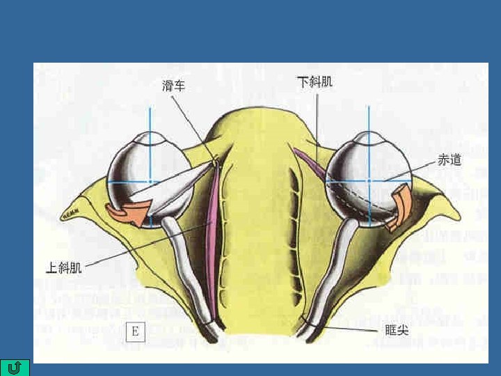
Accessory organs of eye Ⅳ. Extraocular m. 眼外肌: 7 p levator palpebrae superioris上睑提肌: elvates the upper eyelid. p Rectuses直肌: 4 superior rectus 上直肌 inferior rectus 下直肌, medial rectus 内直肌 lateral rectus 外直肌 p Obliquuses 斜肌: 2 Superior obliquus Inferior obliquus
Accessory organs of eye Muscle Action N. supply levator palpebrae superioris elvates upper eyelid Ⅲ Superior rectus turns eyeball superomedially Ⅲ Inferior rectus turns eyeball inferomedially Ⅲ Medial rectus turns the eyeball medially Ⅲ Lateral retus turns the eyeball laterally Ⅵ Superior obliquus turns eyeball inferolaterally Ⅳ Inferior obliquus turns eyeball superolaterally Ⅲ
Accessory organs of eye
Accessory organs of eye Ⅴ. Connective Tissues in the Orbit 眶内结缔组织 1. adipose body of orbit 眶脂体 lies between sheath of eyeball and the orbit acts as a protective cushion and shock sorber for the eyeball 2. orbital fasciae 眶筋膜 a. periorbita 眶骨膜 b. fascial sheath of eyeball 眼球筋膜鞘 c. sheath of ocular muscles 眼肌筋膜鞘 d. orbital septum 眶隔
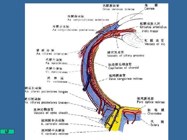
Section 3. The vessels and nerves of eye Ⅰ. Vessels of eye 1. Artery 动脉 (1)Ophthalmic a. 眼动脉: Arises from the internal carotid a. Branches: 1) central a. of retina 视网膜中央 动脉 Enters optic nerve, passes toward the optic disk and then fans out to supply the retina
The vessels and nerves of eye 1) central a. of retina 视网膜中 央动脉 branches: superior nasal arteriole of retina 视网膜鼻侧上小动脉 inferior nasal arteriole of retina 视网膜鼻侧下小动脉 superior temporal arteriole of retina 视网膜颞侧上小动脉 inferior temporal arteriole of retina 视网膜颞侧下小动脉
The vessels and nerves of eye 2) short posterior ciliary a. 睫状后短动脉: 又称脉络膜动脉Choroidal artery 3) long posterior ciliary a . 睫状后长动脉: 又称虹膜动脉; 4) anterior ciliary a. 睫前动脉
The vessels and nerves of eye Ⅱ. Vein 静脉 (1) central v. of retina 视网膜中央静脉 (2) vortex vein 涡静脉 (3)anterior ciliary veins 睫前静脉 (4)眼静脉Ophthalmic v. a)Superior ophthalmic v. b) Inferior ophthalmic v
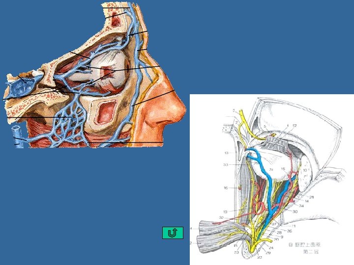
The vessels and nerves of eye Ⅲ. Nerves 神经 视神经 optic nerve: 传导视觉 动眼神经 oculomotor n. 滑车神经 trochlear n. 支配上斜肌 展神经 abducent n. 支配外直肌 眼神经ophthalmic n. 面神经facial n. : 管理泪腺分泌.
superior temporal arteriole of retina superior nasal arteriole of retina inferior temporal arteriole of retina inferior nasal arteriole of retina
Sinus venosus sclerae Ciliary Muscle Iridocorneal angle Dilator Pupillae Sphincter Pupillae Lens ciliary zonule Ciliary Processes
chapter 14 视器The Visual Organ composition 外膜 fibrous tunic : 角膜cornea 巩膜sclera 虹膜iris walls 中膜 vascular tunic: 睫状体ciliary body 脉络膜choroid Eyeball 眼球 内膜retina 脉络膜部choroidal part: pars opticaretinae 视部 视网膜: 睫状体部 pars ciliaris pars caeca 虹膜部 pars iridiac retinae 盲部 房水 aqueous humor 眼内容物 晶状体 lens 玻璃体 vitreous body accessory organs of eyeball 眼附属器: 眼睑、泪器、眼外肌、眶脂体、眶筋膜