Upper Limb Nerves Clinical Anatomy Brachial Plexus Ventral

Upper Limb: Nerves Clinical Anatomy

Brachial Plexus • Ventral rami of C 5‐T 1 • Formed in the posterior triangle of the neck • Organization – Roots • C 5‐T 1 – Trunks • Upper C 5‐C 6 • Middle C 7 • Lower C 8‐T 1 – Divisions • Anterior and posterior divisions – Cords • Lateral – Anterior divisions of middle & upper trunks • Medial – Anterior division of lower trunk • Posterior – All the posterior divisions

Brachial Plexus: Relations • Scalenus anterior and medius • Axillary artery • Axillary sheath

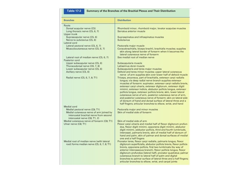
Brachial Plexus: Branches • Roots – – • Upper trunk – – • Lateral pectoral n. Musculocutaneous n. Lateral root of median n. Posterior cord – – • Suprascapular n. Nerve to subclavius Lateral cord – – – • Dorsal scapular n. Long thoracic n. Upper subscapular n. Thoracodorsal n. Lower supscapular n. Axillary n. & radial n. Medial cord – – – Medial pectoral n. Medial cutaneous n. of arm Medial cotaneous n. of forearm Ulnar n. Medial root of median n.
 • Increase the angle between")
Upper lesions of the brachial plexus (Erbo. Duchenne Palsy) • Increase the angle between the head and shoulder • Injury to C 5 & C 6 roots • Affected mm. – Supraspinatus, infraspinatus, subclavius, biceps brachii, coracobrachialis, deltoid, and teres minor • Waiter’s tip position – Limb hang by side – Medially rotated – Pronated forearm • Loss of sensation down lateral side
 – Injury to C 8 &")
Lower lesions of the brachial plexus (Klumpke Palsy) – Injury to C 8 & T 1 – Excessive abduction of the arm – Affected mm. • Small hand muscles – Claw hand • Hyperextension of metacarpophalangeal joints • Flexion of interphalangeal joints – Loss of sensation down medial side

Long thoracic nerve injury • Results from – Injury to posterior triangle of the neck – Injury in the chest wall • Radical mastictomy • Paralysis of serratus anterior m. • Winged scapula – Difficult to raise hand above head – Medial border and inferior angle moves laterally and posteriorly

Musculocutaneous Nerve • Relations – Pierces coracobrachialis m. – Deep to biceps • Branches – Muscular nn. • Innervate the anterior compartment of arm mm. – Lateral cutaneous nerve of the forearm
 • Lateral cutaneous n.")
Musculocutaneous Nerve Block Area: lateral side of the forearm (cutaneous) • Lateral cutaneous n. block – Lateral to the tendon of the biceps m. – On a line between the humeral epicondyles

Median Nerve • Relations – In arm • Brachial a. – In hand • Flexor retinaculum

Median Nerve: branches • Branches in forearm – Muscular branches • Anterior compartment of forearm – Palmar cutaneous branch • Branches in palm – Muscular branches • Thenar muscles – Cutaneous branches

Median nerve injury • Injury at the elbow – Forearm kept in supine position – Wrist flexion is weak and accompanied by adduction – No flexion at interphalangeal joints of 2 nd and 3 rd fingers and weak flexion at the metacarpophalangeal joints of these fingers – Sensory loss at lateral half of the palm

Median nerve injury • Injury at wrist – Paralysis and atrophy of thenar mm. – Loss of opposition movement • Carpal tunnel syndrome – Compression of the median n. by the content of the carpal tunnel – Pain (pins and needles) along the distribution of the median n. to the lateral 3 & ½ fingers

Median Nerve Block Area: lateral side of the palm and palmer 3½ digits and their nail beds • Block at elbow – Brachial a. palpated to the medial side of the biceps tendon on an extended elbow – Medial to the palpated brachial a. • Block at wrist – Between tendons of the palmaris longus and flexor carpi radialis mm. – Proximal to the flexor retinaculum (proximal to distal transverse crease of wrist)

• Relations – In arm • Medial epicondyle – In hand • Flexor retinaculum • Pisiform Ulnar Nerve

Ulnar Nerve: branches • In forearm – Muscular branches – Cutaneous branches • Posterior cutaneous branch • Palmar cutaneous branch • In hand – Superficial terminal branch • Muscular branches (palmaris previs m. ) • Cutaneous branches – Deep terminal branch • Muscular branches

Ulnar nerve injury • Injury at elbow – Common site for ulnar injury is posterior to the medial epicondyle – Flexion of wrist will accompanied by abduction – Inability to adduct and abduct the fingers – Claw deformity – Loss of sensation on the medial ⅓ of the hand • Injury at wrist – Claw hand – Loss of sensation on ⅓ of palmar side

Ulnar Nerve Block • Block at elbow Area: medial side of the hand – Between olecranon process and medial epicondyle of humerus • Block at wrist Area: palmer side of the medial side of the hand – Lateral to the tendon of the flexor carpi ulnaris m. at level of transverse crease of wrist

Radial Nerve • Relations – in arm • Spiral groove • Profunda a. • Lateral epicondyle

Radial Nerve: branches • Muscular branches – Posterior compartments of arm and forearm • Cutaneous branch – Posterior cutaneous n. of arm – Lower lateral cutaneous n. of arm – Posterior cutaneous n. of forearm – Superficial branch of radial n.

Radial nerve injury • Injury in the axilla – Wrist‐drop • Injury in the spiral groove – Wrist‐drop • Injury to the deep branch – No wrist‐drop – Inability to extend the thumb and the metacarpophalangeal joints (test against resistance) – No loss of sensation • Injury to the superficial branch – Limited anasthesia

Radial Nerve Block Area: lateral dorsal side of the hand proximal to the lateral 1½ lateral nail beds • Block at elbow – Halfway between the tendon of the biceps m. and the tip of the lateral epicondyle of humerus in extended elbow • Block at wrist – Lateral to the radial a. at the level of the proximal wrist transverse crease

Axillary Nerve • Relations – Quadrangular space – Posterior circumflex humeral vessels • Branches – Anterior terminal branch – Posterior terminal branch • Upper lateral cutaneous n. of the arm

Axillary Nerve Injury • Results from injury in the quadrangular space – Downward humeral dislocation – Humeral fracture at surgical neck • Deltoid m. paralysis • Deltoid atrophy • Loss of sensation over the lower half of deltoid muscle

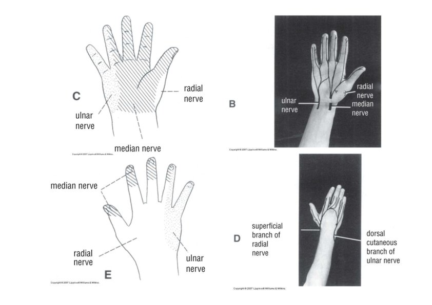
Cutaneous Innervation of the Upper Limb Musculoskeletal Axillary Radial Ulnar Median Medial cord

Dermatomes of the Upper Limb

Dermatomes & Cutaneous Nerves of the Hand

Dermatome Tests for the Upper Limb

Tendon Reflexes & Segmental Innervation of the Upper Limb Muscles • Biceps brachii tendon reflex – C 5 &C 6 – Flexion of the elbow joint – Tapping on the biceps tendon • Triceps tendon reflex – C 6‐C 8 – Extension of the elbow joint – Tapping on the triceps tendon • Brachioradialis tendon reflex – C 5‐C 7 – Supination of the radioulnar joint – Tapping the brachioradialis tendon

The specific neurovascular manifestations of acute cervical disc herniation IV disc level Nerve root level Manifestations Reflexes C 2 C 3 posterior neck numbness and pain radiating to the mastoid and ear reflexes test normal C 3 C 4 posterior neck numbness and pain radiating along the levator scapulae muscle and sometimes to the pectorals reflexes test normal C 4 C 5 lateral neck, shoulder, and arm pain and paresthesia, deltoid weakness and possible atrophy, hypesthesia of C 5 root distribution over middle deltoid area (axillary nerve distribution). reflexes test normal C 5 C 6 pain radiating down the lateral arm and forearm into the thumb and index finger, hypesthesia of the lateral forearm and thumb decreased biceps reflex, biceps and supinator weakness C 6 C 7 pain radiating down the middle forearm to the middle fingers, hypesthesia of the middle fingers decreased triceps and radial reflexes, triceps and grip weakness C 7 C 8 possible pain radiating down the medial forearm and hand, ulnar hypesthesia, intrinsic muscle weakness of the hand. However, these symptoms are uncommon reflexes test normal
 – Supraspinatus")
Myotome Tests for the Upper Limb • Abduction of Arm (C 5) – Supraspinatus –Supraspinatus Nerve – Deltoid‐Axillary Nerve • Arm Adduction (C 7) – Pectoralis Major‐ Pectoral Nerves – Latissimus Dorsi‐ Thoracodorsal Nerve • Forearm Flexion (C 5‐ 6) – Brachialis‐Musculocutaneous – Biceps Brachii‐ Musculocutaneous
 – Triceps Brachii‐Radial")
Myotome Tests for the Upper Limb • Forearm Extension (C 7) – Triceps Brachii‐Radial nerve • Wrist Flexion (C 7, 8 T 1) – Flexor Carpi Radialis‐ Median Nerve – Flexor Carpi Ulnaris‐Ulnar Nerve • Wrist Extension (C 7, 8) – Extensor Carpi Radialis (Longus and Brevis)‐Radial Nerve – Extensor Carpi Ulnaris‐ Posterior Interosseous Nerve
")
Myotome Tests for the Upper Limb • Finger Flexion (C 7, 8 T 1) – Flexor Digitorum Superficialis‐ Median Nerve – Flexor Digitorum Profudus‐Ulnar & Anterior Interosseous branch of Median • Finger Extension (C 7) – Extensor Digitorum‐Posterior Interosseous branch of Radial • Thumb Abduction (C 7, 8 T 1) – Abductor Pollicis Longus‐Radial Nerve – Abductor Pollicis Brevis‐Median Nerve
 – Opponens Pollicis:")
Myotome Tests for the Upper Limb • Thumb Opposition (T 1) – Opponens Pollicis: Median Nerve • Finger Abduction (C 8, T 1) – Dorsal Interossei & Abductor Digiti Minimi‐Ulnar Nerve • Finger Adduction (C 8, T 1) – Palmar interossei‐Ulnar Nerve – http: //www. youtube. com/w atch? v=r. Ki. Twag. LYck
- Slides: 36