Dr Altdorfer The salivary glands Anatomy histology innervation


 anterior: masseter m. anterior: posterior: ramus of")


 → r.")

 serous, compound tubuloalveolar gland")
 and less mucous")
 and less serous")


- Slides: 28
Dr. Altdorfer: The salivary glands – Anatomy, histology, innervation
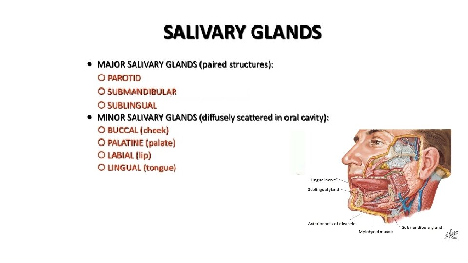
Parotid gland Major salivary glands Parotid duct -Runs paralell with the zygomatic arch - on the external surface of the masseter, Superficial and deep parts - pierces the buccinator, Accessory gland on the parotid duct - opens into the oral vestibule through the parotid papilla, - at the level of the 2 nd upper molar teeth.
Borders of the parotid nest (nidus parotideus) anterior: masseter m. anterior: posterior: ramus of the mandible medial: medial pterygoid m. posterior: sternocleidomastoid m. posterior belly of the digastric medial: stylohyoid m. stylopharyngeus m. styloglossus m.
CROSS SECTION OF THE HEAD PAROTID NEST Structures piercing the parotid gland: - facial nerve (parotid plexus) - auriculotemporal nerve - ext. carotid a. (spf. temp a. ) - retromandibular vein
A B
Parotid gland Blood supply: branches of the maxillary a. - masseteric a. superficial temporal. a Innervation: parasympathetic: IX. sensory: auriculotemporal n. (V/3)
Submandibular gland - main part can be found in the submandibular region, on the external surface of mylohyoid, - smaller part turns around the posterior edge of mylohyoid and lies on its inner surface, - facial artery embedded into the inner surface of the gland, - facial vein runs on its external surface. Blood supply: glandular branches of the facial artery Innervation: parasympathetic (GVM) chorda tympani (VII. ). - hyoglossus mylohyoid
Submandibular gland
Submandibular duct – Wharton’s duct Sublingual caruncula in the lateral sulcus of the tongue first between the lingual and hypoglossal nerves, but than it crosses the lingual nerve and runs anteriorly above it, opens through the sublingual caruncula together with the main duct of the sublingul gland.
Submandibular and sublingual glands Sublingual caruncula Sublingual gland lies on the inner surface of the mylohyoid in the sublingual region, - it causes the sublingual fold, - main duct opens together with the submandibular duct through the sublingual caruncula, - accessory small ducts open along the sublingual fold.
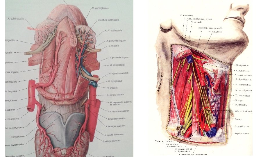
MEDIAL SULCUS OF THE TONGUE AND LATERAL SULCUS OF THE TONGUE Hyoglossus m. Genioglossus m. Mylohyoid m. Lingual a. Hypoglossal n. Submandibular gland
Facial nerve Superior salivatory nucl. GVM nucleus pterygopalatine ggl. greater petr. n. preganglionar parasympathetic fibers V/3 submandibular ggl. . Glossopharyngeal nerve inferior salivatory nucl. GVM postganglionar parasympathetic fibers Otic ganglion lesser petrosal nerve postganglionar parasympathetic fibers V/3 GLANDS chorda tympani V/2
OTIC GANGLION MOTOR ROOT MENINGEAL BR. LINGUAL N. SUBMAND. GGL. CHORDA TYMPANI AURICULOTEMPORAL NERVE INFERIOR ALVEOLAR N.
LATERAL SULCUS OF THE TONGUE, MANDIBULAR NERVE
Minor salivary glands Nuhn-Blandin Glands of the tongue von Ebner Weber mucous serosus mixed
GLANDS OF THE TONGUE von Ebner Weber
Minor salivary glands of palate • Aggregations of mucous acini • No striated duct
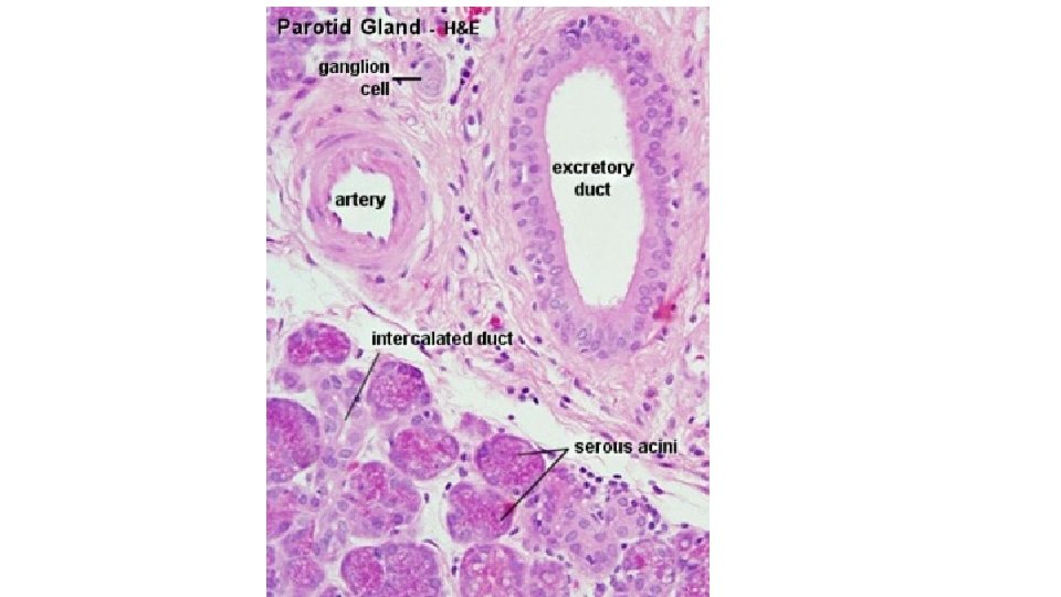
Histology of the salivary glands Merokrine secretion Serous acini Sekretion: protein-rich (amylase) → r. ER, Golgi Basophil cytoplasm (basal), Nuclei: round apical: acidophil (secr. vesicles) Lumen: narrow Mucous acini Sekretion: mucous, viscous! cytoplasm: vacuolizated, foamy nuclei: basal, flattened lumen: elongated
SRTIATED DUCT • Eosinophilic columnar cells • Central nuclei • Basal striation (foldings of the basal plasma membrane)
Parotid gland exclusively (100%) serous, compound tubuloalveolar gland
Submandibular gland mixed seromucous, compound tubuloalveolar gland, with predominating serous (2/3) and less mucous (1/3) acini Ebner / Gianuzzi-demilunes
Sublingual gland Compound tubuloalveolar gland, Mixed muco-serous, with predominating mucous (2/3) and less serous (1/3) acini Ebner / Gianuzzi-demilunes
Pathology TUMOR
Pathology Sialolith+abscess
Literature: • Anatomy, Histology atlases • Dr. Gallatz’s, Dr. Székely’s lecture images