Chapter 15 Cranial Nerves l Chris Rorden University


- Slides: 69

Chapter 15: Cranial Nerves l Chris Rorden University of South Carolina Norman J. Arnold School of Public Health Department of Communication Sciences and Disorders University of South Carolina 1

Functional Classification of CN l Spinal Nerve classification – l General Efferent or Afferent: serve general motor, sensory. Cranial Nerves classification – Receptor type: l l – Signal type l l – General - just like spinal nerves Special –Use special receptors and neurons to serve additional specialized functions Efferent – Motoric Afferent Sensory Voluntary or reflexive? l l Somatic. Innervate somatic muscles (muscles that arise from the soma in the embryological stage – voluntary muscle control) Visceral. Innervate visceral structures. 2

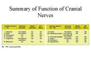
7 Functional Types 1. 2. 3. 4. 5. 6. 7. General Somatic Efferent (GSE) Activates Muscles from Somites (Skeletal, Extraocular, Glossal) General Visceral Efferent (GVE) Activates Visceral Organs Special Visceral Efferent (SVE) Activates Muscles of face, palate, mouth, pharynx and larynx Excludes eye and tongue muscles Special Visceral Afferent (SVA) Mediates visceral sensation of taste from tongue Olfaction from Nose General Visceral Afferent (GVA) Mediates sensory innervation from visceral organs General Somatic Afferent (GSA) Mediates information from muscles, skin, ligament and joints Special Somatic Afferent (SSA) Mediates special sensations of vision from retina and audition and equilibrium from inner ear 3

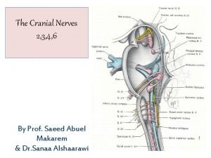
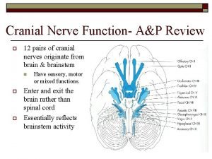
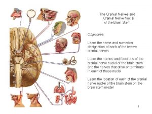
Peripheral Nervous System (PNS) l 12 pairs of cranial nerves. Sensory, motor, or mixed “On Old Olympus Towering Top A Famous Vocal German Viewed Some Hops. ” – “On Old Olympus Towering Top A Finn And German Viewed Some Hops. ”

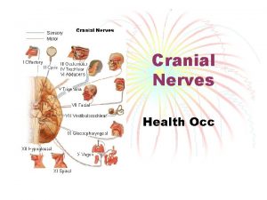
Cranial Nerves (12 pair) I. III. IV. V. VIII. IX. Olfactory: smell Optic: vision Oculomotor: eyelid and eyeball movement Trochlear: motor for vision (turns eye downward and laterally) Trigeminal: chewing, face and mouth touch and pain Abducens: motor to lateral eye muscles Facial: controls most facial expressions , taste, secretion of tears & saliva Vestibulocochlear: sensory for hearing and balance (aka Acoustic, Auditory) Glossopharyngeal: sensory to tongue, pharynx, and soft palate; motor to muscles of the pharynx and stylopharyngeus X. Vagus Nerve: sensory to ear, pharynx, larynx, and viscera; motor to pharynx, larynx, tongue, and smooth muscles of the viscera, 2 parts: superior laryngeal branch and recurrent laryngeal branch XI. Spinal Accessory Nerve: motor to pharynx, larynx, soft palate and neck XII. Hypoglossal Nerve: motor to strap muscles of the neck, intrinsic and extrinsic muscles of the tongue

I: Olfactory l Special Sensory : smell -Injured by shearing (car accident) – unilateral loss of smell rad. usuhs. mil/cranial_nerves/timrad. html 6

II: Optic Special Sensory: Sight l Optic nerve nuclei are located in the lateral geniculate body l Pupil constricts for light to contralateral eye, but not ipsilateral. Unilateral vision loss l 7

III: Oculomotor Somatic Motor: Superior, Medial, Inferior Rectus, Inferior Oblique l Visceral Motor: Sphincter Pupillae l l Pupil asymmetry, no pupil reflex – regardless of which eye observes light. Difficulty with eye movements. 8

IV: Trochlear (Latin for pulley) l Somatic Motor: Superior Oblique l Injury leads to diplopia (due to eye drifting upward), esp when looking down l 9

V: Trigeminal Somatic Sensory: Face l Somatic Motor: Mastication (chewing), Tensor Tympani (reduced ossicle movement), Tensor Palati (soft palate – chewing and eustachion tubes) l l light touch and pain on the forehead (V 1), cheeks (V 2) and chin (V 3). 10

VI: Abducens l Somatic Motor: Lateral Rectus l Damage to the nerve is seen with decreased ability to abduct the eye. (diplopia: affected eye is pulled medially) 11

VII: Facial l l Somatic sensory: Posterior External Ear Canal Special Sensory: Taste (Anterior 2/3 Tongue) Somatic Motor: Muscles Of Facial Expression Visceral Motor: Salivary Glands, Lacrimal Glands Drooping corner of mouth while at rest. Asymmetry of expressions (wrinkle forehead, raise eyebrows, etc) 12

VIII: Vestibulocochlear l Special Sensory: Auditory/Balance l Can patient hear finger rubbing near ear. 13

IX: Glossopharyngeal l l l Somatic Sensory: Posterior 1/3 Tongue, Middle Ear Visceral Sensory: Carotid Body/Sinus Special Sensory: Taste (Posterior 1/3 Tongue) Somatic Motor: Stylopharyngeus Visceral Motor: Parotid Gland Asymmetric palate while saying ‘Aaah’, poor gag reflex (sensory = IX, motor = X) 14

X: Vagus l l l Somatic Sensory: External Ear Visceral Sensory: Aortic Arch/Body Special sensory: Taste Over Epiglottis Somatic Motor: Soft Palate, Pharynx, Larynx (Vocalization and Swallowing) Visceral Motor: Bronchoconstriction, Peristalsis, Bradycardia, Vomitting Asymmetric palate while saying ‘Aaah’, poor gag reflex (sensory = IX, motor = X) 15

XI: Spinal Accessory l Somatic Motor: Trapezius, Sternocleidomastoid l Drooping shoulder. Weakness turning head in one direction, difficult to shrug shoulders against resistance. 16

XII: Hypoglossal l Somatic Motor: Tongue l Observe tongue while on floor of mouth. Twitching can suggest XII injury. 17

Branchial Origin of Speech-Related Muscles l l Speech related muscles = visceral? Six branchial arches present in embryo One disappears during development Some cranial nerves originate from 5 brachial arches and are special visceral efferent nerves Speech related nerves Include – – Trigeminal (V) Facial (VII) Glossopharyngeal (IX) Superior laryngeal and recurrent laryngeal branches of Vagus (X) 18

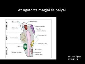
Cranial Nerve Nuclei l Midbrain (3)- Control Eye Muscles – – l Two Motor N. of Oculomotor One Motor N. of Trochlear Pons (6) – Three Sensory N. of Trigeminal l – – – Mesencephalic N. Primary Sensory N. Spinal Trigeminal N. Motor N. of Trigeminal N. Abducens N. Facial Motor N. 19

Cranial Nerve Nuclei: Medulla (9) 1. 2. 3. 4. 5. 6. 7. 8. 9. Cochlear N. (Hearing) Vestibular N. (Equilibrium) Salivary N. (Secretions) Dorsal Motor N. of Vagus (Visceral Motor) Hypoglossal N. (Tongue) Nucleus Solitarius (Visceral Sensory) afferent swallowing Spinal Trigeminal N. (Sensory) Nucleus Ambiguus (Laryngeal & Pharyngeal Motor) efferent swallowing Inferior Olivary N. (Info to Cerebellum) 20

Pathways - Corticobulbar Motor l Corticobulbar tract – l Cross midline at different levels – l Fibers between cortex and brain stem Upper and Lower Motor Neurons Clinical Signs: – Lower Motor Neuron l Paralysis – Upper Motor Neuron l Spasticity l Increased Tendon Reflexes l Contralateral Paresis l Absent Reflexes l Flaccid Muscle Tone l Fibrillation l Fasciculations (twitching) l Atrophy 21

Pathways - Sensory l 3 Major types of sensory pathways – – – l 1 st order - Outside brainstem 2 nd order Cell bodies in gray matter of brainstem 3 rd order - Cell bodies in ventral posterior medial N. of Thalamus projecting to cortex in parietal lobe Smell, hearing and vision are exceptions to rule three 22

Olfactory Nerve (I) Special visceral afferent l Parts l – – – Olfactory Bulb Olfactory Tract Temporal Cortex 23

Olfactory Nerve (I) l l l Fibers pass through the foramina in the cribriform plate to olfactory bulb, olfactory tract to temporal cortex (uncus, amygdaloid N. and parahippocampal gyrus). Connects to limbic system and emotional brain. Olfactory ability decreases with age Anosmia: impaired smell (ask patient to identify odors) 24

Optic Nerve (II) l l l Special somatic afferent Retina to Optic Nerve to Optic Chiasm To Lateral Geniculate Body To Optic Radiations To Visual Cortex in Occipital Lobe Clinically: – – Injury results in visual field loss Common visual field losses in Chapter 8 (ask client to closes one eye and fix gaze straight ahead. Determine when patient can see objects in parts of visual field) 25

Oculomotor Nerve (III) l General somatic efferent – l Innervate extrinsic muscles of eye General visceral efferent – – – Provides parasympathetic projections to constrictor fibers of iris and ciliary muscles Provides motor innervation for iris to adjust to light and lens to focus Edinger-Westphal Nucleus 26

Oculomotor Nerve (III) Ciliary Ganglion Oculomotor Nerve Superior Colliculus Edinger. Westphal Nucleus (Pupil size, lens shape) 27

Left Oculomotor (III) Nerve Paralysis Diplopia Left eye is deviated Does not move laterally 28

Diplopia 29

Clinical Info: Oculomotor Nerve (III) l Ptosis - eyelid droop l Ophthalmoplegia l – – – problems in adjusting to light deviation of eye movements diplopia (double vision) 30

Trochlear IV l l l General somatic efferent Only CN to exit brainstem dorsally Only CN that exits contralaterally Anterior oblique muscle for eye movement is only function Clinical – – Difficulty looking downward and outward when Trochlear is injured eye drifts upward relative to the normal eye 31

Trochlear Nucleus Superior Oblique Muscle Trochlear (IV) Nerve 32

Superior Oblique Muscle Function Right Superior Oblique Muscle Eye ball directed down and out 33

Trigeminal (V) l l l General somatic afferent Principal sensory nerve for head, face, orbit and oral cavity mediate sensations of pain, temperature, proprioception and fine discriminative touch Sensations from anterior 2/3 of tongue Three sensory branches – – – Ophthalmic Maxillary Mandibular 34

Trigeminal (V) 35

Trigeminal (V) l l Special visceral efferent Motor for mastication muscles for chewing and speaking – – – – l Internal and external pterygoid Temporalis Masseter Mylohyoid Anterior belly of digastric Tensor veli palatini Tensor tympani Reflex for jaw jerk reflex (mandibular) 36

Trigeminal (V) Opthalmic Maxillary Mandibular 37

Motor Branch of Trigeminal Nerve Temporalis muscle Mylohyoid Anterior belly Of digastric Tensor palatine Pterygoid muscles Lateral (external) Medial (internal) Tensor tympani Masseter muscle 38

Clinical Info: Trigeminal (V) l Sensory – – – l Test for touch discrimination in different facial zones Check for sneeze and corneal reflexes Tic of douloureux (trigeminal neuralgia) which is excruciating pain Motor – – Check for paralysis or paresis of ipsilateral muscles of mastication Check for absent or exaggerated jaw reflex Look for deviation of jaw toward side of injury Unilateral lesion has mild effect on bite strength while bilateral has severe effect 39

Abducens (VI) l l l General somatic efferent Innervates only a single muscle: lateral rectus muscle which moves eye laterally Clinical Info: – – – Left Abducens (VI) Nerve Paralysis Left eye is deviated medially When injured, medial rectus muscle is unopposed – eye shifts medially Susceptible to disruption Check for medial strabismus l l Turns in medially Double vision 40

Left Abducens (VI) Nerve Paralysis l Diplopia Disappears on Eye Movement to the Right 41

Abducens (VI) Nucleus Abducens (VI) Nerve Lateral Rectus Muscle 42

Facial Nerve (VII) l General visceral efferent – – l Parasympathetic innervation of lacrimal gland palatal saliva Innervation of mucous membrane secretions in mouth and pharynx Special visceral afferent – Gustatory sensations from anterior 2/3 of tongue 43

Facial Nerve (VII) l l l Special visceral efferent Primary motor nerve for facial muscles Extrinsic Muscles of ear – l Stapedius Muscle – l Contraction attenuates sound Swallowing – – l Cats can rotate outer ear Stylohyoid Muscle Posterior Belly of Digastric Muscle Lacrimal secretion - Tears 44

Clinical Info: Facial Nerve (VII) l Upper Motor Neuron Disease – – l Why is it hard to only raise one eyebrow? Unilateral paresis of muscles of lower half of face Muscles above bilaterally innervated Bilateral lesion cause paralysis of upper and lower muscles bilaterally Lower Motor Neuron Disease – – Injury near pons can cause lower motor neuron disease Unilateral Paralysis of all facial muscles, stapedial muscle and taste in 2/3 of tongue 45

Clinical Examples: Facial Nerve UMN LMN 46

Clinical Examples: Facial Nerve 47

Clinical Info: Facial Nerve (VII) l Bell’s Palsy – – LMN syndrome with sudden onset of paralysis of ipsilateral facial muscles Inflammatory injury, infection or degenerative disease 48

Vestibulo-acoustic Nerve (VIII) Special somatic afferent l Vestibular Nerve l – l Acoustic Nerve – l Gives feedback about position of head in space and balance Hearing Clinical Info – Tests for equilibrium, vertigo or dizziness, nystagmus and hearing loss 49

Glosso-pharyngeal Nerve (IX) l General visceral afferent – l General visceral efferent – l Secretion from parotid gland (salivary gland) Special visceral afferent – l Mediates general visceral sensation from soft palate, palatal arch, posterior 1/3 of tongue and carotid sinus Taste sensation form posterior 1/3 of tongue Special visceral efferent – Contributes to swallowing through stylopharyngeus and upper pharyngeal constrictor fibers 50

Clinical Info: Glosso-pharyngeal (IX) May be evident in dysphagia or loss of taste to posterior 1/3 of tongue l Loss of gag reflex l Excessive oral secretions l Dry mouth l Need bilateral damage of nerve to have strong clinical signs l 51

Vagus Nerve (X) l General visceral afferent – – l General visceral efferent – l Innervates glands, cardiac muscles, trachea, bronchi, esophagus, stomach and intestine Special visceral afferent – l Sensation from pharynx, larynx, thorax, abdomen Regulates nausea, oxygen intake, lung inflation Mediates taste sensation from posterior pharynx and epiglottis Special visceral efferent – Controls muscles of larynx, pharynx, soft palate for phonation, swallowing and resonance 52

Clinical Info: Vagus Nerve (X) l l l Bilateral lesion of the brainstem can be fatal due to respiratory involvement Unilateral lesion can result in ipsilateral paresis or paralysis of soft palate, pharynx and larynx Pharyngeal Branch – – l Pharynx and soft palate involvement Uvula pulled to unaffected side, bilateral soft palate droops Recurrent Laryngeal Branch – – Unilateral: Paralysis of vocal folds Bilateral: Inspiratory stridor and aphonia 53

Clinical Info: Vagus Nerve (X) Normal Soft Palate Unilateral Paralysis Bilateral Paralysis 54

Clinical Info: Vagus Nerve (X) Autonomic reflexes reduced l Anesthesia of pharynx and loss of taste l Superior Laryngeal Branch l – Loss of ability to change pitch 55

Spinal Accessory Nerve (XI) l General visceral efferent – l Controls head position by controlling trapezius and sternocleidomastoid muscles Clinical Information – – Affects ability to control head movements Ask patient to rotate head and note control 56

Hypoglossal Nerve (XII) l General somatic efferent – – – Controls tongue movement Controls extrinsic and intrinsic muscles of tongue except palatoglossal (X) Eating, sucking and chewing reflexes 57

Clinical Info: Hypoglossal (XII) LMN unilateral lesion cause wrinkling and flaccidity of tone with atrophy over time l Dysarthria and Dysphagia l Unilateral UMN lesions do not have much affect as tongue is bilaterally innervated l Ask patient to complete oral motor movements l 58

Clinical Info: Hypoglossal (XII) Unilateral Tongue Paralysis Bilateral Tongue Paralysis 59

Innervation of the tongue General Special (tactile, etc. ) (taste) Glossopharyngeal (IX) Nerve Trigeminal (V) Nerve Glossopharyngeal (IX) Nerve Facial (VII) Nerve 60

Cranial Nerve Combinations More than one nerve involved with some structures l Eyes muscle control l Sensory fibers to tongue l – – Anterior 2/3 special and general sensation: Facial and Trigeminal, Posterior 1/3 special and general sensation: Glossopharyngeal 61

Cranial Nerve Combinations l Motor Nerve Supply to Soft Palate and Pharynx – l Vagus, Trigeminal and Glossopharyngeal Sensory Nerve Supply to Soft Palate and Pharynx – Glossopharyngeal, Vagus and Trigeminal 62

Nerve Classifications This division give rise to a classification based on whether a nerve is: l Afferent, efferent, or both l Somatic or visceral, or both l Special, general, or both l The only combination that does not exist is: Special, somatic, efferent. l 63

Case # 1 l l 1. 2. 3. 4. 5. Setting: Neonatal intensive care unit (NICU) Patient: Pt. is a two-day old male. Delivery was complex but completed with cesarean section, neurological exam suggests a right facial paralysis /s other prominent symptoms. What cranial nerve(s) is/are involved? Discuss the probable cause of the right facial paralysis In what cases will the symptoms resolve? What are some possible current functional problems that may be present? What are some possible future functional 64

Case # 2 l l 1. 2. 3. 4. 5. 6. Setting: Out-patient clinic Patient: 64 y. o. male. Pt. is 18 months post-stroke. Neurological exam revealed: aphasia, dilated left pupil, left eye deviated downwards and lateral. Left eyelid droop. What cranial nerve is involved? What kind of a visual problem would this patient have? What can the patient do to compensate for the visual problem? Will this condition persist? In the long run, how will the brain compensate for this problem? Is it probable that the same lesion resulted in the visual problem and the aphasia? 65

Case #3 l l 1. 2. 3. 4. 5. Setting: Nursing home Patient: Pt. is a 78 y. o. female who has been residing at the nursing home for the last 3 years. She was originally admitted to the nursing home following amputation of both legs below the knee. This was necessary secondary to diabetes that results in gradual neuropathy and loss of vascular circulation in the extremities. A recent visit by the primary care physician revealed loss of sensation in the face secondary to progressive neuropathy. Her jaw is slightly deviated to the left. What cranial nerve is involved? How can you determine which afferent part of this cranial nerve is affected? What would cause the jaw to deviate to one side? Is this an upper or lower motor neuron problem? Will she improve? Why/why not? 66

Case #4 l l 1. 2. 3. 4. Setting: ICU Patient: 42 y. o. female. Patient was brought to the ER following a motor vehicle accident. She was comatose for 4 days but is now alert but not oriented. Pt. has multiple fractures including the: left tibia, left humerus and clavicle. Extensive facial bruising. MRI showed scattered bruising of the cortex and possible brain stem involvement. The neuro exam revealed severe aphonia, stridor, absent swallow reflex, drooping soft palate, no gag reflex. What cranial nerve is most likely affected? Is this an upper or lower motor problem? What are some other neurological symptoms that could be present? Would you recommend an oral diet for this patient? Why/why not? 67

Case #5 l l 1. 2. 3. Setting: Nursing home (SNF) Patient: Pt. is a 71 y. o. male who recently suffered a stroke. The MRI revealed multiple infarctions at the level of the basal ganglia and perhaps the brain stem. The neuro report from the hospital suggested that the patient has right lower facial droop, poor movement of most facial muscles, exaggerated smile, and excessive laughter or crying. Does this clinical picture agree with cranial nerve involvement? Why/why not? Is this an upper or lower motor neuron problem? Poor movement of most facial muscles would implicate what cranial nerve? 68

VIII Injury: www. dizziness-and-hearing. com/testing/acoustic_reflexes. htm l Central case example: A 40 year old man was well until he was involved in an auto accident. Two days later he developed diplopia and a rotatory type vertigo. On physical examination he had clear spontaneous nystagmus and mildly decreased hearing on the left side. Audiometry documented mildly impaired hearing on the left, but acoustic reflexes were abnormal with very rapid decay on the left side. Brainstem auditory evoked responses were abnormal on the left (neural response times to sounds). An MRI scan documented a lesion resembling an MS placque in his left cerebellar peduncle area, just behind the 8 th nerve (see figure to right). His symptoms resolved spontaneously and he has had not further neurological complaints in 5 years of followup. 69
 Chris rorden
 Chris rorden
 Chris rorden
 Optic chiasm
 Spinal nerves labeled
 Cranial nerves mixed
 Optic nerve names
 Cranial nerves labeled diagram
 Cranial nerves
 Abducens
 Oculomotor
 Cranial nerves classification
 Smile cranial nerve
 Cranial nerves labeled with roman numerals
 Figure 14-2 cranial nerves labeled
 Cranial nerves labeled with roman numerals
 Cranial nerves 346
 First and second cranial nerves
 Agyidegek
 Old opie occasionally tries
 On old olympus towering
 Vestibulocochlear nerve pathway
 Vertical
 Tractus corticomesencephalicus
 Carnial nerve
 Ipsilateral cranial nerves
 Edinger westphal nucleus
 Cranial nerves labeled
 Cranial nerves
 Cranial nerve
 Cn iiiv - vestibulocochlear nerve
 Cranial cavity anatomy
 Cranial nerve mnemonic
 Cranial nerves
 Cranial nerves sensory and motor
 Cranial nerves motor and sensory
 Motor efferent division
 Testing cranial nerves
 Cranial nerves
 Crematic reflex
 Sheep brain labeled
 Lateral venous lacunae
 Meatus of nose
 Cranial fermenters
 Theories of craniofacial growth
 Cranial nerve 9
 Cn 8 test
 Median and lateral apertures
 Ethmiod
 Vertical
 Grey commissure
 Multiple cranial nerve palsy adalah
 Caudal ventral
 Nükleos
 Pterygoid plexus
 Bone remodeling facial exercise
 Pyrimidal system
 Trigeminal nerve which cranial nerve
 Abducens nerve palsy
 What is meningitis
 Nerve supply of face
 érző homunculus
 Cranial caudal folding
 Vestibulocochlear nerve nuclei
 2012 pearson education inc anatomy and physiology
 What does the suffix malacia mean
 Oblongata
 Dysmetria
 Angular artery
 Cranial fossa
