Brain Cranial Nerves Dr Michael P Gillespie Major

Brain & Cranial Nerves Dr. Michael P. Gillespie

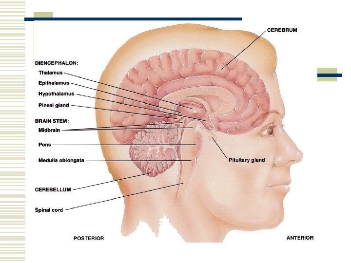
Major Parts of the Brain w Brain stem – continuous with the spinal cord. n n n Medulla oblongata. Pons. Midbrain. w Cerebellum – posterior to the brain stem.

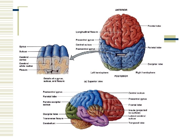
Major Parts of the Brain w Diencephalon – superior to the brain stem. n n Thalamus. Hypothalamus. w Cerebrum – supported on the diencephalon and brain stem. n Largest part of the brain.

Brain Blood Supply w Arteries n n Internal carotid arteries Vertebral arteries w Veins n Internal jugular veins

Brain Blood Flow w The brain consumes about 20% of the oxygen and glucose used at rest. w A brief slowing of blood flow may cause unconsciousness.

Brain Blood Flow w An interruption of blood flow for 1 to 2 minutes impairs neural function. w Total deprivation of oxygen for 4 minutes causes permanent injury. w If the blood entering the brain has a low level of glucose, mental confusion, dizziness, convulsions, and loss of consciousness may occur.
 protects the brain from harmful substances")
Blood Brain Barrier w The blood-brain barrier (BBB) protects the brain from harmful substances and pathogens. w It prevents the passage of many substances from the blood to the brain tissue. w Tight junctions seal together endothelial cells of brain capillaries. w Astrocytes selectively allow some substances through and not others.

Breaching the BBB w The BBB prevents the passage of harmful substances into the brain, but it also prevents the passage of useful drugs. w Drugs are injected in a concentrated sugar solution to facilitate passage. n The high osmotic pressure causes cells lining the barrier to shrink and makes the membrane “leaky”.

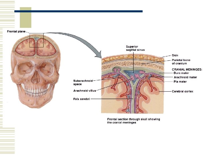
Protective Coverings w Cranial Meninges. n n n Dura mater. Arachnoid mater. Pia mater.
 w Clear colorless liquid. w Protects the brain and spinal cord")
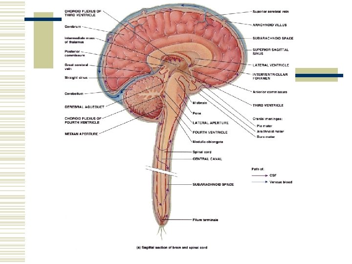
Cerebrospinal Fluid (CSF) w Clear colorless liquid. w Protects the brain and spinal cord from chemical and physical injuries. w Carries oxygen, glucose, and other needed chemicals from the blood to the neurons and neuroglia. w Circulates in the subarachnoid space (between the arachnoid mater and pia mater).

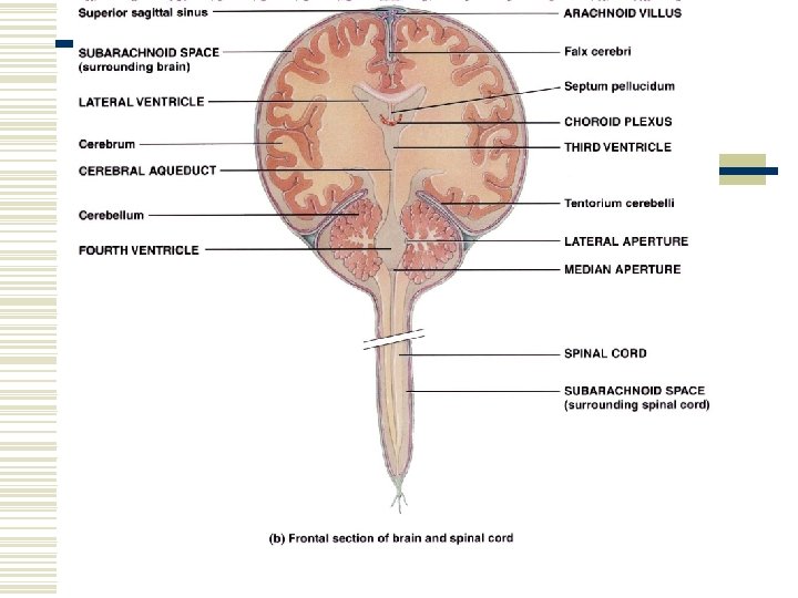
Protective Coverings w Extensions of the dura mater separate the parts of the brain. n n n Falx cerebri – separates the two hemispheres of the cerebrum. Falx cerebelli – separates the two hemispheres of the cerebellum. Tentorium cerebelli – separates the cerebrum from the cerebellum.

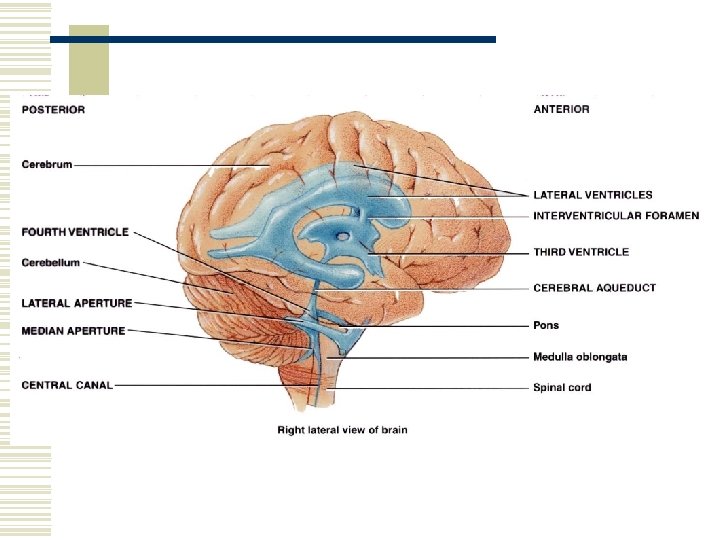
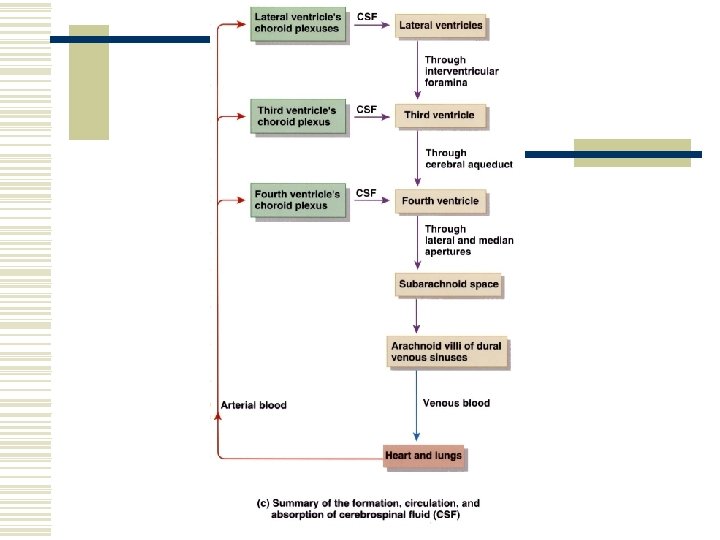
Formation of CSF in the Ventricles w CSF is formed in the ventricles. n Formed by ependymal cells that cover the choroid plexuses of the ventricles.

Formation of CSF in the Ventricles w There are 4 ventricles. w Functions of CSF. n Mechanical protection. l l n n Shock absorption. Buoys the brain. Chemical protection – optimal chemical environment. Circulation – medium of exchange for wastes and nutrients.

Hydrocephalus w Abnormalities of the brain can interfere with drainage of CSF from the ventricles and subarachnoid space. w CSF pressure increases causing hydrocephalus. w In infants this causes the fontanels to budge.

Hydrocephalus w Tumors, inflammation, developmental malformations can all cause hydrocephalus. w Pressure buildup can damage the delicate nervous tissue. w A surgeon can implant a drain line called a shunt to divert CSF. w In adults, hydrocephalus may occur after head injury, meningitis, or subarachnoid hemorrhage.

Brain Stem w Between the brain and spinal cord. w 3 regions. n n n Medulla oblongata. Pons. Midbrain.
 tracts and")
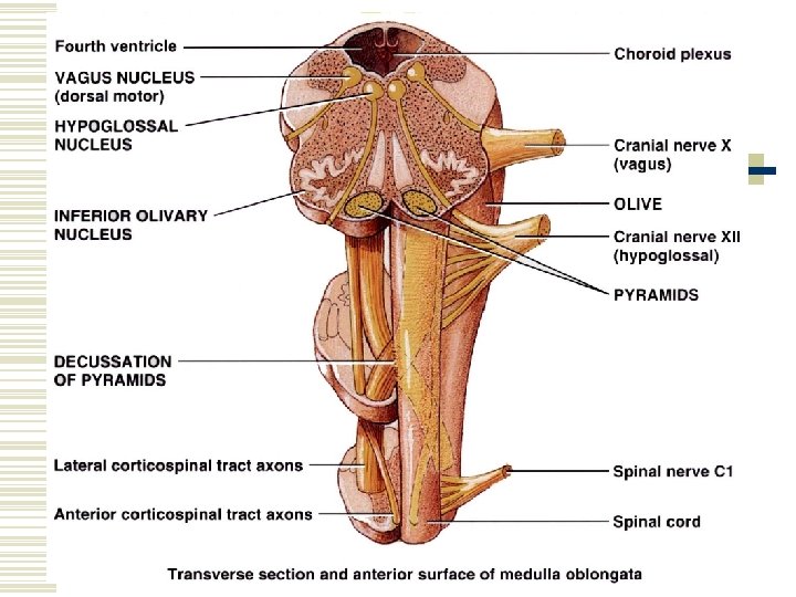
Medulla Oblongata w A continuation of the spinal cord. w Sensory (ascending) tracts and motor (descending) tracts travel through the white matter of the medulla. w Many nerves decussate (cross over) in the medulla.

Medulla Oblongata w Cardiovascular center regulates the heartbeat and the diameter of the blood vessels.

Medulla Oblongata w The medullary rhythmicity area adjusts the rhythm of the breathing and controls reflexes for vomiting, coughing, and sneezing.

Medulla Oblongata w The nuclei for the following cranial nerves reside in the medulla: n n n VIII (vestibulocochlear). IX (glossopharyngeal). X (vagus). XI (accessory). XII (hypoglossal).

Pons w Pneumotaxic area and apneustic area regulate breathing. w Nuclei for cranial nerves V (trigeminal), VI (abducens), VII (facial), and VIII (vestibulocochlear).
 and inferior")
Midbrain w The midbrain or mesencephalon contains the superior colliculi (visual actvities) and inferior colliculi (auditory pathways). w The midbrain contains the substantia nigra which release dopamine to help control subconscious muscle activities. Loss of these neurons results in Parkinson disease. w Cranial nerves III (oculomotor) and IV (trochlear) originate here.

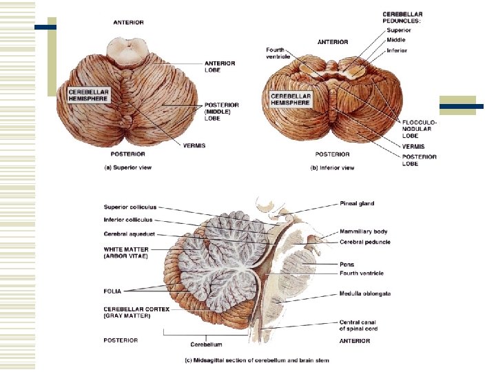
Cerebellum w The second largest part of the brain. w A main function of the cerebellum is to evaluate how well movements are being carried out and correct for discrepancies. This helps to “smooth out” movements.

Diencephelon w Epithalamus. n Contains the pineal gland which secretes melatonin. w Thalamus. n n Relays sensory information to the cortex. Provides crude perception of touch, pressure, pain, and temperature.
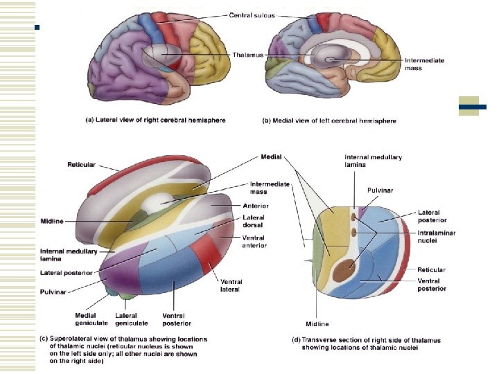

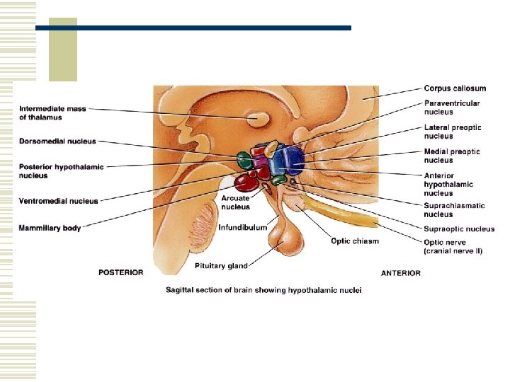
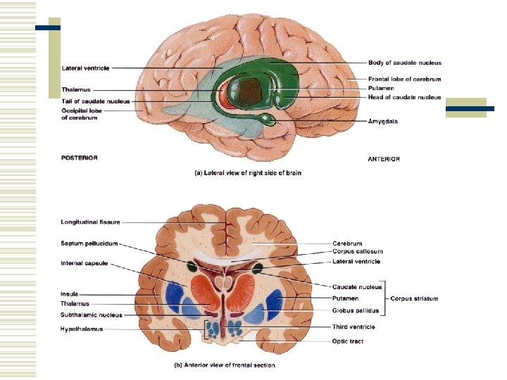
Diencephelon w Subthalamus. n Controls body movements. w Hypothalamus. n n n Controls and integrates activities of the ANS. Regulates emotional and behavioral patterns. Regulates cicadian rhythms. Regulates eating and drinking behavior. Produces hormones oxytocin and ADH.

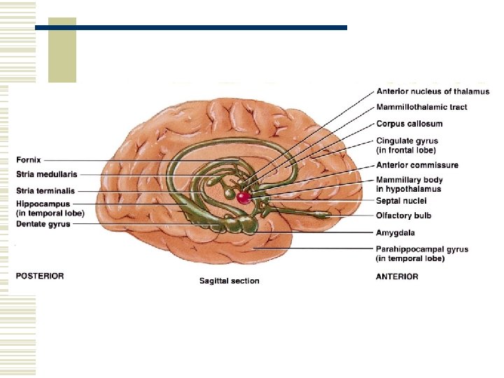
Cerebrum w Sensory areas interpret sensory impulses. w Motor areas control muscular movement. w Association areas function in emotional and intellectual processes. w Basal areas regulate gross muscle movements and regulate muscle tone. w Limbic system functions in survival behaviors.

Brain Injuries w Concussion – an abrupt, temporary loss of consciousness following a blow to the head. n n Most common brain injury. Signs – headache, drowsiness, lack of concentration, confusion, amnesia.

Brain Injuries w Contusion – bruising of the brain due to trauma and includes leakage of blood. n Signs - immediate loss of consciousness, transient cessation of respiration, decreased blood pressure.

Brain Injuries w Laceration – tear of the brain usually from a skull fracture or gunshot wound. n n Rupture of large blood vessels. Consequences – cerebral hematoma (localized pool of blood, usually clotted), edema, and increased intracranial pressure.

Cerebral Cortex Areas and Functions w Sensory areas – receive and interpret sensory information.

Cerebral Cortex Areas and Functions w Motor areas – initiate movements. w Association areas – deal with integrative functions: n n n n Memory. Emotions. Reasoning. Will. Judgement. Personality. Intelligence.

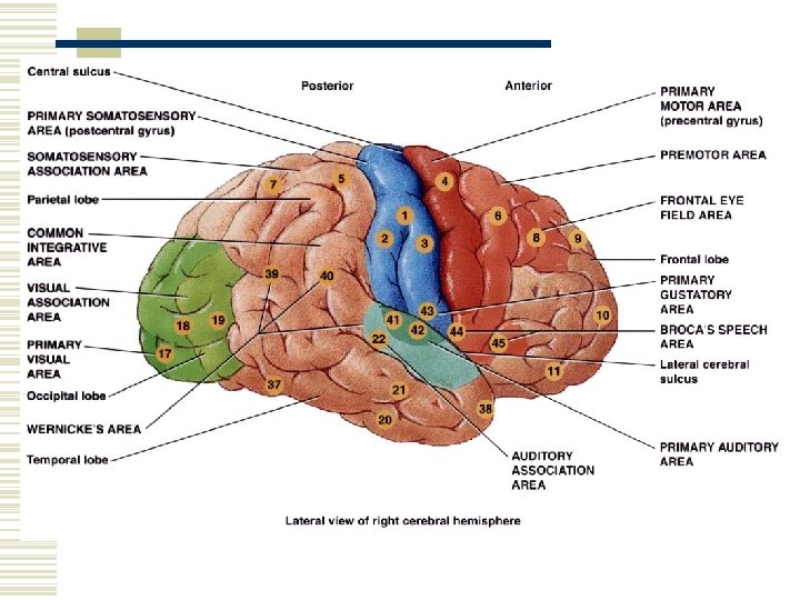
Sensory Areas w Primary somatosensory area – receives sensations for touch, proprioception, pain, itching, tickle, and thermal sensations. n w w Located in the postcentral gyrus of the parietal lobes. Primary visual area. Primary auditory area. Primary gustatory area – taste. Primary olfactory area.

Motor Areas w Primary motor area – located in the precentral gyrus of the frontal lobe. w Broca’s speech area – coordinates the contractions of speech and breathing muscles.

Association Areas w Somatosensory association area – integrates and interprets sensations. w Visual association area – evaluates what is seen. w Auditory association area – evaluates sounds.
 area – interprets the meaning of speech. w")
Association Areas w Wernicke’s (posterior language) area – interprets the meaning of speech. w Common integrative area. w Premotor area – controls learned skilled movements. w Frontal eye field area – controls voluntary scanning movements of the eyes.

Aphasia w An inability to use or comprehend words.

Aphasia w Damage to Broca’s area results in nonfluent aphasia. n n Inability to properly articulate to form words. These people know what they wish to say, but cannot speak.

Aphasia w Damage to the auditory association area results in fluent aphasia. n Faulty understanding of spoken words. Word deafness – inability to understand spoken words. l Word blindness – inability to understand written words. l

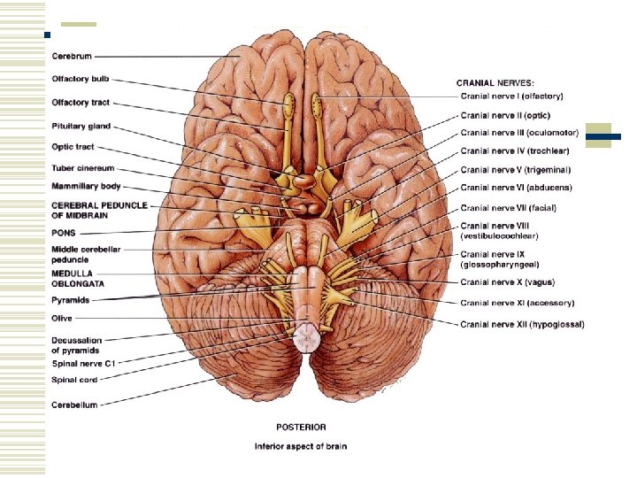
Cranial Nerve I - Olfactory w Type: sensory. w Function: smell. w Anosmia – loss of sense of smell.

Cranial Nerve II – Optic Nerve w Type: sensory. w Function: vision. w Anopia – blindness in one or both eyes.
. w Function: movement of")
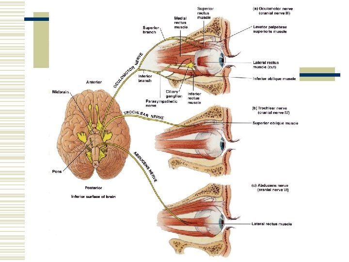
Cranial Nerve III - Oculomotor w Type: mixed (mainly motor). w Function: movement of the upper eyelid and eyeball. Accomodation of the lens for nearn vision and constriction of the pupil. w Strabismus – deviation of the eye in which both eyes don’t focus on the same object. w Ptosis – drooping of the upper eyelid. w Diploia – double vision.
. w Function: movement")
Cranial Nerve IV – Trochlear Nerve w Type: mixed (mainly motor). w Function: movement of the eyeball. w Diplopia and strabismus occur with trochlear nerve damage.


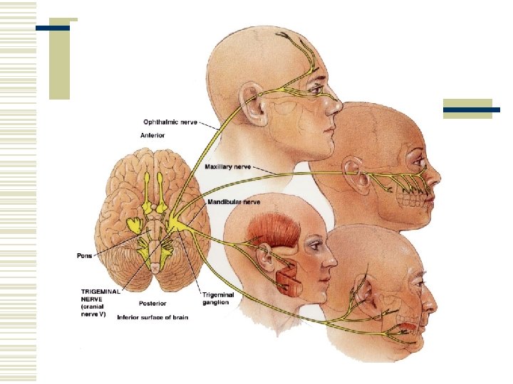
Cranial Nerve V – Trigeminal Nerve w Type: mixed. w Function: conveys impulses for touch, pain, temperature and proprioception. Chewing. w Trigeminal neuralgia (tic douloureux) – pain to branches of the trigeminal nerve. w Dentists apply anesthetic to branches of this nerve.
. w Function: movement of")
Cranial Nerve VI - Abducens w Type: mixed (mainly motor). w Function: movement of the eyeball. w With damage to this nerve the eye cannot move laterally beyond the midpoint and usually points medially.

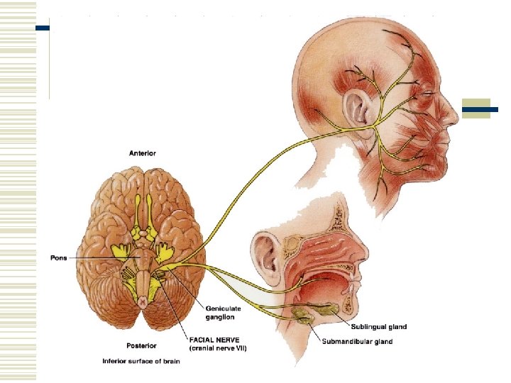
Cranial Nerve VII – Facial Nerve w Type: mixed. w Function: Propriception and taste. Facial expression. Secretion of saliva and tears. w Injury produces bell’s palsy (paralysis of facial muscles).
. w Function: conveys")
Cranial Nerve VIII – Vestibulocochlear Nerve w Type: mixed (mainly sensory). w Function: conveys impulses for equilibrium and hearing. w Injury can cause vertigo, ataxia (muscular incoordination), nystagmus (rapid movement of the eyeball), and tinnitus.

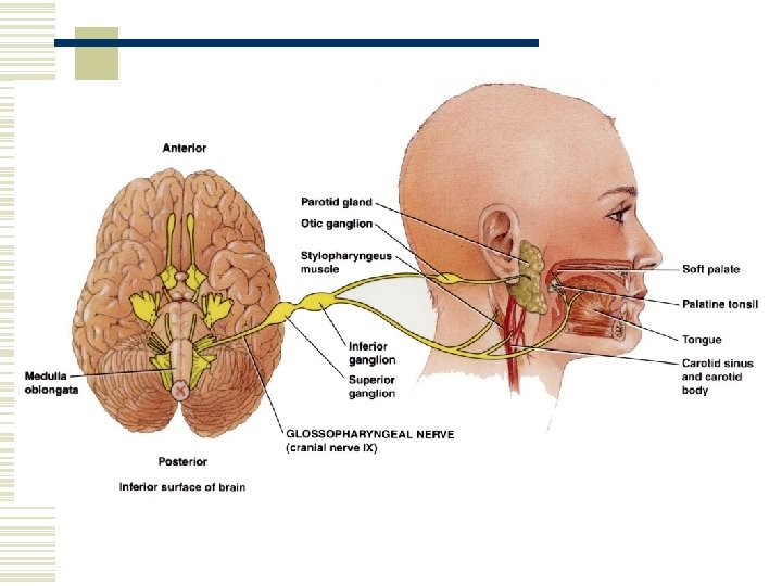
Cranial Nerve IX – Glossopharyngeal Nerve w Type: mixed. w Function: taste and somatic sensations from the posterior 1/3 of the tongue. Elevates the pharynx during swallowing and speech. Stimulates the secretion of saliva. w Injury causes decreased salivary secretion, loss of taste, and difficulty swallowing.

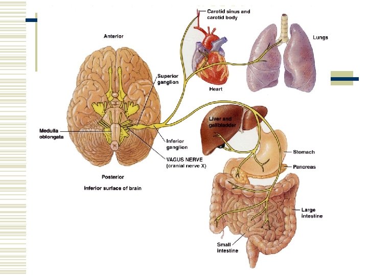
Cranial Nerve X – Vagus Nerve w Type: mixed. w Function: taste and somatic sensations. Swallowing, coughing, and voice production. Regulates GI tract and heart rate. w Injury interferes with swallowing, paralyzes vocal cords, and causes the heart rate to increase.
. w Function: Proprioception.")
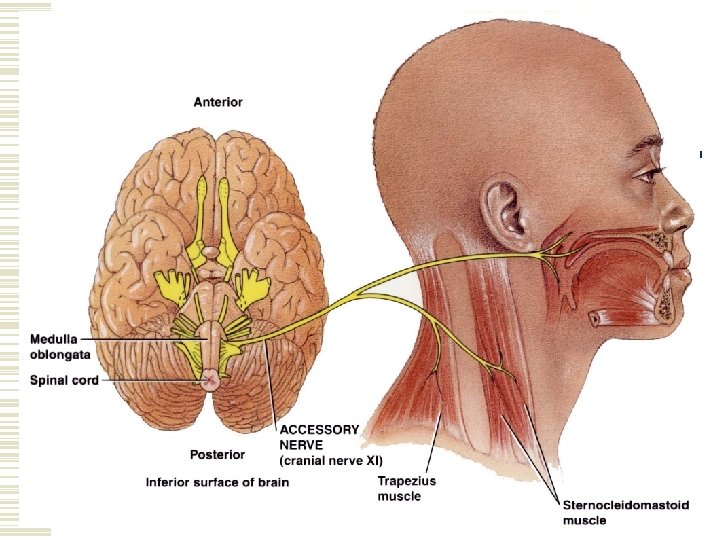
Cranial Nerve XI – Accessory Nerve w Type: mixed (mainly motor). w Function: Proprioception. Swallowing, movement of head and shoulders. w If the nerves are damaged the SCM and Trapezius become paralyzed.
. w Function: Proprioception.")
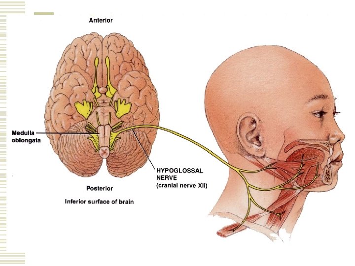
Cranial Nerve XII – Hypoglossal Nerve w Type: mixed (mainly motor). w Function: Proprioception. Movement of the tongue during speech and swallowing. w Injury results in difficulty in chewing, speaking, and swallowing. When protruded, the tongue curls towards the affected side and atrophies on the affected side.

Cranial Nerves w On Old Olympus’ Towering Tops A Fin And German Viewed Some Hops. w This mnemonic device helps you memorize the names of the cranial nerves. w The first letter from each word corresponds to the first letter of each cranial nerve.
- Slides: 75