Ventricles Spinal Cord Cranial Nerves Objectives Identify cranial

Ventricles, Spinal Cord & Cranial Nerves

Objectives • Identify cranial nerves and describe the function of each • Understand the signs and tests for CN dysfunction • Make an accurate model of the ventricle system of the brain • Discuss the clinical implications of dysfunction in the ventricular system • Describe the basic organization of the spinal cord

Cranial Nerves and Brain Stem Please refer to Cranial Nerve Handout

A few things first • 12 pairs • All ipsilateral symptoms and signs except IV Trochlear (which is also the only CN that exits brainstem on the dorsal surface) • The optic and olfactory nerves are CNS, the rest have axons and receptors that are considered PNS • Can carry motor, sensory or mixed information ipsilateral = same side

testing each cranial nerve more or less separately is helpful in diagnostics but not a good way to test function! Reality is more complex than that!
 Receptors>nerve> olfactory bulb> olfactory tract (axon): • • • Amygdala (fear)")
1 Olfactory (S) Receptors>nerve> olfactory bulb> olfactory tract (axon): • • • Amygdala (fear) Orbitofrontal cortex (food!) Thalamus (declarative memory) Hypothalamus (visceral) Hippocampus (memory)

Olfactory Test • Baby powder • Chocolate • Cinnamon • Coffee • Mothballs • Peanut Butter • Soap
")
2 Optic Nerve (S)

Rods and Cones> optic nerve> optic chiasm> optic tract> thalamus > superior colliculi > thalamus> occipital lobe

Optic Pathway Rods and Cones> optic nerve> optic chiasm> optic tract> thalamus > superior colliculi > thalamus> occipital lobe Test: • acuity • pupillary light reflex • visual field Superior colliculi
 All movements of eye, except: • Down and medial (trochlear) •")
3 Oculomotor (M) All movements of eye, except: • Down and medial (trochlear) • Lateral (abducens) Directions of each CN and muscles

Oculomotor Reflexes • Pupillary Reflex • Consensual • Accommodation

Oculomotor Test with abducens and trochlear: basically move stimulus in shape of an H Signs of Lesion: 1. Lateral Strabismus (complains of double vision) 2. Ptosis (droopy eyelid) 3. Nystagmus (in absence of stimulus)
 • Starts in and around oculomotor nucleus, crosses at midbrain,")
4 Trochlear Nerve (M) • Starts in and around oculomotor nucleus, crosses at midbrain, exits out the contralateral side of the dorsal brain stem and then back into the orbit (eewwwhhh!) Test for Lesion: can’t look down and in (trouble descending stairs)
 Sensory: face, mouth, cornea Motor: chewing muscles Reflexes: Masseter Corneal")
5 Trigeminal Nerve (Mx) Sensory: face, mouth, cornea Motor: chewing muscles Reflexes: Masseter Corneal 3 Branches: 1. Ophthalmic 2. Maxillary 3. Mandibular

Test Trigeminal 1. Sensation of face 2. Clamp down jaw, try to open 3. Palpate masseter and temporal mm
 Abducts the eye (moves it laterally) Think about it- adduct would")
6 Abducens (M) Abducts the eye (moves it laterally) Think about it- adduct would be medial
 Sensory: taste tip of tongue Motor: facial expression, lacrimal & salivary")
7 Facial (Mx) Sensory: taste tip of tongue Motor: facial expression, lacrimal & salivary glands Supplies the motor response to the corneal reflex (when the eye is touched) Test- facial expression and taste on tip of tongue
 Tests: • Audition- tuning fork • Turn head and see if")
8 Vestibulocochlear (S) Tests: • Audition- tuning fork • Turn head and see if eyes move slowly in opposite direction • Test for balance by gently pushing them
 • Sensory: soft palate and pharynx, taste/sensation from posterior tongue,")
9 Glossopharyngeal Nerve (Mx) • Sensory: soft palate and pharynx, taste/sensation from posterior tongue, carotid sinus (blood pressure sense) • Motor: parotid gland (salivary gland) and 1 pharyngeal mm • Sensory portion of the gag and swallowing reflexes so that’s how it is tested
 • Sensory: larynx & pharynx • Motor: autonomic functions of gut,")
10 Vagus (Mx) • Sensory: larynx & pharynx • Motor: autonomic functions of gut, cardiac (inhibition), vocal cords, swallowing • Motor component of the gag and swallow reflexes • Test: reflexes, hoarseness, uvula (thing in the back of your throat)

11 Spinal accessory nerve • Innervates shoulder and neck mm • Test sternocleidomastoid and trapezius

12 Hypoglossal Nerve • Movement of the tongue • Test: stick tongue out, it will point to the side of the lesion, make sure they can push equally on both checks with tongue

Basic Organization of the Spinal Cord

8 12 5 5 1 Cervical Thoracic Lumbar Sacral Coccygeal

S 1
 Action C 5 Deltoid Shoulder Abduction C 5 Infraspinatus Humeral external rotation")
Root Muscle(s) Action C 5 Deltoid Shoulder Abduction C 5 Infraspinatus Humeral external rotation C 5, 6 Biceps Flexion of supinated forearm C 6 Extensor carpi radialis, ulnaris Wrist extension C 7 Extensor digitorum, triceps Finger extension, forearm extension at elbow C 8, T 1 Interossei, lumbricals Finger ab and ad (spread and close against force) L 2 -L 4 Quadriceps, iliopsoas, adductors Knee extension, thigh on hip flexion, thigh adduction L 5 Anterior tibial, extensor halluces Ankle and big toe dorsiflexion (walk on heels) S 1 Gastrocnemius Ankle plantar flexion (walk on tip toes)

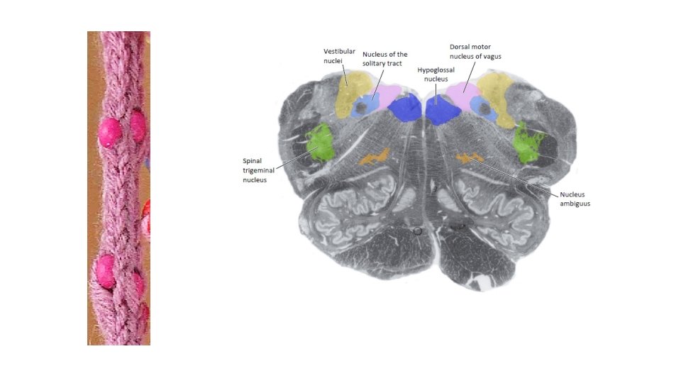
Same magnification, cross sections White matter is dark, grey is light Dorsal on top, ventral on bottom (imagine a person laying on stomach) Dorsal column Dorsal horn Lateral column Lateral horn Ventral column

Dorsal=sensory Ventral=motor

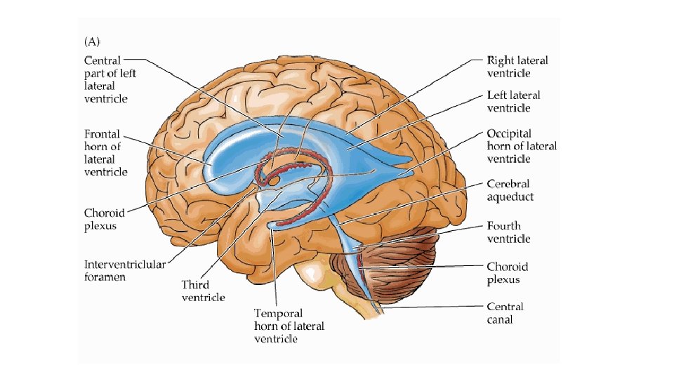
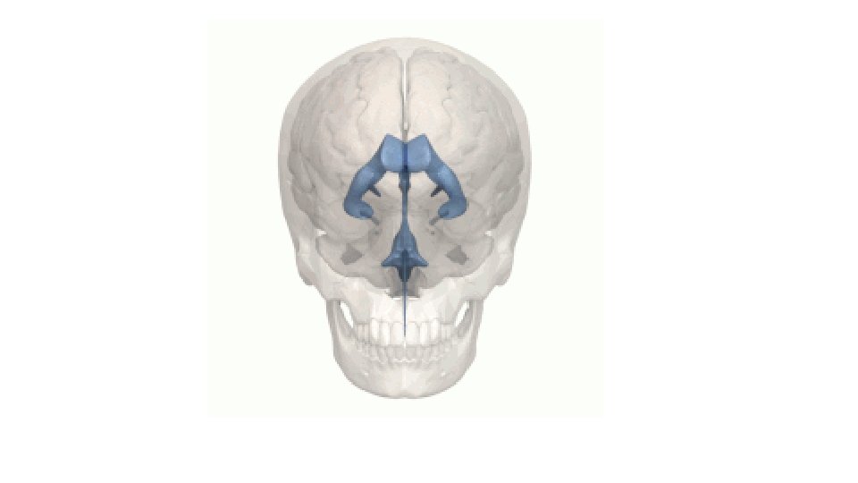
Ventricles
- Slides: 35