Pyogenic Meningitis Dr Ravideep Meningitis is defined as


 or")

")


 Email : ravideepy@gmail. com")
- Slides: 40
Pyogenic Meningitis Dr Ravideep
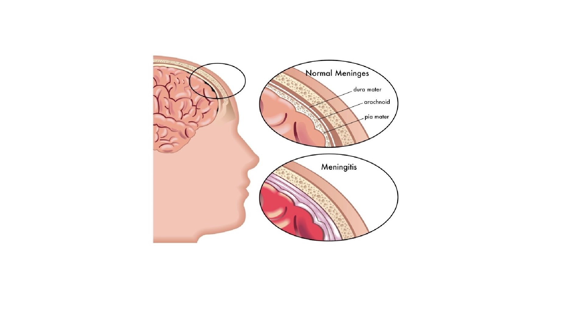
• Meningitis is defined as inflammation of membranes surrounding the brain and spinal cord. • Meningoencephalitis is inflammation of meninges and brain cortex.
Incidence and Etiology • Commonest in Neonates and Infancy because of poor immunity and phagocytic functions. • More frequent in infant Males • Greater risk during 6 -12 months and 95% cases occur between 1 month and 5 years.
Causative organisms Neonates Escherichia coli Streptococcus pneumoniae Salmonella species Pseudomonas aeuroginosa Staphylococcal aureus 3 months - 3 years Hemophilus influenzae type b Streptococcus pneumoniae Meningococci ( Neisseria meningitides ) >2 years Streptococcus pneumoniae Neisseria meningitides Diminished host response , Malignancies , Children on Immunosuppressive drugs More susceptible to develop meningitis by fungi , Listeria monocytogenes and mycoplasma
Pathogenesis • Infection spreads hematogenously to meninges from distant foci , e. g. pneumonia , empyema , pyoderma and osteomyelitis. • Rarely , the infection may extend from contiguous septic foci , e. g. infected paranasal sinuses , mastoiditis , fracture of base of skull. • Anatomic and congenital defect can also cause infection , e. g. myelomeningocele
Pathology • Leptomeninges are infiltrated with inflammatory cells • Cortex of brain shows deem , exudate and proliferation of microglia • Ependymal cells are destroyed and purulent exudate collects at base of brain , most marked in interpeduncular and schismatic cisterns • Exudates may block the foramina of Luschka and Magendie - Hydrocephalus • Thrombophlebitis of cerebral vessels may occur leading to infarction and neurologic sequelae
Pathology • Fulminating illness may occur in case of meningococcal meningitis and death may occur in few hours because of endotoxic shock. • Bacterial pathogens on destruction liberate cell wall and membrane active components ( trichroic acid , endotoxins , peptidoglycans ). • In response host cells produce tumor necrosis factor , cytokines and platelet aggravating factor. Their interaction with BBB and neurons results in extensive host damage. • Cerebral edema ( vasogenic ) occurs due to endothelial cell injury or cytotoxins.
Clinical features Neonates and Infants • Infective illness in mother , prolonged rupture of membranes or difficult delivery put the newborn at risk • Premature infants have low level of antibodies • Predisposing factor is spina bifida or dermal sinus
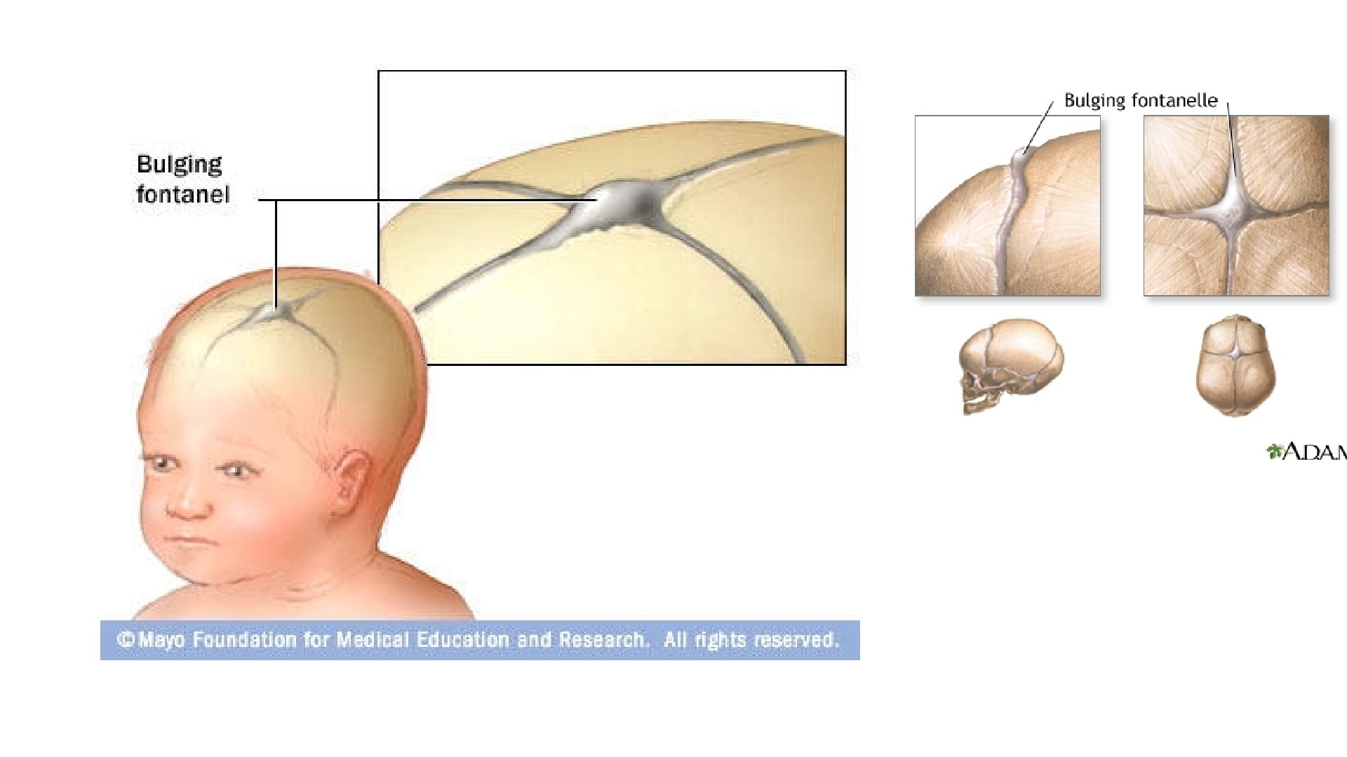
Older children • Classic signs preceded by upper respiratory or GIT symptoms • High grade fever , head ache and projectile vomiting • Seizures are common • Increased CSF pressure leads to bulging fontanelle and diastasis of sutures
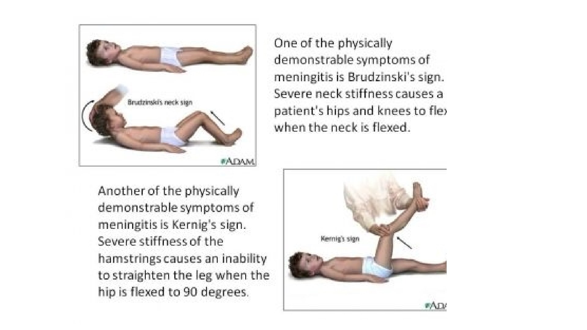
• Neck stiffness , positive kerning’s sign and brudzinski’s sign • Cranial nerve palsies and papilledema • Hemiplegia in cases late reported , ataxia may also be present • Patient may be comatose or semi comatose
• Meningococcal meningitis is characterised by presence of features of Waterhouse Friderichsen syndrome ( adrenal insufficiency , hypotension , shock and coma) occurs due to haemorrhage and necrosis in adrenal glands. • Otitis media and mastoiditis is likely to lead streptococcal and pneumococcal meningitis. Exudates are common on cortex and subdural effusion is usual complication.
Meningococcemia
• Staphylococcal meningitis is likely following umbilical sepsis , pyoderma or septicaemia. In older children it follows otitis media , mastoiditis , sinus thrombosis , pneumonia , arthritis and septic lesions of scalp or skin. • Hemophilus influenzae type b meningitis is frequent between ages 3 and 12 months. Convulsions are common. Residual auditory deficit is a common complication.
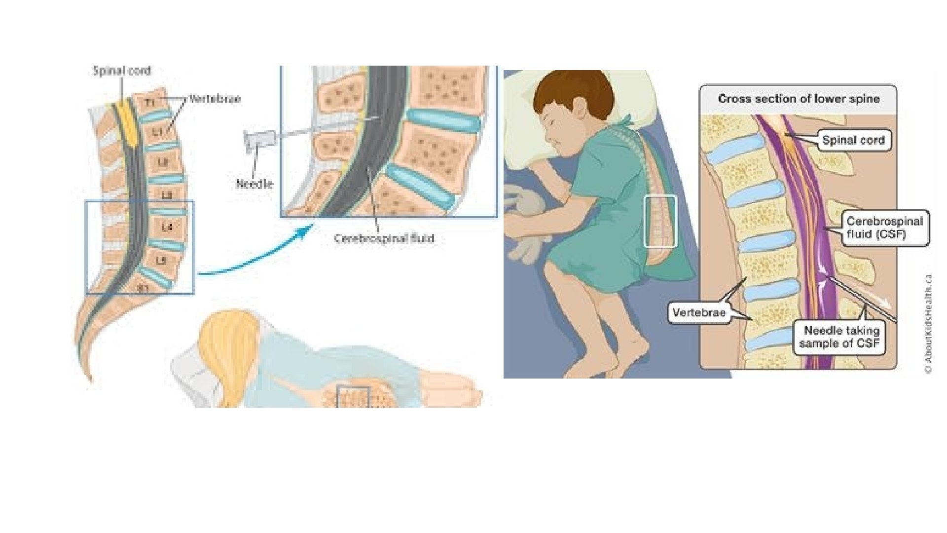
Investigations Lumbar puncture • CSF pressure should be noted , funds checked for papilledema • Xanthochromia due to bilirubin from haemorrhage or increased protein • CSF glucose should be compared to blood glucose , CSF glucose is 2/3 of blood glucose • In CSF of neonates normally there are unto 30 lymphocytes and 150 mg/dl protein • Gram stain is important to recognise the causative agent
Gram staining Meningococci Gram negative intracellular diplococci Pneumococci Gram positive diplococci H. Influenzae Gram negative cocobacilli E. Coli Gram negative bacilli
CSF findings in various CNS disorders Condition Normal Acute Bacterial Meningitis TB meningitis Viral encephalitis Protein (mg/dl) Glucose (mg/dl) 20 -45 >50 or 75% of Blood glucose 100 -500 <40 May be none Opalscent 10 -2000 PMN early but lymphocyte later >50 <40 May be none Clear 5 -500 Mostly lymphocytes PMN early 30 -150 30 -70 Color Leucocytes Clear 0 -5 60 -70% Lymphocytes 100 -20000 Opalscent PMN to purulent predominate
Lumbar puncture Contraindications • Increased ICP especially with focal neurological deficits • Severe cardio pulmonary compromise • Infection of skin overlying the site of LP • Bleeding or clotting disorder
CSF culture • The yield of CSF culture decreases soon after antibiotic therapy has been started • PCR more sensitive in patients treated by antibiotics
Blood culture • H. influenzae and S. pneumoniae Blood counts • Total and DLC- generally there is leucocytosis with predominant polymorphs X ray Chest • To rule out TB and Pneumonia
CT scan Indications • Prolonged comatose condition • Seizures 72 hours after onset of treatment • Continued excessive irritability • Focal neurological findings • Persistently abnormal CSF findings • Relapse or recurrence
Rapid Diagnostic Tests • Concurrent electrophoresis • Latex particle agglutination • ELISA to detect bacteria antigen in CSF • CSF lactate level • Latex agglutination and ELISA have sensitivity and specificity of almost 80% • PCR - Herpes simplex , Enteroviruses , Meningococci & TB.
Differential Diagnosis • Meningism - no neurological signs and CSF normal • Aseptic meningitis - CSF pressure elevated , mild pleocytosis and moderate increase in protein with near normal sugar. No organisms are cultured • TB meningitis • Viral encephalitis • Cryptococcal meningitis- immunocompromised host • Subarachnoid haemorrhage - CT scan
Treatment Initial Empiric Therapy • Third generation cephalosporin such as Ceftriaxone(100 -150 mg/kg/day) or Cefotaxime(200 mg 300 mg/kg/day) • Ampicillin ( 200 mg/kg) and chloramphenicol (100 mg/kg/day) for 10 -14 days is also effective • All antibiotics are administered IV • If fever or Meningeal signs persists after 48 hours of therapy , a LP should be repeated and choice of antibiotics reviewed
Treatment Specific antimicrobial therapy • Meningococcal or Pneumococcal meningitis : Penicillin 400 -500, 000 units/kg/day q 4 hr. Cefotaxime ( 150 -200 mg/kg/day q 8 hr) or ceftriaxone(100 -150 mg/kg/day q 12 hr ) are also effective. • H. influenzae meningitis : ceftriaxone or cefotaxime is used as single agent. Combination of Ampicillin and chloramphenicol less preferred • Staphylococcal : vancomycin ( 15 -20 mg/kg IV q 6 hr ) is the treatment of choice in case of MRSA. Addition of rifampicin increases CSF penetrance and efficacy of these drugs
Treatment Specific antimicrobial therapy • Listeria : Ampicillin ( 300 mg/kg/day q 6 hr) and aminoglycoside (gentamicin , amikacin or netilmycin ) preferred • Gram negative bacilli : Cefotaxime , Ceftazidime or ceftriaxone , or combination of ampicillin and amino glycoside may be used. • Pseudomonas : combination of ceftazidime and amino glycoside is used. Ticarcilin in place of ceftazidime may be used. Meropenem(40 mg/kg IV q 8 hr) or cefepime are effective agents , if above drugs fail.
Treatment Duration • Generally show quick improvement in days • Treatment is for 10 -14 days • Staphylococcal and gram negative infections extended to 3 weeks • Routine LP at the end of therapy not recommended.
Treatment Steroid therapy • No role in neonatal meningitis • Dexamethasone at dose of 0. 15 mg/kg IV q 6 hr for 2 -4 days initiated shortly before or along with first dose of antibiotic • Helps to reduce incidence of residual neurological complications , such as sensorineural deafness , hydrocephalus and behavioural disturbances • Especially useful in H. influenzae meningitis
Treatment Symptomatic Therapy • Cerebral edema & Increased ICP : Head end elevated about 30 degrees. Osmotic diuresis with 0. 5 mg/kg of mannitol as a 20% solution is administered IV every 4 -6 hr for maximum of 6 doses • Convulsions : diazepam 0. 3 mg/kg ( Max 5 mg) IV followed by phenytoin 15 -20 mg/kg loading dose and continued at a dose of 5 mg/kg IV or PO. Antiepileptic drugs can be stopped after 3 months • Fluid and electrolyte homeostasis : Maintanence fluids are given , hypotonic fluids avoided.
Treatment Symptomatic therapy • Inappropriate ADH secretion : Fluid restriction and diuretics • Hypotension : treated with IV fluids and vasopressors such as dopamine and dobutamine • Nursing care : oral cavity , eyes , bladder and bowel should be taken care of. Bed sores are prevented by frequent posture change and by using soft foam mattress or air cushion
Complications Increased ICP Cranial Nerve Palsies Seizures Stroke Ataxia SIADH Subdural effusions Spastic paraparesis Blindness Cerebral infarcts Long term neurologic sequelae Epilepsy Deafness Spasticity
Complications • Subdural empyema is managed by drainage of subdural space along with intensive antibiotic therapy , subdural effusions generally resolve spontaneously • Hydrocephalus may occur in acute phase and generally regresses. Ventriculoatrial or ventriculoperitoneal shunt is rarely required
Subdural effusion
Prevention Vaccination • Vaccines available against S. pneumoniae , N. meningitides and H. influenzae type b • Pneumococcal polysaccharide vaccine available • Meningococcal vaccine for high risk groups
Antibiotic prophylaxis • Meningococcal : Rifampicin at a dose of 10 mg/kg given 12 hourly for 2 days • H. influenzae : Rifampicin at dose of 20 mg/kg/day for 4 days for household contacts and patient • S. pneumoniae : no prophylaxis
Follow Up • Auditory evaluation should be carried out at the time of discharge and 6 weeks later
THANK YOU Dr Ravideep (+917416878888) Email : ravideepy@gmail. com