Cranial nerves and their nuclei Cranial Nerves Figure


 • Optic (2) • Vestibulocochlear (8)")
 • Trochlear (4) • Abducens (6) • Accessory (11)")
 • Facial (7) • Glossopharyngeal (9) • Vagus (10)")

")

")

")
 • Motor to ocular muscles: rectus (superior對側, inferior同側and medial同側 ),")


")
 • To contralateral (對側) superior oblique muscle • Located at the")

 • To lateral rectus muscle • Located in the caudal pons")


, maxillary (V")
")


")

 2. Superior salivatory nucleus To chorda")


")

")

 • Branchial motor to stylopharyngeus 2. Inferior salivary nucleus (GVE)")
")

")
 • A hybrid nucleus • Branchial motor to larynx and")
")

")


- Slides: 78
Cranial nerves and their nuclei 鄭海倫 整理
Cranial Nerves Figure 13. 4 a
Location of the cranial nerves • Anterior cranial fossa: C. N. 1– 2 • Middle cranial fossa: C. N. 3 -6 • Posterior cranial fossa: C. N. 7 -12
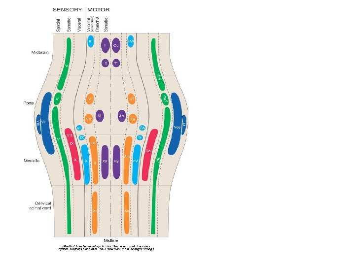
Functional components in nerves • General Somatic Efferent • Special Visceral Afferent • GSE GSA GVE GVA • (SSE) SSA SVE SVA
Neuron columns in the embryonic spinal cord *
The floor of the 4 th ventricle in the embryonic rhombencephalon
Sp: special sensory B: branchial motor Ss: somatic sensory Sm: somataic motor Vi: visceral sensory A: preganglionic autonomic (visceral motor)
• STT: spinothalamic tract • CST: corticospinal tract • ML: medial lemniscus
Sensory nerve • Olfactory (1) • Optic (2) • Vestibulocochlear (8)
Motor nerve • Oculomotor (3) • Trochlear (4) • Abducens (6) • Accessory (11) • Hypoglossal (12)
Mixed nerve • Trigeminal (5) • Facial (7) • Glossopharyngeal (9) • Vagus (10)
Innervation of branchial muscles • Trigemial • Facial • Glossopharyngeal • Vagus
Cranial Nerve I: Olfactory Table 13. 2(I)
Cranial Nerve II: Optic • Arises from the retina of the eye • Optic nerves pass through the optic canals and converge at the optic chiasm • They continue to the thalamus (lateral geniculate body) where they synapse • From there, the optic radiation fibers run to the visual cortex (area 17) • Functions solely by carrying afferent impulses for vision
Cranial Nerve II: Optic Table 13. 2(II)
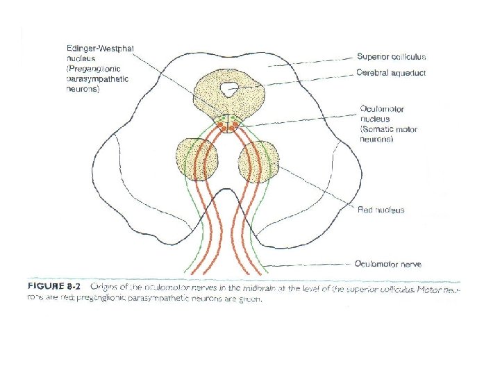
Cranial Nerve III: Oculomotor • Fibers extend from the ventral midbrain, pass through the superior orbital fissure, and go to the extrinsic eye muscles • Functions in raising the eyelid, directing the eyeball, constricting the iris, and controlling lens shape
Cranial Nerve III: Oculomotor Table 13. 2(III)
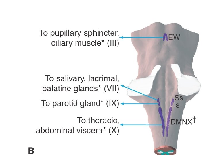
1. Oculomotor nucleus (GSE) • Motor to ocular muscles: rectus (superior對側, inferior同側and medial同側 ), inferior oblique同側, levator palpebrae superioris雙側 2. Edinger-Westphal nucleus (GVE) • to ciliary ganglion ciliarlis and sphincter pupillae muscles
Oculomotor nucleus: a series of cell columns or subnuclei M: medial longitudinal fasciculus PAG: periaqueductal gray
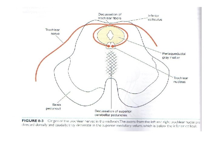
Cranial Nerve IV: Trochlear • Fibers emerge from the dorsal midbrain and enter the orbits via the superior orbital fissures; innervate the superior oblique muscle • Primarily a motor nerve that directs the eyeball
Cranial Nerve IV: Trochlear Table 13. 2(IV)
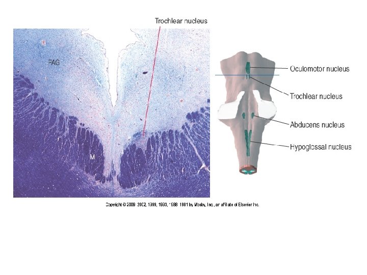
Trochlear nucleus (GSE) • To contralateral (對側) superior oblique muscle • Located at the level of the inferior colliculus • It indents the medial longitudinal fasciculus
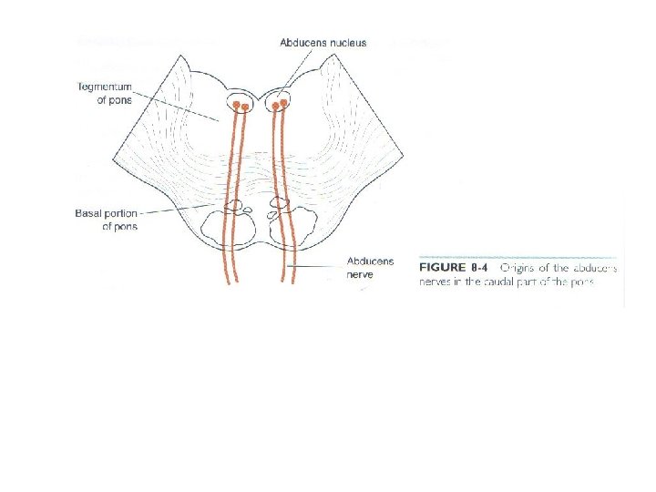
Cranial Nerve VI: Abducens • Fibers leave the inferior pons and enter the orbit via the superior orbital fissure • Primarily a motor nerve innervating the lateral rectus muscle Table 13. 2(VI)
Abducens nucleus (GSE) • To lateral rectus muscle • Located in the caudal pons beneath the floor of the 4 th ventricle
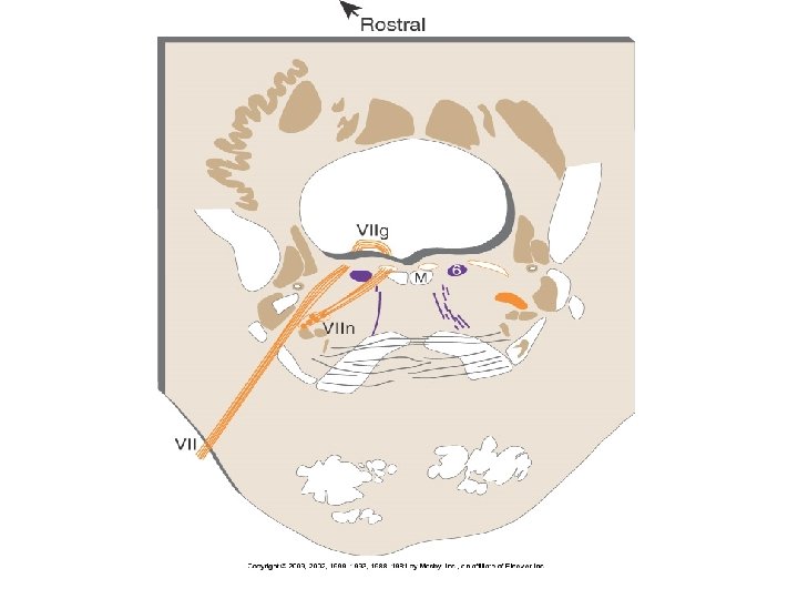
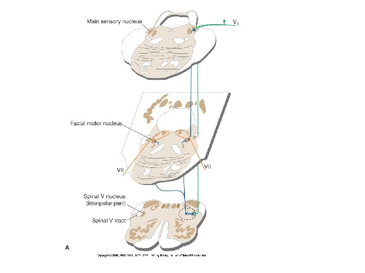
ICP: inferior cerebellar peduncle ML: medial lemniscus Sp. Vt: spinal trigeminal tract VII: facial nerve VIIg: internal genu of the facial nerve VIIn: facial motor nucleus
DMN X: dorsal motor nucleus of the vagus ST: solitary tract Sol: nucleus of the solitary tract 4 V: 4 th ventricle
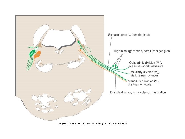
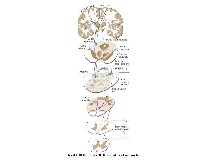
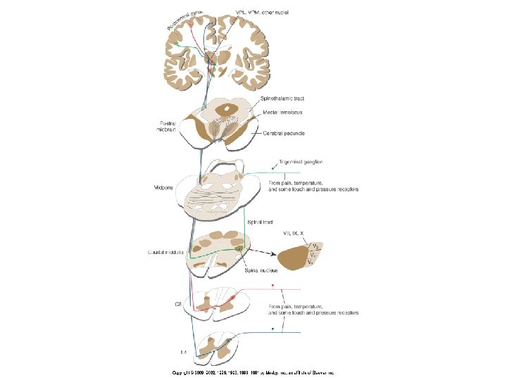
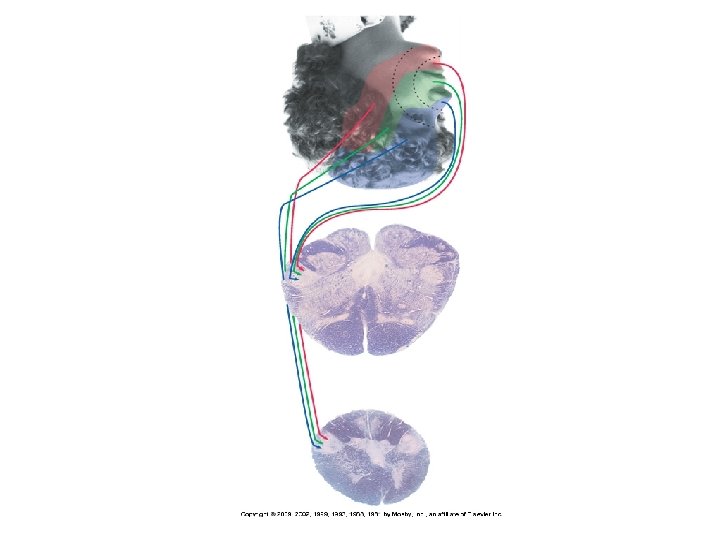
Cranial Nerve V: Trigeminal • Composed of three divisions: ophthalmic (V 1), maxillary (V 2), and mandibular (V 3) • Fibers run from the face to the pons via the superior orbital fissure (V 1), the foramen rotundum (V 2), and the foramen ovale (V 3) • Conveys sensory impulses from various areas of the face (V 1) and (V 2), and supplies motor fibers (V 3) for mastication
Cranial Nerve V: Trigeminal Table 13. 2(V)
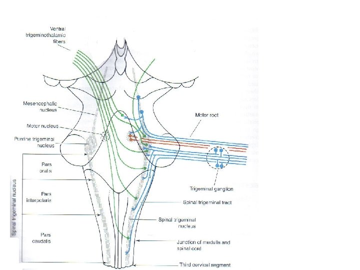
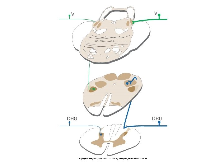
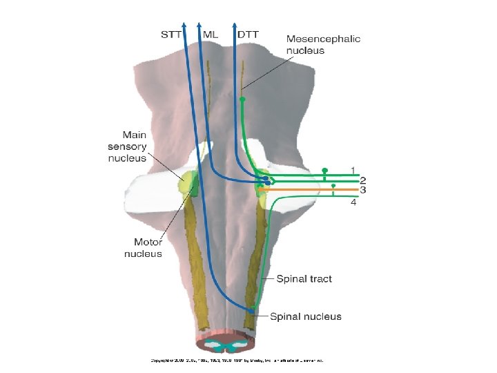
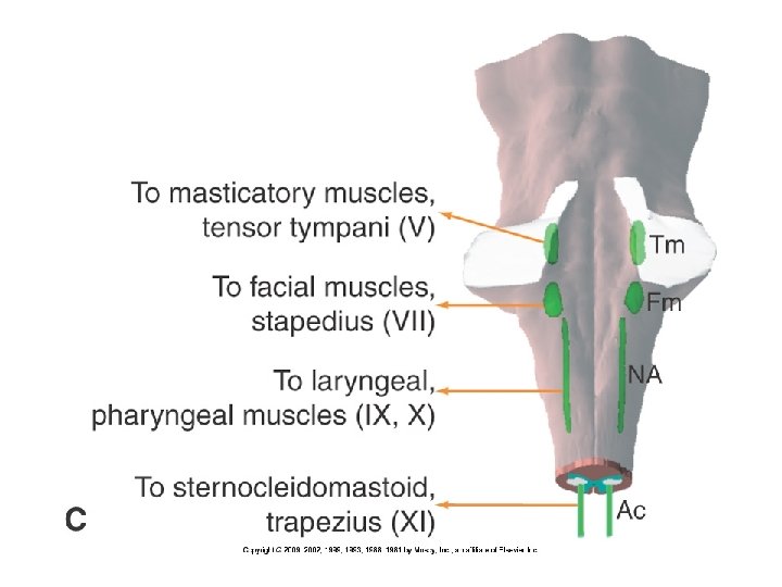
1. Main sensory nucleus 2. Nucleus of the spinal trigeminal: receives information of pain and temperature 3. Mesencephalic nucleus Central processes Peripheral processes motor nuclei of trigeminal mandibular division 4. Trigeminal motor nucleus (SVE): inervates muscles of mastication
SCP: superior cerebellar peduncle MCP: middle cerebellar peduncle V: trigeminal nerve
P: pyramid FC: fasciculus cuneatus NC: nucleus cuneatus
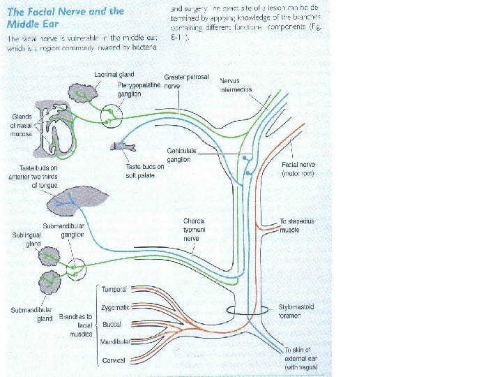
Cranial Nerve VII: Facial • Fibers leave the pons, travel through the internal acoustic meatus, and emerge through the stylomastoid foramen to the lateral aspect of the face • Mixed nerve with five major branches • Motor functions include facial expression, and the transmittal of autonomic impulses to lacrimal and salivary glands • Sensory function is taste from the anterior two-thirds of the tongue
Cranial Nerve VII: Facial Table 13. 2(VII)
Facial colliculus
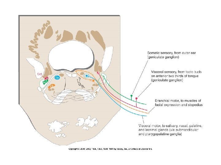
1. Motor nucleus of facial nerve (facial nucleus) 2. Superior salivatory nucleus To chorda tympani branch and join the lingual branch of C. N. V 3 3. Lacrimal nucleus To greater petrosal branch and terminate the pterygopalatine ganglion
1. Geniculate ganglion Central processes Peripheral processes nervus intermedius chorda tympani, greater petrosal and lesser palatine (some fibers join the auricular branch of the vagus) 2. Gustatory nucleus (The large-celled rostral part of the nucleus of the tractus solitarius) Enter the brain in the nervus intermedius and turn in the tractus solitarius, then terminate in the nucleus of the tractus solitarius 3. Nucleus of spinal trigeminal tract From the nervus intermedius
Taste
Taste sensation • Facial • Glossopharyngeal • Vagus
Dorsum of the tongue Around the circumvallate papillae Root of the tongue
Cranial Nerve VIII: Vestibulocochlear • Fibers arise from the hearing and equilibrium apparatus of the inner ear, pass through the internal acoustic meatus, and enter the brainstem at the pons-medulla border • Two divisions – cochlear (hearing) and vestibular (balance) • Functions are solely sensory for the sense of equilibrium and of hearing
Cranial Nerve VIII: Vestibulocochlear Table 13. 2(VIII)
Cranial Nerve IX: Glossopharyngeal • Fibers emerge from the medulla, leave the skull via the jugular foramen, and run to the throat • Nerve IX is a mixed nerve with motor and sensory functions • Motor – innervates part of the tongue and pharynx, and provides motor fibers to the parotid gland • Sensory – fibers conduct taste and general sensory impulses from the tongue and pharynx
Cranial Nerve IX: Glossopharyngeal Table 13. 2(IX)
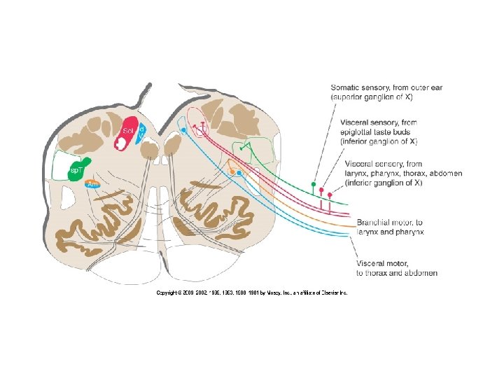
1. Nucleus ambiguus (SVE) • Branchial motor to stylopharyngeus 2. Inferior salivary nucleus (GVE) • to parotid gland (via otic ganglion)
3. Spinal trigeminal nucleus • Somatic sensory from outer ear (superior ganglion of IX) 4. Nucleus of the solitary tract • Visceral sensory from carotid body and sinus, mucosa of pharynx, posterior tongue, middle ear (inferior ganglion of IX) • Visceral sensory from taste buds on posterior third of tongue (inferior ganglion of IX)
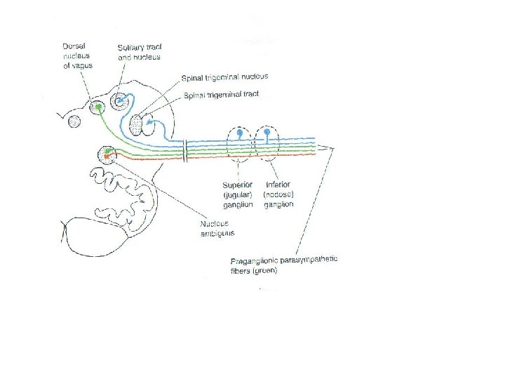
Cranial Nerve X: Vagus • The only cranial nerve that extends beyond the head and neck • Fibers emerge from the medulla and emerge via the jugular foramen • The vagus is a mixed nerve • Most motor fibers are parasympathetic fibers to the heart, lungs, and visceral organs • Its sensory function is in taste
Cranial Nerve X: Vagus Table 13. 2(X)
1. Nucleus ambiguus (SVE) • A hybrid nucleus • Branchial motor to larynx and pharynx and autonomic motor to thorax and abdomen 2. dorsal motor nucleus (GVE) • autonomic motor to thorax and abdomen
3. Spinal trigeminal nucleus • Somatic sensory from outer ear (superior ganglion of X) 4. Nucleus of the solitary tract • Visceral sensory from larynx , pharynx , thorax and abdomen (inferior ganglion of X)
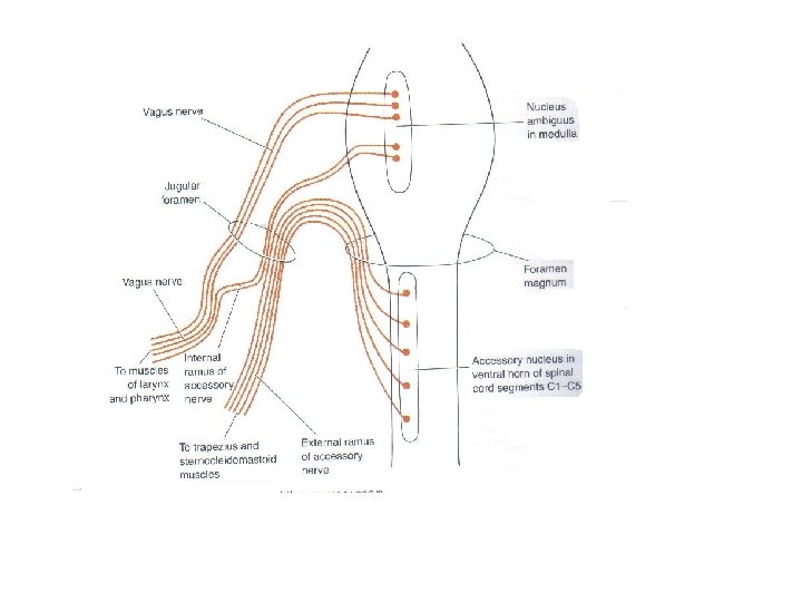
Cranial Nerve XI: Accessory • The spinal root passes upward into the cranium via the foramen magnum • The accessory nerve leaves the cranium via the jugular foramen • Primarily a motor nerve supplying: – Fibers to the larynx, pharynx, and soft palate – Innervates the trapezius and sternocleidomastoid, which move the head and neck
Cranial Nerve XI: Accessory Table 13. 2(XI)
• Formed from a cranial root emerging from the medulla and a spinal root arising from the superior region of the spinal cord 1. Cranial root Nucleus ambiguus (SVE) 2. Spinal root Spinal accessory nuclei (GSE)
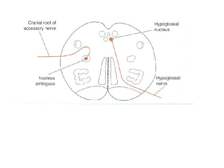
Cranial Nerve XII: Hypoglossal • Fibers arise from the medulla and exit the skull via the hypoglossal canal • Innervates both extrinsic and intrinsic muscles of the tongue, which contribute to swallowing and speech Table 13. 2(XII)
Hypogolssal nucleus • Lying between the dorsal nucleus of the vagus and the midline of the medulla • Sents off GSE fibers
Parasympathetic ganglion • • Ciliary ganglion Pterygopalatine ganglion Submandibular ganglion Otic ganglion