The Cranial Cavity CONTENTS 1 The brain and

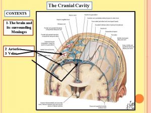
The Cranial Cavity CONTENTS 1 -The brain and its surrounding Meninges 2 Arteries 3 Veins

4 -Venous sinuses

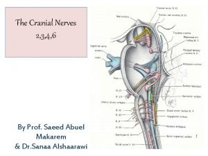
Olfactorynerves. Ill 0 t knic ne<Ve[V, 1 Ocu motorn rve 1 [111 Abduc ntn rve [_VI] _, , . IV 2 l [V Troe learn rve ( I V ] - -. J J. i I nef. V [IIIJ Vestibul och ar nerve [VIII] Accessory nerve [XIJ Inerv IXIIJ

VAULT OF THE SKULL The internal surface of the vault presents: 1 - The coronal 2 - Sagittal 3 -Lambdoid sutures 4 In the midline is a shallow sagittal groove containing the SUPERIOR SAGITTAL SINUS 5 On each side of the groove are several small pits, called GRANULAR PITS? What for (see next slide) 6 Grooves for the middle meningeal artery

Opening of superi or cerebral v ein Ara chnoi d granulations Anterior meningeal bran ch of anteri or ethmoidal artery Mastoid bran ch of occipital artery Frontal (anteri or) and parietal (posterior ) bran ches of middle men ingeal artery Middle meningeal artery

The Meninges The brain in the skull is surrounded by three membranes or meninges: 1 -THE DURA MATER 2 -THE ARACHNOID MATER 3 -THE PIA MATER


1 -THE DURA MATER 2 -THE ARACHNOID MATER 3 -THE PIA MATER

1 -DURA MATER OF THE BRAIN Made of two layers: a-The endosteal layer b-The meningeal layer These are closely united except along where they separate to form VENOUS SINUSES

A-The endosteal layer Is the ordinary periosteum covering the inner surface of the skull bones It does not extend through the foramen magnum to become continuous with the dura mater of the spinal cord Around the margins of all the foramina in the skull it becomes continuous with the periosteum on the outside of the skull bones At the sutures it is continuous with the sutural ligaments.

B-The meningeal layer Is the dura mater proper It is a dense, strong, fibrous membrane covering the brain and is continuous through the foramen magnum with the dura mater of the spinal cord It provides tubular sheaths for the cranial nerves as the latter pass through the foramina in the skull Outside the skull the sheaths fuse with the epineurium of the nerves

The meningeal layer sends inward FOUR SEPTA 1 -THE FALX CEREBRI 2 -THE TENTORIUM CEREBELLI 3 -THE FALX CEREBELLI 4 -THE DIAPHRAGMA SELLAE

The meningeal layer sends inward SEPTA

The meningeal layer sends inward SEPTA

1 -THE FALX CEREBRI

Is a sickle-shaped fold of dura mater that lies in the midline between the two cerebral hemispheres Its narrow end in front is attached to the THE CRISTA GALLI Its broad posterior part blends in the midline with the upper surface of the Tentorium cerebelli The superior sagittal sinus runs in its upper fixed margin the inferior sagittal sinus runs in its lower concave free margin The straight sinus runs along its attachment to the tentorium cerebelli.

THE TENTORIUM CEREBELLI Is a crescent-shaped (or tent-shaped) fold of dura mater • Roofs over the posterior cranial fossa It covers the upper surface of the cerebellum and supports the occipital lobes of the cerebral hemispheres. In front is a gap, the tentorial notch, for the passage of the midbrain It has: an inner free border an outer attached or fixed border Divides the cranial cavity into: 1 SUPRATENTORIAL 2 INFRATENTORIAL We were dividing the cranial cavity into: anterior , middle and posterior cranial cavity. At present it is well established that we divide the cranial cavity into supratentorial and infratentorial regions.

The fixed border is attached to: the posterior clinoid processes The superior borders of the petrous bones The margins of the grooves for the transverse sinuses on the occipital bone

The free border runs forward at its two ends: Attached to the anterior clinoid process on each side. At the point where the two borders cross, the third and fourth cranial nerves pass forward to enter the lateral wall of the cavernous sinus

Remember that the dura is a tough structure and its tentorium as well, thus one should think about it as a real septa Which really separates the structures above it from those below it. Any intracranial mass inside the skull (tumor, bleeding…) may force its neighboring structures to herniate ? For example

(Temporal Lobe) Herniation Consequences 1 -Compression of cranial nerve III. The ipsilateral third nerve, The first clinical sign is ipsilateral pupil dilation To be explained later in the eye section since the parasympathetic fibers that supply the constrictor pupil are located on the outside of the nerve (III ) and are inactivated first by compression. 2 -Compression of midbrain cerebral peduncles: resulting in contralateral hemiparesis or hemiplegia 3 -Brainstem compression The patient becomes comatose and may develop bradycardia secondary to increasing brainstem compression


The falx cerebri and the falx cerebelli are attached to the upper and lower surfaces of the tentorium, respectively The straight sinus runs along its attachment to the falx cerebri the superior petrosal sinus along its attachment to the petrous bone the transverse sinus along its attachment to the occipital bone

3 -THE FALX CEREBELLI is a small, sickle-shaped fold of dura mater that is attached to the internal occipital crest and projects forward between the two cerebellar hemispheres. Its posterior fixed margin contains the occipital sinus. 4 -THE DIAPHRAGMA SELLAE Is a small circular fold of dura mater that forms the roof for the sella turcica Attached to the tuberculm sellae anteriorly Attached to the dorsum sellae posteriorly A small opening in its center allows passage of the stalk of the pituitary gland

The Venous Blood Sinuses are blood-filled spaces situated between the layers of the dura mater They are lined by endothelium Their walls are thick and composed of fibrous tissue They have no muscular tissue The sinuses have no valves They receive tributaries from the brain, the diplo « ﺃ of the skull, the orbit, and the internal ear

The superior sagittal sinus

lies in the upper fixed border of the falx cerebri It becomes continuous with the right transverse sinus. The sinus communicates on each side with the VENOUS LACUNAE Numerous arachnoid villi and granulations project into the lacunae The superior sagittal sinus receives THE SUPERIOR CEREBRAL VEINS

THE INFERIOR SAGITTAL SINUS • lies in the free lower margin of the falx cerebri It runs backward and joins the great cerebral vein to form the straight sinus It receives cerebral veins from the medial surface of the cerebral hemisphere. THE STRAIGHT SINUS lies at the junction of the falx cerebri with the tentorium cerebelli Formed by the union of the inferior sagittal sinus with the great cerebral vein it drains into the left transverse sinus

THE RIGHT TRANSVERSE SINUS begins as a continuation of the superior sagittal sinus; (the left transverse sinus is usually a continuation of the straight sinus ) Each sinus lies in the lateral attached margin of the tentorium cerebelli, and they end on each side by becoming the sigmoid sinus

The sigmoid sinuses Are a direct continuation of the transverse sinuses Each sinus turns downward behind the mastoid antrum of the temporal bone and then leaves the skull through the jugular foramen Become the internal jugular vein The occipital sinus lies in the attached margin of the falx cerebelli It communicates with the vertebral veins through the foramen magnum and the transverse sinuses

Superior sagittal sinus Ophthalmic artery Internal carotid artery Olfactory bulb Oculomotor nerve (IIQ Optic nerve 01) Trochlear nerve OV) Anterior intercavemous sinus Ophthalmic nerve (V 1) Stalk of pituitary gland Posterior intercavernous sinus Trigeminal ganglion / Middle meningeal artery Trigeminal n. (V)Superior petrosal sinu_s - -- - -. J : . . . u. - - - Inferior petrosal sinus : . . . : : ; ; -1 - L/44 ---, , k-'i, L. sr - - - . . . , _: ! - - - Trigeminal nerve (V} -; - -r - -'c: Abducentnerve {VI) - Abducent n. {VI) Facial nerve (VIQ Superior petrosal sinus Vestibulocochlear nerve {VIII) Glossopharyngea l nerve(IX) Labyrinthine artery Si moid sinus Hypoglossal nerve (XIQ Accessory nerve (XI) Cerebellar tentorium Vertebral artery Great cerebral vein Cerebral falx (cut) Straight sinus Inferior sagittal sinus Superior sagittal sinus

CAVERNOUS SINUS lies on the lateral side of the body of the sphenoid bone Anteriorly, the sinus receives 1 -The inferior ophthalmic vein 2 -The central vein of the retina The sinus drains posteriorly into: the transverse sinus through the superior petrosal sinus Intercavernous sinuses

Important Structures Associated With the Cavernous Sinuses 1 The internal carotid artery 2 The sixth cranial nerve on the lateral wall 1 The third 2 Fourth cranial nerves 3 The ophthalmic and maxillary divisions of the fifth cranial nerve 4 -The pituitary gland, which lies medially in the sella turcica

5 -The veins of the face, which are connected with the cavernous sinus via a-The facial vein b-Inferior ophthalmic vein and are an important route for the spread of infection from the face 6 -The superior and inferior petrosal sinuses, which run along the upper and lower borders of the petrous part of the temporal bone

Pituitary Gland (Hypophysis Cerebri) The pituitary gland is a small, oval structure attached to the undersurface of the brain by the infundibulum The gland is well protected in the sella turcica of the sphenoid bone

Dural Nerve Supply Branches of the trigeminal, vagus, and first three cervical nerves and branches from the sympathetic system pass to the dura. Numerous sensory endings are in the dura. Stimulation of the sensory endings of the trigeminal nerve above the level of the tentorium cerebelli produces referred pain to an area of skin on the same side of the head. Stimulation of the dural endings below (posterior cranial fossa) the level of the tentorium produces referred pain to the back of the neck and back of the scalp along the distribution of the greater occipital nerve

&nsonerveendinsarerestrictedtoilieduramaterandcerebralblood vessels, andarenotfoundineitherthebrainitself, orin ilie arachnoidorpiamater. Stimulationofthesenerveendinscausespainandis thebasisofcertainformsofheadache. I • C 2 • · • • C 3


Dural Arterial Supply Numerous arteries supply the dura mater For example, the internal carotid, Maxillary vertebral arteries. However!!!!


The middle meningeal artery is the main artery that supplies the dura mater arises from the maxillary artery in the infratemporal fossa it passes through the foramen spinosum to lie between the meningeal and endosteal layers of dura Branches The anterior (frontal) branch deeply grooves or tunnels the anteroinferior angle of the parietal bone, and its course corresponds roughly to the line of the underlying precentral gyrus of the brain. The posterior (parietal) branch curves backward and supplies the posterior part of the dura mater

2 -Arachnoid Mater of the Brain The arachnoid mater is a delicate membrane covering the brain and lying between THE PIA MATER INTERNALLY THE DURA MATER EXTERNALLY It is separated from the dura by a potential space THE SUBDURAL SPACE and from the pia by THE SUBARACHNOID SPACE which is filled with cerebrospinal fluid

in certain situations the arachnoid and pia are widely separated to form THE SUBARACHNOID CISTERNAE In certain areas the arachnoid projects into the venous sinuses to form ARACHNOID VILLI The arachnoid villi are most numerous along the superior sagittal sinus. Aggregations of arachnoid villi are referred to as arachnoid granulations Arachnoid villi serve as sites where the cerebrospinal fluid diffuses into the bloodstream. All the cerebral arteries, the cranial nerves and veins lie in the space

The arachnoid fuses with the epineurium of the nerves at their point of exit from the skull For example THE OPTIC NERVE the arachnoid forms a sheath for the nerve that extends into the orbital cavity through the optic canal and fuses with the sclera of the eyeball Thus, the subarachnoid space extends around the optic nerve as far as the eyeball

Papilledema Because the optic nerve sheath is continuous with the subarachnoid space of the brain, increased pressure is transmitted through to the optic nerve. the anterior end of the optic nerve stops abruptly at the eye.

The cerebrospinal fluid is produced by THE CHOROID PLEXUSES Within THE LATERAL THIRD and FOURTH VENTRICLES OF THE BRAIN. It escapes from the ventricular system of the brain through the three foramina in the roof of the fourth ventricle and so enters the subarachnoid space. It now circulates both upward over the surfaces of the cerebral hemispheres and downward around the spinal cord The spinal subarachnoid space extends down as far as the second sacral vertebra Eventually, the fluid enters the bloodstream by passing into the arachnoid villi and diffusing through their walls.

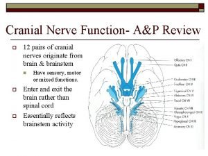
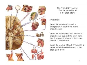
THE CRANIAL NERVES IN THE CRANIAL CAVITY THE 12 PAIRS OF CRANIAL NERVES ARE NAMED AS FOLLOWS: I. OLFACTORY (SENSORY) II. OPTIC (SENSORY) III. OCULOMOTOR (MOTOR) IV. TROCHLEAR (MOTOR) V. TRIGEMINAL (MIXED) VI. ABDUCENT (MOTOR) VII. FACIAL (MIXED) VIII. VESTIBULOCOCHL EAR (SENSORY) IX. GLOSSOPHARYNGEAL (MIXED) X. VAGUS (MIXED) XI. ACCESSORY (MOTOR) XII. HYPOGLOSSAL (MOTOR)

Origin of the 12 cranial nerves CEREBRUM 1&2 BRAINSTEM MIDBRAIN 3&4 PONS 5, 6, 7, & 8 MEDULLA 9, 10, 11 & 12 Accessory nerve (11 th) has dual origin – Cranial & spinal root Only one nerve arise from dorsal aspect – Trochlear nerve (4 th)

Cribriform plate of ethmoid bone Olfactory tract Olfactory bulb Posterior supe ri or lateral nasal branches from maxill ary nerve (V 2) Lateral internal nasal bran ch of anterior ethmoidal nerve (V 1) Maxill ary nerve (V 2) (sphen opalatine foramen disse cted away) External nasal bra nch of anterior ethmoidal nerve (V 1) Greater petrosal nerve Nerve (Vidi an) of pterygoid canal Nasopala tine nerve (V 2) passing t o septum (cut) Pterygopa. Iatine gang. Ii on Pharyngeal bran ch of maxill ary nerve (V 2) Posteri or i nferi or lateral nasal branch from greater palatine nerve (V 2) Greater palatine nerves (V 2) Lesser palatine nerves (V 2)

Clinical Features of the Neonatal Skull FONTANELLES Palpation of the fontanelles enables the physician to determine 1 -The progress of growth in the surrounding bones, 2 -the degree of hydration of the baby if the fontanelles are depressed below the surface THE BABY IS DEHYDRATED a bulging fontanelle indicates RAISED INTRACRANIAL PRESSURE

Samples of cerebrospinal fluid can be obtained by passing a long needle obliquely through the anterior fontanelle into the subarachnoid space CLOSES anterior after 18 months, because the frontal and parietal bones have enlarged to close the gap.

Intracranial Hemorrhage Intracranial hemorrhage may result from trauma or cerebral vascular lesions. Four varieties are considered here: EXTRADURAL SUBARACHNOIDl Cerebral

Extradural hemorrhage results from injuries to the meningeal arteries or veins. The most common artery to be damaged is the anterior division of the middle meningeal artery Bleeding occurs and strips up the meningeal layer of dura from the internal surface of the skull. The intracranial pressure rises, and the enlarging blood clot exerts local pressure on the underlying motor area in the precentral gyrus.

Epidural Hemorrhage is a medical emergency. The blood vessel involved is the middle meningeal artery. Clinical features include: A CT scan shows a lens-shaped (biconvex) hyperdensity adjacent to bone arterial blood is located between the skull and dura lucid interval (no symptoms) for a few hours followed by death (“talk and die syndrome”) CT-Brain

(Temporal Lobe) Herniation

: pl ir I I l I mt m

Lucid interval lucid interval is a temporary improvement in a patient's condition after a traumatic brain injury, after which the condition deteriorates It occurs after the patient is knocked out by the initial concussive force of the trauma, then lapses into unconsciousness again after recovery when bleeding causes the hematoma to expand past the point at which the body can no longer compensate A lucid interval is especially indicative of an epidural hematoma. An estimated 20 to 50% of patients with epidural hematoma experience such a lucid interval. It can last minutes or hours To stop the hemorrhage, the torn artery or vein must be ligated or plugged. The burr hole through the skull wall should be placed about 1 to 1. 5 in. (2. 5 to 4 cm) above the midpoint of the zygomatic arch.

Subdural Hemorrhage A subdural hemorrhage is caused by a violent shaking of the head (e. g. , child abuse or car accident) and commonly occurs in alcoholics and elderly. . The blood vessels involved are the superior cerebral veins (“bridging veins”). Clinical features include: A CT scan shows a thin, crescent-shaped hyperdensity that hugs the contours of the brain; venous blood is located between the dura and arachnoid; blood accumulates slowly (days to weeks after trauma); no blood in the CSF after lumbar puncture.

A Cerebralvein Duralborder hemao tma 4 . , , , A '/ Arachnoid. , , B. Subdural Hematoma*

Epidural above the dura V sua 1 Jy arterial

A subarachnoid hemorrhage is caused by a contusion or laceration injury to the brain or a berry aneurysm. Subarachnoid Hemorrhage The blood vessels involved are the cerebral arteries or the anterior or posterior communicating arteries. Clinical features include: A CT scan shows a hyperdensity in the cisterns, fissures, and sulci of the brain; thickening of the falx cerebri; arterial blood with the subarachnoid space; irritation of the meninges causes a sudden onset of the “worst headache of my life”; stiff neck; vomiting; decreased mentation; early “herald headache” may occur; and blood within the CSF after lumbar puncture.

Cerebral hemorrhage is generally caused by rupture of the thin-walled a branch of the middle cerebral artery. The hemorrhage involves the vital corticobulbar and corticospinal fibers in the internal capsule and produces hemiplegia on the opposite side of the body. The patient immediately loses consciousness, and the paralysis is evident when consciousness is regained
- Slides: 63

