Topographical anatomy of the viscerocranium External carotid artery


: The superior thyroid artery arising from the external carotid artery")


")


 becomes")

 lacrimal gland middle meningeal nerve ORBIT sphenopalatine foramen rotundum trigeminal")


- Slides: 34
Topographical anatomy of the viscerocranium: External carotid artery, trigeminal nerve Supraorbital margin NEUROCRANIUM VISCEROCRANIUM External acustic meatus Mark Kozsurek, M. D. , Ph. D. mark@kozsurek. hu EM II. . , 10/02/2020
External carotid artery
superficial temporal maxillary neck of mandible ICA ECA top of thyroid cartilage External carotid artery extends from the superior margin of thyroid cartilage to the neck of the mandible. CCA
Dessie, Meselech Ambaw (2018): The superior thyroid artery arising from the external carotid artery and EBSLN near the upper pole of the thyroid gland; CCA = Common carotid artery, ECA = External carotid artery, ICA = Internal carotid artery, IJV = Internal jugular vein, SLA = Superior laryngeal artery, EBSLN = External branch of the superior laryngeal nerve. . PLOS ONE. Figure. https: //doi. org/10. 1371/journal. pone. 0197075. g 001 Note that ICA is behind ECA and has no extracranial branches!
ANTERIOR BRANCHES OF ECA superficial temporal angular maxillary nasal branches ascending palatine ICA ECA lingual sublingual facial sup. laryngeal sup. thyroid CCA submental labial branches
ECA branches in the submandibular region Facial artery branches off where ECA is crossed by the post. belly of digastric (po. D) and the stylohyoid (SH), then it touches the submandibular gland turns onto the face along the anterior margin of masseter. 3. po. D SH Lingual artery arises at the level of the greater horn of hyoid bone and runs behind the hyoglossus (HG) muscle. Superior thyroid artery is the only branch of the ECA running downward. Before reaching the thyroid gland, gives off a branch entering the larynx through the thyrohyoid membrane. 2. 2. HG 1. superior thyroid - sup. laryngeal 2. lingual artery 3. facial artery
ECA branches around the tongue and larynx HG 1. superior thyroid sup. laryngeal 2. lingual artery GG 2. 1. Note the lingual artery in the medial lingual sulcus bounded by the hyoglossus (HG) and genioglossus (GG) muscles.
2. lingual artery - sublingual artey Hyoglossus m. Lingual artery runs medial to the hyoglossus muscle. This muscle isolates the lateral and the medial lingual sulcus.
2. lingual artery 3. facial artery Facial artery terminates as the angular artery at the root of the nose, but before reaching that point, it gives branches to the lower and upper lips and to the nose 3. . ECA branches on the face 2. 3. .
POSTERIOR BRANCHES OF ECA superficial temporal angular stylomastoid occipital transverse facial maxillary post. auricular nasal branches ascending palatine ICA ECA lingual sublingual facial sup. laryngeal sup. thyroid CCA submental labial branches
2. lingual artery 3. facial artery --------------4. occipital artery 5. post. auricular 5. 4. 3. . 2.
MEDIAL BRANCH OF ECA superficial temporal angular post. meningeal stylomastoid occipital transverse facial maxillary post. auricular nasal branches ascending palatine ICA ECA ascending pharyngeal Ascending pharyngeal artery arises from ECA on its medial side and ascends along the wall of pharynx, then enters the skull through the jugular foramen and supplies the dura as the posterior meningeal artery. lingual sublingual facial sup. laryngeal sup. thyroid CCA submental labial branches
Maxillary artery and its branches Arises from the external carotid artery right behind the neck of mandible and runs as a wavy structure in the infratemporal fossa. Supplies: - muscles of mastication (masseteric, deep temporal and pterygoid branches) - reaches all the three big compartments of the facial skeleton - ORBIT (infraorbital artery) - NASAL CAVITY (sphenopalatine artery) - ORAL CAVITY (inferior alveolar, buccal, descending palatine arteries) - middle zone of the dura mater (middle meningeal artery)
T III II I LP g lin ian l sm MP I. : mandibular (or bony) part Runs along the inferior border of lateral pterygoid muscle (LP) and passes between the neck of mandible and the sphenomandibular lig (sml). , crosses the inferior alveolar nerve (ian). II. : pterygoid (or muscular) portion Runs obliquely upward and forward on the outer surface of lateral pterygoid muscle and is covered by the temporalis muscle (T). Passes between the superior and inferior bellies of lateral pterygoid and reaches the pterygomaxillary fissure III. : pterygomaxillary part Terminal portion within the pterygopalatine fossa.
Branches of 3 rd part reach all the compartments of the viscerocranium!
3 rd part branches in the orbit (Do not forget: contents of orbit are mainly supplied by the ophthalmic artery from ICA!) Infraorbital artery supplies upper incisors and canine teeth and the middle region of the face (lower eyelids, nose, upper lip). Upper premolars and molars get blood from posterior superior alveolar artery.
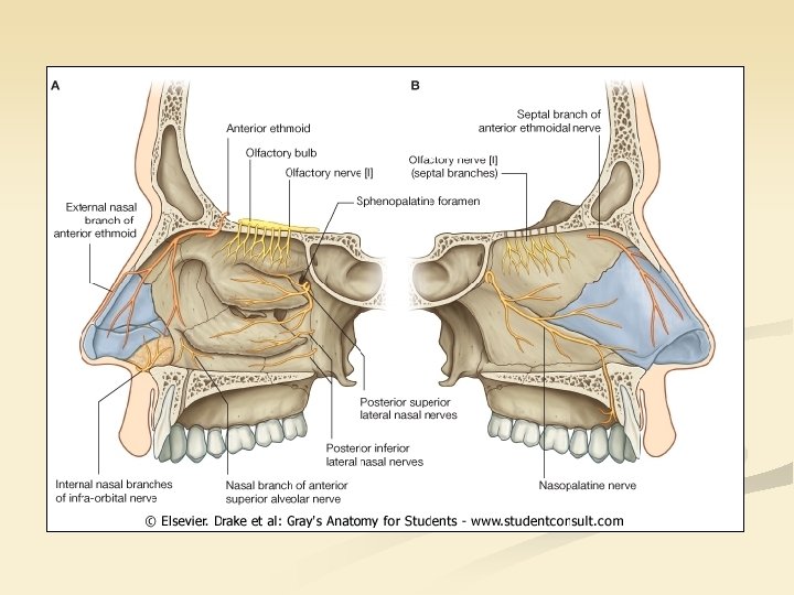
3 rd part branches in the nasal cavity (Anterior and posterior ethmoidal arteries come from the orbit and are the branches of ophthalmic artery. Sphenopalatine artery enters through the sphenopalatine foramen and supplies the inferior-posterior part of the nasal cavity. Its strongest branch is the nasopalatine which reaches the hard palate via the incisive foramen.
3 rd part branches in the oral cavity Anastomosis on the palate: n incisive artery (maxillary-spenopalatine-nasopalatine arteries) n descending palatine artery (directly from maxillary artery) Lower teeth are supplied by the inferior alveolar artery.
Trigeminal nerve (CN 5)
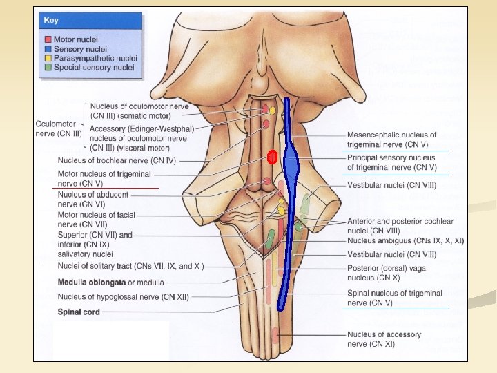
n Trigeminal nerve has distinct nuclei for all the three modalities of sensory information: n protopathic (pain, heat): spinal trigeminal nucleus n epicritic (fine touch, vibration, two-point discrimination): chief (or principal or pontine) sensory trigeminal nucleus n proprioceptive (position and movement of joints and muscles): mesencephalic trigeminal nucleus (Must be noted that the mesencephalic trigeminal nucleus is rather an internalized sensory ganglion with pseudounipolar cells then a real brainstem nucleus!) n Trigeminal nerve is the sensory nerve of the facial skeleton (including the skin, the mucous membrane of the nasal and oral cavities, the teeth and the structures of the orbit). n Trigeminal nerve innervates those muscles developing from the first pharyngeal arch (muscles of mastication; Mylohyoid, anterior belly of Digastric, Tensor tympani and Tensor veli palatini). Get your smartphones ready! QR code is comming!
r n. ochlea ant. ethmoid for. zygomatic n. inf. orbital fissure y ar ili c so na post. ethmoid for. lacrimal gland supraorbital foramen lo cilianrg nerv y es frontal n. lac rim al n. ant. ethm. n. post. ethmoidal n. frontal notch infratr ant. sup. bran nasal ches ant. meningeal nerve supratrochlear n. supraorbital n. med. et lat. branches n. sup. orbital fissure tentorial branches trigeminal ggl. pterygopalatine ggl. in the pterygopalatine fossa for. rotundum greater petrosal n. from CN VII. Ophthalmic nerve (V/1. )
LPS SO lac fr 4 The first muscle you observe following the removal of the roof of the orbit is the Levator palpebrae superioris (LPS) and medially the Superior oblique (SO). From medial to lateral direction the trochlear (4), frontal (fr) and lacrimal (lac) nerves appear.
LPS SO Following the removal of the Superior oblique the Medial rectus (MR) becomes visible. SR Close to the medial wall the nasociliary nerve (nc) is seen. MR nc cil 6 3 LR Note the ciliary ganglion (cil) on the lateral side of the optic nerve and the abducens nerve (6) on the surface of Lateral rectus muscle (LR). The most of the muscles of the eyeball are innervated by the branches of oculomotor nerve (3).
Ophthalmic artery: roughly has the same branches as the ophthalmic nerve!
Maxillary nerve (V/2. ) lacrimal gland middle meningeal nerve ORBIT sphenopalatine foramen rotundum trigeminal ggl. zygomatic nerve inf. orbital fissure infraorbital foramen sup. alveolar branches infraorbital nerve * pterygoid canal greater petrosal nerve from CN VII. pharyngeal and esophageal branches greater and lesser palatine canals lesser palatine nerves * fibres to the pterygopalatine ganglion nasal br. NASAL CAVITY nasopalat ine n. n i t a l a rp greate ORAL CAVITY pharyngeal, palatine, nasal glands palatine taste buds incisive canal
Posterior, middle and anterior superior alveolar nerves constitute the superior alveolar plexus. From this the dental branches enter through the root canals of the superior teeth and innervate the pulp, while gingival branches arrising also from this plexus supply the buccal gingiva. (Palatine gingiva is innervated by the incisive nerve – terminal branch of the nasopalatine –, and the greater palatine nerve. )
mandibular meningeal nerve trigeminal ggl. foramen spinosu m ale for anterior motor group parotid gland chorda tympani from CN VII. otic ggl. buccal branches (!) auriculotemporal nevre branches of the motor group: masseteric nerve deep temporal nerve submandibular medial pterygoid nerve gland lateral pterygoid nerve tensor tympani nerve tensor veli palatini nerve buccal nerves (!) Mandibular nerve (V/3. ) lesser petrosal nerve from CN IX. n ov e m a lin gua ln erv e taste buds of tongue sublingual inf. alveolargland nerve submandibular ggl. mylo hyoid nerve (!) Mylohyoid Digastric, ant. belly
Infratemporal region - medial and lateral pterygoid muscles - maxillary artery - mandibular nerve
Pterygomandibular space It is enclosed by the internal surface of the ramus of the mandible, the medial pterygoid and the lateral pterygoid muscle. Contains: inferior alveolar artery, vein and nerve, lingual nerve
Lingual sulci
Any feed-back is appreciated! Thank you!