Cranial Cavity Anterior Cranial Fossa Middle Cranial Fossa


- Slides: 26
Cranial Cavity
Anterior Cranial Fossa Middle Cranial Fossa Posterior Cranial Fossa
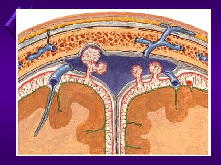
emissary v. diploic veins
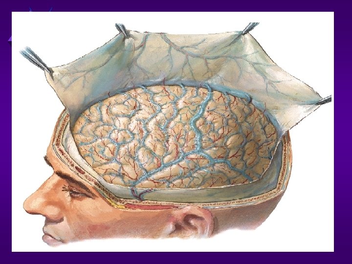
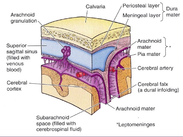
The meninges The brain & spinal cord are surrounded by 3 membranes the dura , arachnoid and pia maters. Dura mater of brain: It is formed from 2 layers. which are united except along certain lines, where they separate to form sinuses. The endosteal layer is the ordinary periosteum covering the inner surface of the skull bones. The meningeal layer: is the dura mater proper. It is a dense, strong, fibrous membrane covering the brain.
Cerebral
DURAL FOLDS Definition: inward reduplication of inner layer of dura mater that form membranous folds between different parts of the brain. Types of dural folds: 1 - vertical folds: - falxi cerebri. - falx cerebelli. 2 - horizontal folds: - diaphragma sellae -tentorium cerebella. - cavum trigeminale.
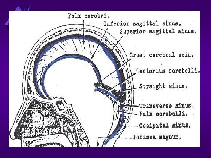
The falx cerebri It is a sickle- shaped fold of dura mater that lies in the midline between the 2 cerebral hemispheres. Its narrow end in front is attached to the internal frontal crest & crista galli. Its broad posterior part blends in the midline with the upper surface of the tentorium cerebelli. Sinuses related to falx cerebri: -superior sagittal sinus runs in its upper fixed margin. -inferior sagittal sinus runs in lower concave free margin. -straight sinus runs along its attachment to tentorium cerebelli.
Tentorium cerebelli
The tentorium cerebelli -It is crescent- shaped fold of dura mater that roofs over the posterior cranial fossa. -It covers the upper surface of the cerebellum & supports the occipital lobes of the cerebral hemispheres. -The falx cerebri & the falx cerebelli are attached to the upper & lower surfaces of the tentorium. Sinuses related to tentorium cerebelli: The superior petrosal sinus runs along its attachment to the petrous bone, and the transverse sinus along its attachment to the occipital bone. The straight sinus runs along its attached to falx cerebri.
Falx cerebelli It is a small sickle- shaped fold of dura mater that is attached to the internal occipital crest and projects forward between the 2 cerebellar hemispheres. Its posterior margin contains the occipital sinus. Sinuses related to falx cerebelli: 1 - straight sinus: 2 - occipital sinus:
Diaphragma sellae It is a small cicular fold of dura mater that forms the roof for sella turcica. A small opening in its center allows passage of the stalk of the hypophysis cerebri.
Dural Venous Sinuses Definition: venous channels between the 2 layers of dura mater inside the Cranial cavity. - Classification: Single sinuses - superior sagittal sinus. - straight sinus. - inferior sagittal sinus. - occipital sinus. - basilar sinus. paired sinuses - sphenoparietal sinus. - petrosquamous sinus. - cavernous sinus. - superior petrosal sinus. - inferior petrosal sinus. - transverse sinus. - sigmoid sinus.
Superior Sagittal Sinus Sphenoparietal Cavernous Superior Petrosal Straight Confluence Basilar Sigmoid Transverse
Superior Sagittal Dural Sinuses Inferior Sagittal Great Cerebral Sup/Inf Petrosal Straight Confluence Occipital Transverse Sigmoid
DURAL VENOUS SINUSES
CAVERNOUS SINUS Site: The cavernous sinuses are two large venous plexuses that lie on either side of the body of the sphenoid bone, extending from the superior orbital fissure to the apex of the petrous temporal bone, with an average length of 2 cm and width of 1 cm.
Relations: - Anteriorly: superior orbital fissure. - Posteriorly: trigeminal ganglion and apex of pterous temporal bone. - Superiorly: ICA. - Inferiorly: sphenoidal air sinus and body of sphenoid. - Medially: pituitary gland, sphenoidal air sinus and body of sphenoid - Laterally: uncus of the temporal lobe.
Contents: - structures in the lateral wall of the sinus: - oculomotor N. - trochlear N. - ophthalmic N. - maxillary N. - structures inside the sinus: - abducent N. - internal carotid artery.
Tributaries & communications of the cavernous sinus - ophthalmic veins, sphenoparietal vein and central retinal vein: opens anteriory. - superficial middle cerebral vein and inferior cerebral veins: open into its upper surface. - emissary vein: from pterygoid plexus through foramen ovale and foramen lacerum. - the facial vein via the superior ophthalmic vein Driange of the sinus: to the transverse sinus via the superior petrosal sinus; to the internal jugular vein via the inferior petrosal sinus
CAVERNOUS SINUS & CONTENTS Pituitary Gland CN III CN IV CN VI CN V 1 CN V 2 Internal Carotid
Cavernous sinus Superior ophthalmic vein Angular vein Inferior ophthalmic vein Facial vein Deep facial vein Pterygoid venous plexus Maxillary vein Retromandibular vein