Essentials of Human Anatomy Physiology Elaine N Marieb

Essentials of Human Anatomy & Physiology Elaine N. Marieb Seventh Edition Chapter 7 The Nervous System. The Peripheral Nervous System This presentation contains copyright protected materials. Lecture Slides in Power. Point by Jerry L. Cook Copyright © 2003 Pearson Education, Inc. publishing as Benjamin Cummings

Functions of the Nervous System · Sensory input – gathering information · To monitor changes occurring inside and outside the body · Changes = stimuli · Done by a sensory receptor (Ex. - Rods and cones of eye, olfactory neurons of nose, touch receptors in integument…)

Functions of the Nervous System · Integration · To process and interpret sensory input and decide if action is needed · Done in brain or spinal cords · Motor output · A response to integrated stimuli · The response activates muscles or glands (effectors)
 · Brain ·")
Structural Classification of the Nervous System · Central nervous system (CNS) · Brain · Spinal cord Copyright © 2003 Pearson Education, Inc. publishing as Benjamin Cummings Slide 7. 2
 · Nerves outside")
Structural Classification of the Nervous System · Peripheral nervous system (PNS) · Nerves outside the brain and spinal cord Copyright © 2003 Pearson Education, Inc. publishing as Benjamin Cummings Slide 7. 2

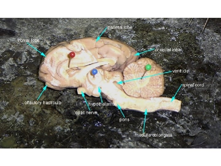
Regions of the Brain · Cerebral hemispheres · Diencephalon · Brain stem · Cerebellum Copyright © 2003 Pearson Education, Inc. publishing as Benjamin Cummings
 · Paired (left and right) superior parts of the brain ·")
Cerebral Hemispheres (Cerebrum) · Paired (left and right) superior parts of the brain · Include more than half of the brain mass cwx. prenhall. com/. . . /custom 1/deluxe-content. html
 · The surface is made of ridges (gyri) and grooves (sulci)")
Cerebral Hemispheres (Cerebrum) · The surface is made of ridges (gyri) and grooves (sulci) 1 - Central sulcus 2 - Precentral gyrus 3 - Postcentral gyrus 10 - Lateral sulcus 16 - Parieto-occipital sulcus Figure 7. 13 a Slide 7. 28 b
 divide the cerebrum into lobes ·")
Lobes of the Cerebrum · Fissures (deep grooves) divide the cerebrum into lobes · Surface lobes of the cerebrum · Frontal lobe · Parietal lobe · Occipital lobe · Temporal lobe Slide 7. 29 a

Specialized Areas of the Cerebrum · Somatic sensory area – receives impulses from the body’s sensory receptors · Primary motor area – sends impulses to skeletal muscles · Broca’s area – involved in our ability to speak Copyright © 2003 Pearson Education, Inc. publishing as Benjamin Cummings Slide 7. 30

Specialized Area of the Cerebrum · Cerebral areas involved in special senses · Gustatory area (taste) · Visual area (sight) · Auditory area (hearing) · Olfactory area (smell) Copyright © 2003 Pearson Education, Inc. publishing as Benjamin Cummings Slide 7. 32 a

Specialized Area of the Cerebrum · Interpretation areas of the cerebrum · Speech/language region · Language comprehension region · General interpretation area Copyright © 2003 Pearson Education, Inc. publishing as Benjamin Cummings Slide 7. 32 b

Layers of the Cerebrum · Gray matter · Outer layer · Composed mostly of neuron cell bodies Figure 7. 13 a Copyright © 2003 Pearson Education, Inc. publishing as Benjamin Cummings Slide 7. 33 a

Layers of the Cerebrum · White matter · Fiber tracts inside the gray matter · Example: corpus callosum connects hemispheres Figure 7. 13 a Copyright © 2003 Pearson Education, Inc. publishing as Benjamin Cummings Slide 7. 33 b

Layers of the Cerebrum · Basal nuclei – internal islands of gray matter Figure 7. 13 a Copyright © 2003 Pearson Education, Inc. publishing as Benjamin Cummings Slide 7. 33 c

Diencephalon · Sits on top of the brain stem · Enclosed by the cerebral heispheres Slide 7. 34 a

Diencephalon · Made of three parts · Thalamus · Hypothalamus · Epithalamus Slide 7. 34 a

Thalamus · Surrounds the third ventricle · The relay station for sensory impulses · Transfers impulses to the correct part of the cortex for localization and interpretation Copyright © 2003 Pearson Education, Inc. publishing as Benjamin Cummings Slide 7. 35

Hypothalamus · Under the thalamus · Important autonomic nervous system center · Helps regulate body temperature · Controls water balance · Regulates metabolism
 · The pituitary gland")
Hypothalamus · An important part of the limbic system (emotions) · The pituitary gland is attached to the hypothalamus Copyright © 2003 Pearson Education, Inc. publishing as Benjamin Cummings

Epithalamus · Forms the roof of the third ventricle · Houses the pineal body (an endocrine gland) · Includes the choroid plexus – forms cerebrospinal fluid Copyright © 2003 Pearson Education, Inc. publishing as Benjamin Cummings Slide 7. 37

Brain Stem · Attaches to the spinal cord · Parts of the brain stem · Midbrain · Pons · Medulla oblongata Copyright © 2003 Pearson Education, Inc. publishing as Benjamin Cummings Slide 7. 38 a

Midbrain · Mostly composed of tracts of nerve fibers · Has two bulging fiber tracts – cerebral peduncles · Has four rounded protrusions – corpora quadrigemina · Reflex centers for vision and hearing Slide 7. 39

Pons · The bulging center part of the brain stem · Mostly composed of fiber tracts · Includes nuclei involved in the control of breathing Copyright © 2003 Pearson Education, Inc. publishing as Benjamin Cummings Slide 7. 40

Medulla Oblongata · · The lowest part of the brain stem Merges into the spinal cord Includes important fiber tracts Contains important control centers · Heart rate control · Blood pressure regulation · Breathing · Swallowing · Vomiting Copyright © 2003 Pearson Education, Inc. publishing as Benjamin Cummings Slide 7. 41

Reticular Formation · Diffuse mass of gray matter along the brain stem · Involved in motor control of visceral organs · Reticular activating system plays a role in awake/sleep cycles and consciousness Copyright © 2003 Pearson Education, Inc. publishing as Benjamin Cummings Slide 7. 42 a

Cerebellum · Two hemispheres with convoluted surfaces · Provides involuntary coordination of body movements Copyright © 2003 Pearson Education, Inc. publishing as Benjamin Cummings Slide 7. 43 a

The Limbic System - Evolutionarily primitive structures -Located on top of the brainstem and buried under the cortex http: //www. umm. edu/images/ency/fullsize/limbic_system_19244. jpg

The Limbic System Involved in : - Emotions and motivations, particularly those related to survival (fear, anger, and emotions related to sexual behavior) - Feelings of pleasure related to survival, such as eating and sex. http: //files. bsc. edu/CLASSES/Terry%20 Goodrick/limbic_system 2. jpg
- a very powerful biological force for survival. If you")
The Pleasure Circuit Pleasure (reward)- a very powerful biological force for survival. If you do something pleasurable, the brain is wired such that you tend to do it again. Life sustaining activities, such as eating, activate a circuit of specialized nerve cells devoted to producing and regulating pleasure.

The Pleasure Circuit One important set of nerve cells, which uses a chemical neurotransmitter called dopamine, sits at the very top the brainstem in the ventral tegmental area (VTA) Dopamine-containing neurons relay messages about pleasure through their nerve fibers to nerve cells in the nucleus accumbens. http: //www. niaaa. nih. gov/publications/arh 26 -2/26_2 images/brain. gif

The Pleasure Circuit Other fibers reach to a related part of the frontal region of the cerebral cortex. http: //abdellab. sunderland. ac. uk/Lectures/Reward/Pics/DAsystem. png

SUBSTANTIA NIGRA Another part of the brain effected by drugs is the substantia nigra. -Found in the midbrain -Controls movement, attention, releases dopamine, produces GABA, controls eye movement (along with frontal eye field) -One of the two primary output nuclei of the brain's basal ganglia http: //www. memorylossonline. com/glossary/substantianigra. jpg

Cerebrospinal Fluid · Similar to blood plasma composition · Formed by the choroid plexus · Forms a watery cushion to protect the brain Copyright © 2003 Pearson Education, Inc. publishing as Benjamin Cummings Slide 7. 46

Location of the Cerebrospinal Fluid Circulated in arachnoid space, ventricles, and central canal of the spinal cord Figure 7. 17 b Copyright © 2003 Pearson Education, Inc. publishing as Benjamin Cummings Slide 7. 47 b

Protection of the Central Nervous System · Scalp and skin · Skull and vertebral column · Meninges Figure 7. 16 a Copyright © 2003 Pearson Education, Inc. publishing as Benjamin Cummings Slide 7. 44 a

Protection of the Central Nervous System · Cerebrospinal fluid · Blood brain barrier Figure 7. 16 a Copyright © 2003 Pearson Education, Inc. publishing as Benjamin Cummings Slide 7. 44 b

Meninges · Dura mater · Double-layered external covering · Periosteum – attached to surface of the skull · Meningeal layer – outer covering of the brain · Folds inward in several areas Slide 7. 45 a

Meninges · Arachnoid layer · Middle layer · Web-like · Pia mater · Internal layer · Clings to the surface of the brain Copyright © 2003 Pearson Education, Inc. publishing as Benjamin Cummings Slide 7. 45 b

The Blood-Brain Barrier - Capillaries of the brain with tight junctions between their endothelial cells. - Normally, glucose, salt, water, and certain nutrients are actively passed into the brain. -Alcohol, respiratory gases, nicotine, and anesthesia may cross. -Fat-soluble molecules travel through much faster. -For example, morphine takes about 20 -30 minutes to cross the barrier. If it is converted to the fat-soluble heroin, it will

Spinal Cord · Extends from the medulla oblongata to the region of T 12 · Below T 12 is the cauda equina (a collection of spinal nerves) · Enlargements occur in the cervical and lumbar regions Figure 7. 18 Copyright © 2003 Pearson Education, Inc. publishing as Benjamin Cummings Slide 7. 52

Spinal Cord Anatomy · Exterior white mater – conduction tracts Figure 7. 19 Copyright © 2003 Pearson Education, Inc. publishing as Benjamin Cummings Slide 7. 53 a
")
Spinal Cord Anatomy · Internal gray matter - mostly cell bodies · Dorsal (posterior) horns · Anterior (ventral) horns Figure 7. 19 Copyright © 2003 Pearson Education, Inc. publishing as Benjamin Cummings Slide 7. 53 b

Spinal Cord Anatomy · Central canal filled with cerebrospinal fluid Figure 7. 19 Copyright © 2003 Pearson Education, Inc. publishing as Benjamin Cummings Slide 7. 53 c

Spinal Cord Anatomy · Meninges cover the spinal cord · Nerves leave at the level of each vertebrae · Dorsal root- contains sensory (afferent) neurons · Associated with the dorsal root ganglia – collections of cell bodies outside the central nervous system · Ventral rootcontains motor (efferent) neurons Copyright © 2003 Pearson Education, Inc. publishing as Benjamin Cummings Slide 7. 54

Corpus Callosum Grey Matter Cerebrum Cerebellum White Matter Spinal Cord Hypothalamus Thalamus Midbrain Medulla

Pituitary Meninges Cerebrum Olfactory bulb Pineal Glad Pons
 · Nerves outside")
Structural Classification of the Nervous System · Peripheral nervous system (PNS) · Nerves outside the brain and spinal cord Copyright © 2003 Pearson Education, Inc. publishing as Benjamin Cummings Slide 7. 2

Why did the neuron get sent to the principal’s office?

It had trouble controlling its impulses. http: //www. unm. edu/~lkravitz/MEDIA 2/Action%20 Potential. jpg

Peripheral Nervous System · Nerve = bundle of neuron fibers Copyright © 2003 Pearson Education, Inc. publishing as Benjamin Cummings Slide 7. 55

Structure of a Nerve · Neuron fibers are bundled by connective tissue · Endoneurium surrounds each fiber · Groups of fibers are bound into fascicles by perineurium · Fascicles are bound together by epineurium Copyright © 2003 Pearson Education, Inc. publishing as Benjamin Cummings Figure 7. 20 Slide 7. 56
 nerves – carry impulses toward the CNS Copyright")
Classification of Nerves · Afferent (sensory) nerves – carry impulses toward the CNS Copyright © 2003 Pearson Education, Inc. publishing as Benjamin Cummings Slide 7. 57
 nerves – carry impulses away from the CNS")
Classification of Nerves · Efferent (motor) nerves – carry impulses away from the CNS Copyright © 2003 Pearson Education, Inc. publishing as Benjamin Cummings Slide 7. 57

Classification of Nerves · Some nerves are strictly sensory OR motor · Mixed nerves – both sensory and motor fibers Copyright © 2003 Pearson Education, Inc. publishing as Benjamin Cummings Slide 7. 57

When does a brain get afraid?

When it loses its nerve.

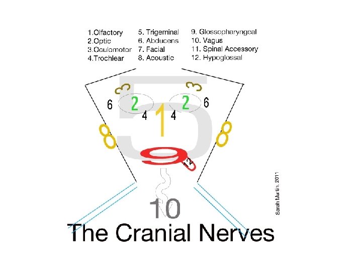
Cranial Nerves · 12 pairs of nerves that mostly serve the head and neck · Numbered I-XII (1 -12) from front to back · Also named Copyright © 2003 Pearson Education, Inc. publishing as Benjamin Cummings Slide 7. 58

Cranial Nerves · I Olfactory nerve – sensory for smell Copyright © 2003 Pearson Education, Inc. publishing as Benjamin Cummings Slide 7. 60

Cranial Nerves · II Optic nerve – sensory for vision Copyright © 2003 Pearson Education, Inc. publishing as Benjamin Cummings Slide 7. 60

Cranial Nerves · III Oculomotor nerve – motor fibers to eye muscles Copyright © 2003 Pearson Education, Inc. publishing as Benjamin Cummings Slide 7. 60

Cranial Nerves · IV Trochlear – motor fiber to eye muscles Copyright © 2003 Pearson Education, Inc. publishing as Benjamin Cummings Slide 7. 60

Cranial Nerves · V Trigeminal nerve – sensory for the face; motor fibers to chewing muscles Copyright © 2003 Pearson Education, Inc. publishing as Benjamin Cummings Slide 7. 61

Cranial Nerves · VI Abducens nerve – motor fibers to eye muscles Copyright © 2003 Pearson Education, Inc. publishing as Benjamin Cummings Slide 7. 61

Cranial Nerves · VII Facial nerve – sensory for taste; motor fibers to the face Copyright © 2003 Pearson Education, Inc. publishing as Benjamin Cummings Slide 7. 61

Cranial Nerves · VIII Vestibulocochlear nerve – sensory for balance and hearing Copyright © 2003 Pearson Education, Inc. publishing as Benjamin Cummings Slide 7. 61

Cranial Nerves · IX Glossopharyngeal nerve – sensory for taste; motor fibers to the pharynx Copyright © 2003 Pearson Education, Inc. publishing as Benjamin Cummings Slide 7. 62

Cranial Nerves · X Vagus nerves – sensory and motor fibers for pharynx, larynx, and viscera Copyright © 2003 Pearson Education, Inc. publishing as Benjamin Cummings Slide 7. 62

Cranial Nerves · XI Accessory nerve – motor fibers to neck and upper back Copyright © 2003 Pearson Education, Inc. publishing as Benjamin Cummings Slide 7. 62

Cranial Nerves · XII Hypoglossal nerve – motor fibers to tongue Copyright © 2003 Pearson Education, Inc. publishing as Benjamin Cummings Slide 7. 62

Distribution of Cranial Nerves O O O, To Touch And Feel Very Good Velvet, AH! OLd OPie OCCasionally TRies TRIgonometry And Feels VEry GLoomy, VAGUe, Acutely HYPOactive On Occasion Our Trusty Truck Acts Funny. Very Good Vehicle Any How. On Old Olympus' Towering Top A Finely Vested German Viewed A Hawk On Old Olympus' Towering Top A Finn And German Viewed A Hops Oh Once One Takes The Anatomy Final Very Good Vacations Are Heavenly

Cranial Nerves · Most are mixed nerves, but three are sensory only · Some Say Money Matters, But My Brother Says Big Brains Matter More. Copyright © 2003 Pearson Education, Inc. publishing as Benjamin Cummings Slide 7. 58

Spinal Nerves · There is a pair of spinal nerves at the level of each vertebrae for a total of 31 pairs · Spinal nerves are named for the region from which they arise Copyright © 2003 Pearson Education, Inc. publishing as Benjamin Cummings Slide 7. 63

Spinal Nerves · Spinal nerves are formed by the combination of the ventral and dorsal roots of the spinal cord Copyright © 2003 Pearson Education, Inc. publishing as Benjamin Cummings Slide 7. 63
 division · Nerve fibers")
Functional Classification of the Peripheral Nervous System · Sensory (afferent) division · Nerve fibers that carry information to the central nervous system Figure 7. 1 Copyright © 2003 Pearson Education, Inc. publishing as Benjamin Cummings Slide 7. 3 a
 division · Nerve fibers")
Functional Classification of the Peripheral Nervous System · Motor (efferent) division · Nerve fibers that carry impulses away from the central nervous system Figure 7. 1 Copyright © 2003 Pearson Education, Inc. publishing as Benjamin Cummings Slide 7. 3 b
 division · Two subdivisions")
Functional Classification of the Peripheral Nervous System · Motor (efferent) division · Two subdivisions · Somatic nervous system = voluntary · Autonomic nervous system = involuntary Figure 7. 1 Copyright © 2003 Pearson Education, Inc. publishing as Benjamin Cummings Slide 7. 3 c

Autonomic Nervous System · The involuntary branch of the nervous system · Consists of only motor nerves Copyright © 2003 Pearson Education, Inc. publishing as Benjamin Cummings Slide 7. 67

Autonomic Nervous System · Divided into two divisions · Sympathetic division · Parasympathetic division Copyright © 2003 Pearson Education, Inc. publishing as Benjamin Cummings Slide 7. 67

Autonomic Nervous System · Divided into two divisions · Sympathetic division · Parasympathetic division Copyright © 2003 Pearson Education, Inc. publishing as Benjamin Cummings Slide 7. 67

Differences Between Somatic and Autonomic Nervous Systems · Nerves · Somatic – one motor neuron · Autonomic – preganglionic and postganglionic Copyright © 2003 Pearson Education, Inc. publishing as Benjamin Cummings

Differences Between Somatic and Autonomic Nervous Systems · Effector organs · Somatic – skeletal muscle · Autonomic – smooth muscle, cardiac muscle, and glands Copyright © 2003 Pearson Education, Inc. publishing as Benjamin Cummings Slide

Differences Between Somatic and Autonomic Nervous Systems · Neurotransmitters · Somatic – always use acetylcholine · Autonomic – use acetylcholine, epinephrine (adrenaline), or norepinephrine (noradrenaline) Slide 7. 68 b

Types of Reflexes and Regulation · Autonomic reflexes · Smooth muscle regulation · Heart and blood pressure regulation · Regulation of glands · Digestive system regulation Copyright © 2003 Pearson Education, Inc. publishing as Benjamin Cummings Slide 7. 25

Types of Reflexes and Regulation · Somatic reflexes · Activation of skeletal muscles Copyright © 2003 Pearson Education, Inc. publishing as Benjamin Cummings Slide 7. 25

The Reflex Arc · Reflex – rapid, predictable, and involuntary responses to stimuli. A protective response. Figure 7. 11 a Copyright © 2003 Pearson Education, Inc. publishing as Benjamin Cummings Slide 7. 23

Simple Reflex Arc · Reflex arc – direct route from a sensory neuron, to an interneuron, to an effector Figure 7. 11 b, c Figure 7. 11 a Copyright © 2003 Pearson Education, Inc. publishing as Benjamin Cummings Slide 7. 24 Slide 7. 23

Autonomic Functioning · Sympathetic – “fight-or -flight” · Response to unusual stimulus · Takes over to increase activities · Remember as the “E” division = exercise, excitement, emergency, and embarrassment Copyright © 2003 Pearson Education, Inc. publishing as Benjamin Cummings Slide 7. 74 a

Chapter Thirty Three of Harry Potter and the Order of the Phoenix “Fight-or-Flight”

Anatomy of the Sympathetic Division · Originates from T 1 through L 2 Copyright © 2003 Pearson Education, Inc. publishing as Benjamin Cummings Slide 7. 70

Anatomy of the Sympathetic Division · Ganglia are at the sympathetic trunk (near the spinal cord) Copyright © 2003 Pearson Education, Inc. publishing as Benjamin Cummings Slide 7. 70

Anatomy of the Sympathetic Division · Short pre-ganglionic neuron and long postganglionic neuron transmit impulse from CNS to the effector Copyright © 2003 Pearson Education, Inc. publishing as Benjamin Cummings Slide 7. 70

Anatomy of the Sympathetic Division · Norepinephrine and epinephrine are neurotransmitters to the effector organs Figure 7. 26 Copyright © 2003 Pearson Education, Inc. publishing as Benjamin Cummings Slide 7. 70 7. 71

Autonomic Functioning · Parasympathetic – housekeeping activites · Conserves energy · Maintains daily necessary body functions · Remember as the “D” division - digestion, defecation, and diuresis Copyright © 2003 Pearson Education, Inc. publishing as Benjamin Cummings Slide 7. 74 b

Anatomy of the Parasympathetic Division · Originates from the brain stem and S 1 through S 4 · Terminal ganglia are at the effector organs · Always uses acetylcholine as a neurotransmitter Copyright © 2003 Pearson Education, Inc. publishing as Benjamin Cummings Slide 7. 72
 · CNS develops from the embryonic neural tube · The")
Central Nervous System (CNS) · CNS develops from the embryonic neural tube · The neural tube becomes the brain and spinal cord · The opening of the neural tube becomes the ventricles · Four chambers within the brain · Filled with cerebrospinal fluid http: //jan. ucc. nau. edu/~brc/sph 405/class/gross/cns 1/ntube. jpg Slide 7. 26 http: //socrates. berkeley. edu/~jmp/neurocc/5. 4 a. jpg

Development Aspects of the Nervous System · The nervous system is formed during the first month of embryonic development · Any maternal infection can have extremely harmful effects · The hypothalamus is one of the last areas of the brain to develop Copyright © 2003 Pearson Education, Inc. publishing as Benjamin Cummings Slide 7. 75 a

Development Aspects of the Nervous System · No more neurons are formed after birth, but growth and maturation continues for several years- many neuronal connections are made · The brain reaches maximum weight as a young adult · The frontal lobe, associated with judgment and reasoning, is the last to finish developing- at age 21 Copyright © 2003 Pearson Education, Inc. publishing as Benjamin Cummings Slide 7. 75 b

Nervous System Disorders This presentation contains copyright protected materials.

Concussions- Causes http: //www. rosenkilde. com/Concussions. html

Concussions • About 502, 000 children ages 8 to 19 went to emergency rooms with concussions in 2001 to 2005, and about half the injuries were sports -related. About a quarter of the 8 - to 13 -year-olds, generally elementary to middle school, got hurt during organized team sports. Football and ice hockey had the highest concussion rates. Maybe the most alarming finding, though, was that when the researchers looked at a decade's worth of ER visits for sports-related concussions, they found a huge increase even though sports participation declined: The number for 14 - to 19 -year-olds jumped to 22, 000 in 2007 from 7, 000 in 1997; and for 8 - to 13 -year-olds it went to 8, 000 from 3, 800. The numbers don't include concussions treated by trainers and coaches, in family doctors' offices or at urgent care clinics; those that didn't result in formal medical attention; or those involving athletes who hid their symptoms.

Concussions- Symptoms • • • Headache Impaired vision Nausea and vomiting Dizziness Lack of energy Sensitivity to light or sound Balance problems Memory/concentration problems Overly emotional

Concussions- Treatment • Stop the activity causing it! • Get plenty of sleep, minimize physical activity • Minimize strenuous mental activity • Use ice • Pain medication
 • http: //www. bu. edu/today/2010/head-trauma-linked-to-als-like-disease/")
Chronic Traumatic Encephalophathy (CTE) • http: //www. bu. edu/today/2010/head-trauma-linked-to-als-like-disease/
/ Lou Gehrig’s Disease • Progressive degeneration of CNS due to increased")
Amyotrophic Lateral Sclerosis(ALS)/ Lou Gehrig’s Disease • Progressive degeneration of CNS due to increased glutamate • Because of neuron destruction, initiation of muscle movement and muscle control become more and more difficult • About 30, 000 Americans have this- 60% male, 93% caucasian, usually between ages 40 -70

Cause of ALS • Not known for all • For some rare forms- inherited (SOD 1 gene mutation- SOD 1 causes production of an antioxidant enzyme). • Autoimmune responses? —which occur when the body's immune system attacks normal cells. Some scientists theorize that antibodies may directly or indirectly impair the function of motor neurons, interfering with the transmission of signals between the brain and muscles. • Environmental factors? such as exposure to toxic or infectious agents. Other research has examined the possible role of dietary deficiency or trauma. However, as of yet, there is insufficient evidence to implicate these factors as causes of ALS.
/ Lou Gehrig’s Disease- Symptoms • muscle weakness in one or more")
Amyotrophic Lateral Sclerosis(ALS)/ Lou Gehrig’s Disease- Symptoms • muscle weakness in one or more of the following: hands, arms, legs or the muscles of speech, swallowing or breathing • twitching (fasciculation) and cramping of muscles, especially those in the hands and feet • impairment of the use of the arms and legs • "thick speech" and difficulty in projecting the voice • in more advanced stages, paralysis, shortness of breath, difficulty in breathing and swallowing
/ Lou Gehrig’s Disease- Treatment • Riluzole, the first treatment to alter")
Amyotrophic Lateral Sclerosis(ALS)/ Lou Gehrig’s Disease- Treatment • Riluzole, the first treatment to alter the course of ALS, was approved by the FDA in late 1995. This antiglutamate drug was shown scientifically to prolong the life of persons with ALS by at least a few months. More recent studies suggest Riluzole slows the progress of ALS, allowing the patient more time in the higher functioning states when their function is less affected by ALS.

ALS/ Stephen Hawking • http: //videos. howstuffworks. com/discovery/ 30901 -stephen-hawkings-speaking-eyesvideo. htm
- Causes ? Most often in women between the ages of 20")
Multiple Sclerosis (MS)- Causes ? Most often in women between the ages of 20 and 40 • MS is caused by damage to the myelin sheath, the protective covering that surrounds nerve cells. When this nerve covering is damaged, nerve signals slow down or stop. • The nerve damage is caused by inflammation. Inflammation occurs when the body's own immune cells attack the nervous system (autoimmune). This can occur along any area of the brain, optic nerve, and spinal cord. • It is unknown what exactly causes this to happen. The most common thought is that a virus or gene defect, or both, are to blame. Environmental factors may play a role. • You are slightly more likely to get this condition if you have a family history of MS or live in an part of the world where MS is more common http: //www. rosenkilde. com/Multiple. Sclerosis. html

MS- Symptoms • • • Loss of balance Muscle spasms Numbness Coordination problems Vision problems Lack of concentration Depression Slurred speech Trouble swallowing Fatigue

MS- Treatment • Interferon- inhibit some immune cells while stimulating others • Immunosuppresants- decreases immune cells • Steroids- antiinflammatory • Medications for the symptoms

Huntington’s Disease- Cause • Hereditary • Autosomal dominant- a person with the disorder has a 50% chance of passing it on to a child • Symptoms usually don’t start until age 35 • Genetic testing is available http: //vimeo. com/5539318

Huntington’s Disease- Symptoms • Changes in personality and cognition • Increasing lack of balance and coordination • Seizures • Tremor • Chorea- uncontrollable, jerky movements • Dementia • Difficulty with speech

Huntington’s Disease- Treatment • Physical, occupational, and speech therapy • Use of some anticonvulsant medications (antidipressants, antipsychotics)

Strokes- Causes http: //www. womenshealth. gov/publications/ our-publications/fact-sheet/stroke. cfm

Strokes- Symptoms http: //stroke. ahajournals. org/content/30/7/1326/F 2. expansion

Strokes- Treatment • • Rehabilitation after stroke Blood thinners Medications to reduce blood pressure Stopping smoking! Eating healthy!

Cerebral Palsy- Causes • Brain abnormalities or injury prior to or during birth • May be caused by low oxygen levels My cousin, Ross. Now 18 Born at 26 weeks (14 weeks early!)
 Now 18")
Cerebral Palsy My cousin, Ross. Born at 26 weeks (14 weeks early!) Now 18 Impaired movement, especially in legs. Could not see well, so he memorized many children’s books. He has done audio narration for multiple books. Legally blind, but is now an artist. Has sold over 400 paintings.

Cerebral Palsy- Symptoms • Muscle spasticity or weakness, may be localized • Contracture (tightening around joints) • Speech, vision coordination issues • Sometimes decreased mental capacity • Seizures

Cerebral Palsy- Treatment • • Physical, occupational, speech therapy Anticonvulsants Muscle relaxants Botulinum http: //www. pvhmc. org/Physical-

Alzheimer’s Disease http: //www. alz. org/braintour/alzheimers_ch anges. asp Alzheimers tutorial http: //www. examiner. com/holistic-health-in-national/is-it-alzheimer-s-disease-how-to-prepare-for- http: //www. alz. org/braintour/healthy_vs_alzheimers. asp
 before it")
Migraines • Severe headaches • May get an aura (warning symptomsvisual problems) before it begins • Nausea, vomiting, sensitivity to light, numbness, tingling • May last hours to several days • Treated with pain killers or anti-migraine medications http: //images. search. yahoo. com/search/images; _ylt=Al. Bi. VARcdy. JXm 1 d. HEyx. Hs. Debv. Zx 4? p=mig raines&toggle=1&cop=mss&ei=UTF-8&fr=fp-yie 9

Epilepsy • Epilepsy affects nearly 3 million Americans and 50 million people worldwide • Abnormal electrical activity in the brain possibly caused by injury to or changes in the brain. • Periods of disturbed brain function that may include changes in attention or behavior and seizures. • Treated with anticonvulsant medications. If that is unsuccessful, surgery may be necessary.

Meningitis and Encephalitis Meningitis is a viral or bacterial infection of the meninges. It will cause swelling of the meninges, headache, fever, seizure, brain damage, and if untreated, death. Swelling of the brain, usually caused by a virus that a person is exposed to from an insect bite, food, or beverages. It has the same symptoms as meningitis. Meninges http: //www. expeditionzone. com/Storiefiles/thumb/meningitis. jpg http: //www. stjohn. org/healthinfolib

Phineas Gage http: //www. slate. com/articles/health_and_science/2014/05/phin eas_gage_neuroscience_case_true_story_of_famous_frontal_lobe_pati ent. html

Brain Tumor Surgery http: //video. nationalgeographic. com/video/ science/health-human-body-sci/humanbody/brain-tumor-sci/

Best Brain Show EVER!!!!! http: //braingames. nationalgeographic. com/ episode/0/

Remember: He who laughs last thinks slowest…
- Slides: 132