Axial and Appendicular Skeleton Chapters 7 8 Axial

 has 206")


")


 that")


- Slides: 69
Axial and Appendicular Skeleton Chapters 7 & 8
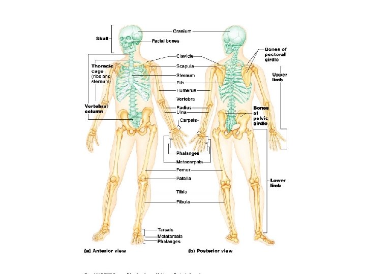
• Axial and Appendicular Skeleton • The skeleton (greek=dried up body) has 206 bones. The axial skeleton is composed of the skull, vertebral column, and rib cage. The appendicular skeleton is composed of the upper and lower limbs and the shoulder and pelvic girdle. • *** see page 147 for the definition of certain bone land marks.
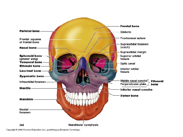
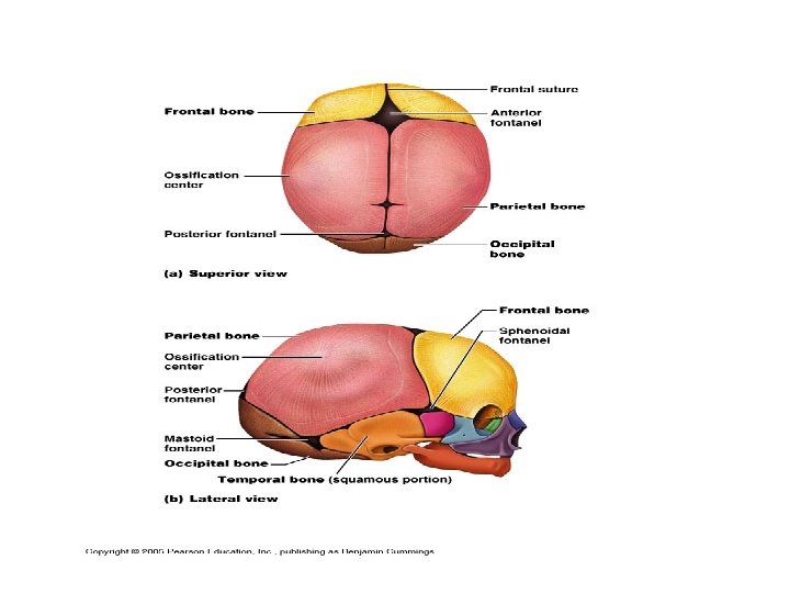
• I. The skull-contains cranial bones that protect the brain, these are joined at sutures. The facial bones make up the rest of the skull and serve several functions. See the section on the fetal skull pg 173 -174 and learn the regions. • A. The skull is composed of many cavities and openings which allow nerves, blood vessels, and organs to function. • B. Cranium- composed of eight bones, two are paired • 1. parietal (2) 3. frontal 5. ethimoid • 2. temporal (3) 4. occipital 6. sphenoid • C. Facial bones- there are 14 facial bones • 1. mandible 4. nasal (2) 7. zygomatics • 2. maxillae (2) 5. vomer 8. lacrimal (2) • 3. palantine(2) 6. inferior nasal conchae (2)
Supracilliary Arch Orbital Part
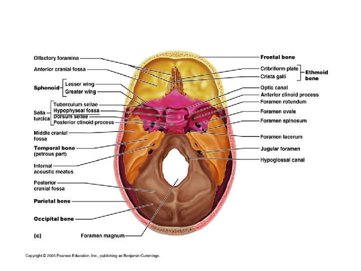
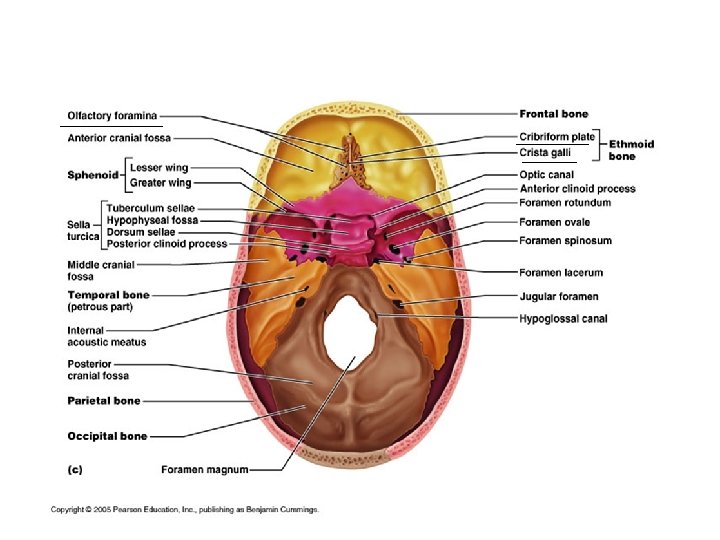
Anterior Cranial Fossa
Parietal Bone and Sutures
Occipital Bone
Occipital Bone
Occipital Bone Groove for sigmoid sinus Groove for transverse dural sinus
Temporal Bone and Zygomatic bone
Temporal Bone
Petrous Part
Temporal Bone Groove for sigmoid sinus Groove for transverse dural sinus
Cranial Fossa
SPHENOID BONE
Sphenoid Bone
Sphenoid Bone
Ethmiod bone Superior Nasal Conchae
Facial Bones Alveolar Margin Alveolar sockets (where teeth insert)
Mandible and Maxillae
Facial Bones
Cleft Palate is a congenital condition in which the two halves of the maxillary and palate bone fuse. The opening affects the roof of the mouth and the nasal cavity and upper lip. It is treated with surgery and can be easily prevented by taking folic acid during pregnancy.
• D. Special parts of the skull • 1. Orbits- cavities that hold the eye and all tissues related to the eye. It is formed by the union of seven bones which also form the lacrimal fossa, optic foramen, and orbital fissures. • 2. Nasal Cavity- made up of bone and cartilage the top is made by the cibriform plate and the bottom by the palantine. There are three conchae (shelfs) two made by the ethmoid bone and one by the inferior conchae bone. The nasal septum divides the nose down the middle. • 3. Paranasal sinuses- air filled cavities around the nasal area that connect to the nasal cavity and help in treating the air as well as in voice resonance. • 4. Hoyd bone- inferior to mandible, only bone that does not have a joint, it is an attachment site for the tongue and other neck muscles.
Bones of the nasal cavity
Bone of the Eye Orbit
Sinuses
Hyoid Bone
Vertebral Column • II. The vertebral column- made up of 26 (33 technically but 9 are fused) bones, forms the body’s axis, it protects the spinal cord. It articulates with the ribs and provides attachment for dorsal muscles and rib muscles. Anterior and posterior longitudinal ligaments, the supraspinous and interspinous ligaments, and the ligamentum flavum stabilize the vertebral column.
Intervertebral Disc • A. Intervertebral discs- composed of fibrocartilage it endures compression and shock absorption. It has two regions the annulus fibrosus and the nucleus pulpusus. They make up 25% of the height of the column and flatten throughout the day.
Vertebral sections and curves • B. Regions and normal curvatures- The spinal column in made up of the cervical (7), throracic (12), lumbar (5), sacrum (5 fused), and coccyx (4 fused). Each region has a curve: concave for cervical and lumbar and convex for thoracic and sacral.
Ligaments of the vertebrae
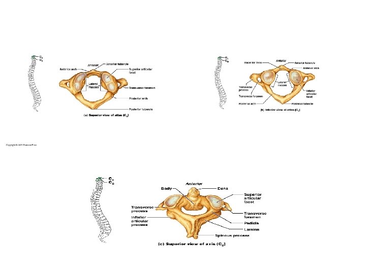
Common vertebral landmarks C. General structure of vertebrae- All vertebrae except the first two cervical vertebrae (atlas and axis) share common characteristics that differ slightly according to region. All vertebrae have a centrum (body), vertebral arch, vertebral foramen, pedicles, laminae, spinous process, tansverse process, and inferior/superior articulating facets.
Axis and Cervical Vertebrae D. Regional vertebral characteristics- each region has vertebrae that serve different functions thus they vary slightly: 1. Cervical vertebrae- rectangle like, bifid spinous process, triangular vertebral foramen, has transverse foramen, superior/anterior facets are located in slightly opposite positions.
Thoracic and Lumbar Vertebrae 2. Thoracic vertebrae- body is slightly heart shape, has demifacets (for ribs), long spinous process, circular vertebral foramen, transverse processes articulate with ribs, provides most of the rotation. 3. Lumbar vertebrae-bears most of the weight so it has a massive body, short thick pedicles and laminae, triangular vertebral foramen, articulating facets prevent rotation, and short thick spinous process.
Anterior and Posterior Sacrum 4. Sacrum-five fused vertebrae, articulates with the bones of pelvis, center of gravity for the human is within this region 5. Coccyx- 4 (or 3 -5) fused vertebrae, also known as the tail bone.
Spinal Abnormalities • E. Abnormalities of the spinal column- These present themselves when the curvatures of the spinal column are exaggerated. • 1. Scoliosis- lateral curvature, most often in thoracic region. The cause is sometimes unknown. Sometimes the vertebrae are deformed and or the one side of muscles is stronger pulling the vertebral column to that side. In some cases scoliosis can cause compression of the lung. • 2. Kyphosis- a bent on a saggital plane along the thoracic region causing a hunchback. Happens most often in women as a result of vertebral fractures due to osteoporosis. • 3. Lordosis – an over curvature of the lumbar region causing a swayed back. This often occurs in people carrying a large long infront or spinal tuberculosis or osteomalacia (boen softening)
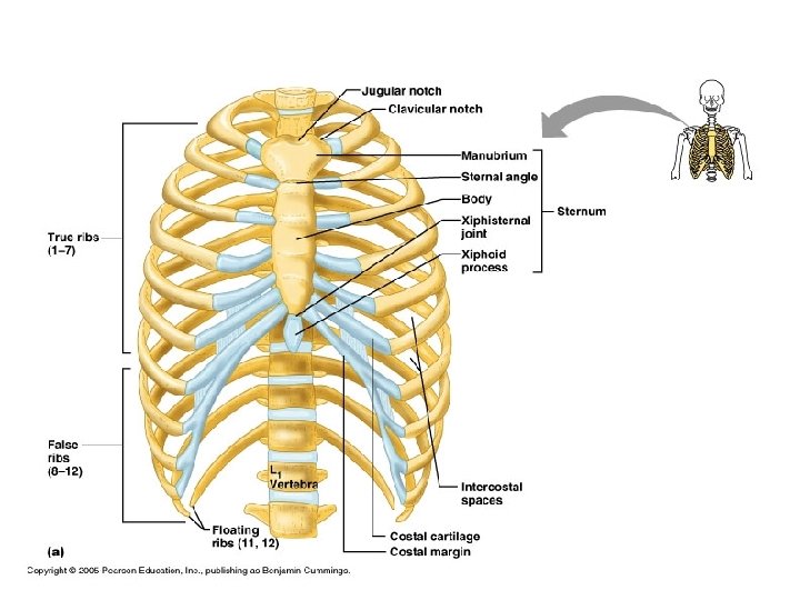
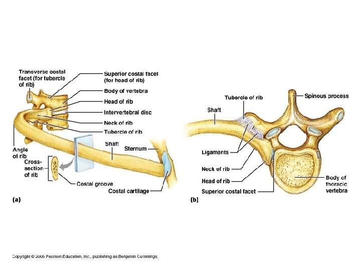
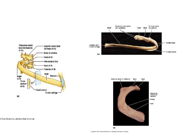
Bones of the Anterior Thorax • III. The bony thorax- the rib cage is also part of the axial skeleton and assist in breathing as well as protect internal organs. • A. Sternum- has three sections: manubrium, body, xyphoid process (cartilage) • B. Ribs- there are twelve pairs of ribs, the first 7 are true ribs (attach to sternum) and the last 5 are false ribs (floating).
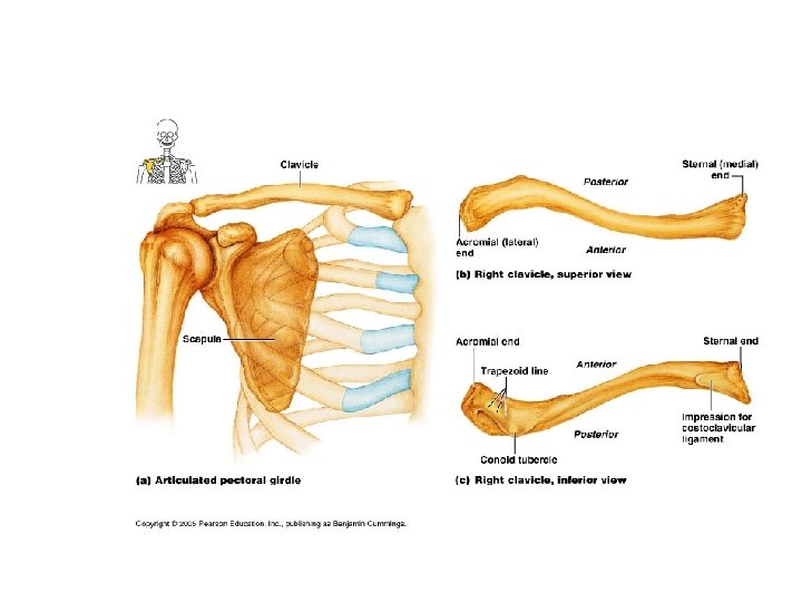
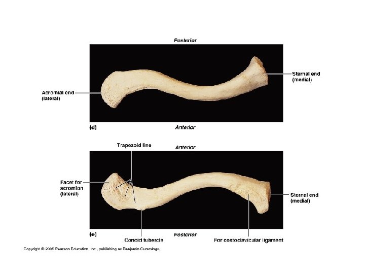
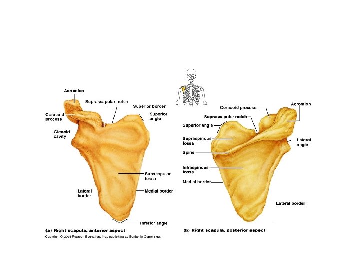
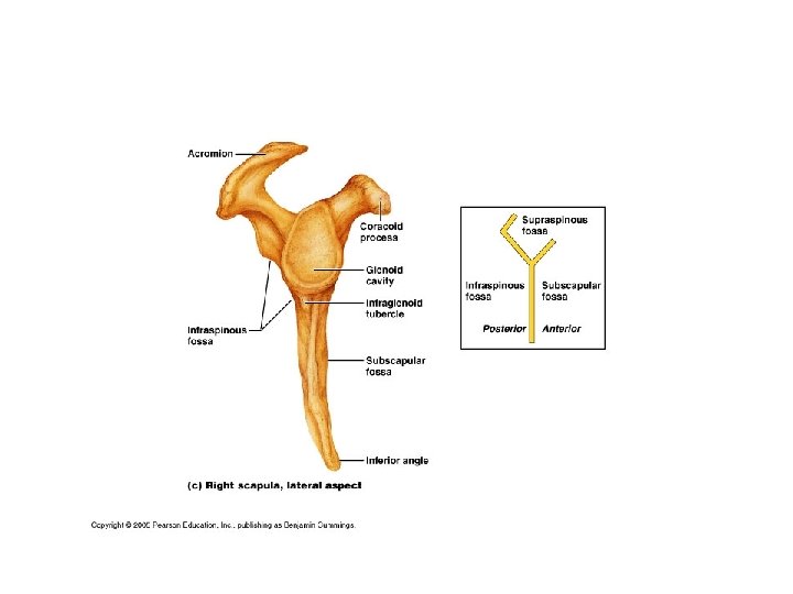
Bone of the upper appendicular Skeleton • IV. The pectoral girdle- consist of the scapula and clavicle, attaches upper limbs to axial skeleton, and provides attachment sites for muscles • A. Clavicles-also called the collarbone, they are long bones. • B. Scapulae-also called shoulder-blades, they are flat bones with three borders.
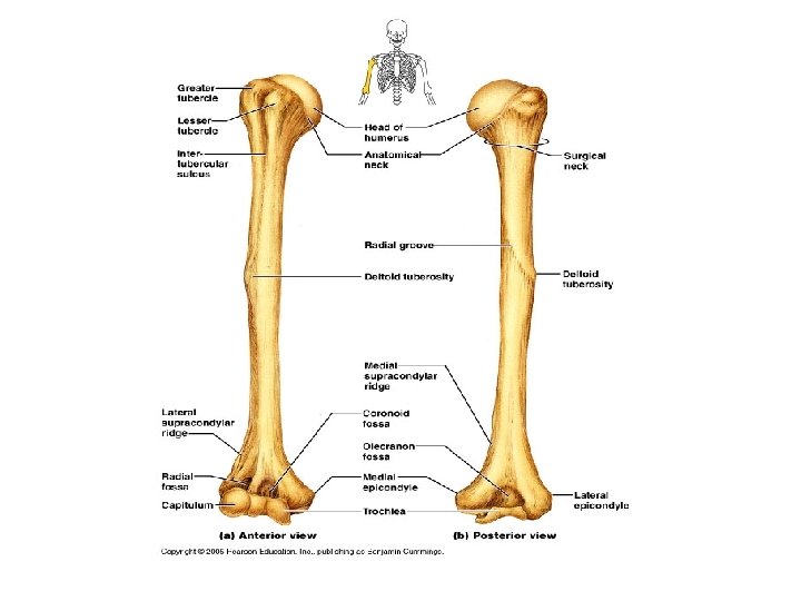
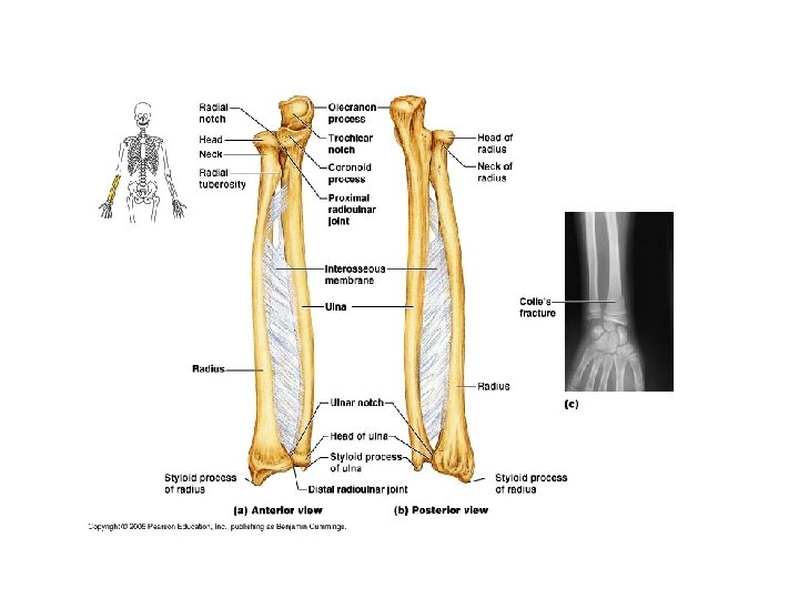
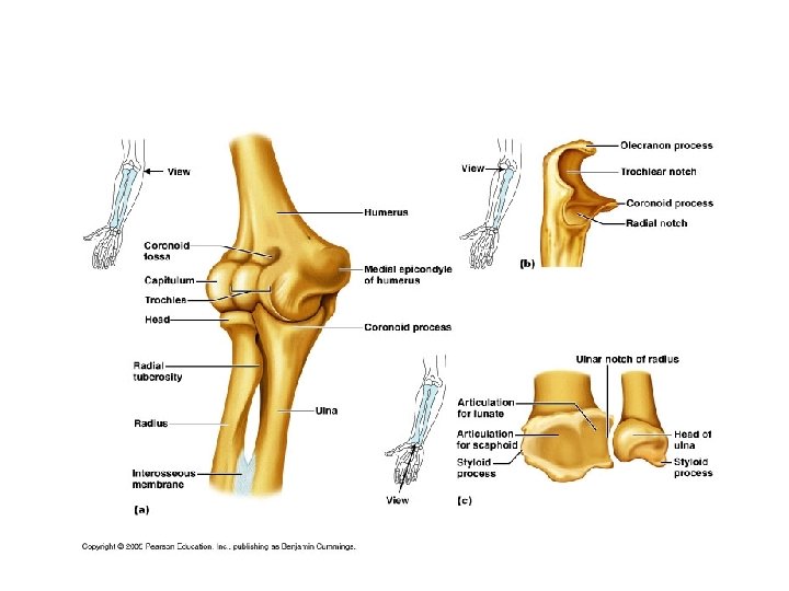
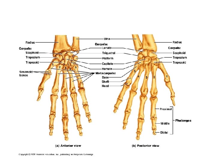
• V. The upper limb- 30 bones make up these three sections • A. Arm- applies to area between shoulder and elbow: the humerus. • B. Forearm- applies to area from elbow to writst: radius and ulna. These bones articulate with the humerus, the wrist, and each other. They are attached to each other by the interosseous membrane. • C. Hand- includes the carpals (wrist bones) metacarpals (palm) and phalanges (finger digits). Phalanges are numbered 1 -5 beginning with the thumb (pollex)
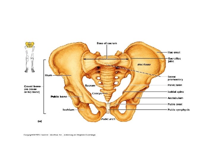
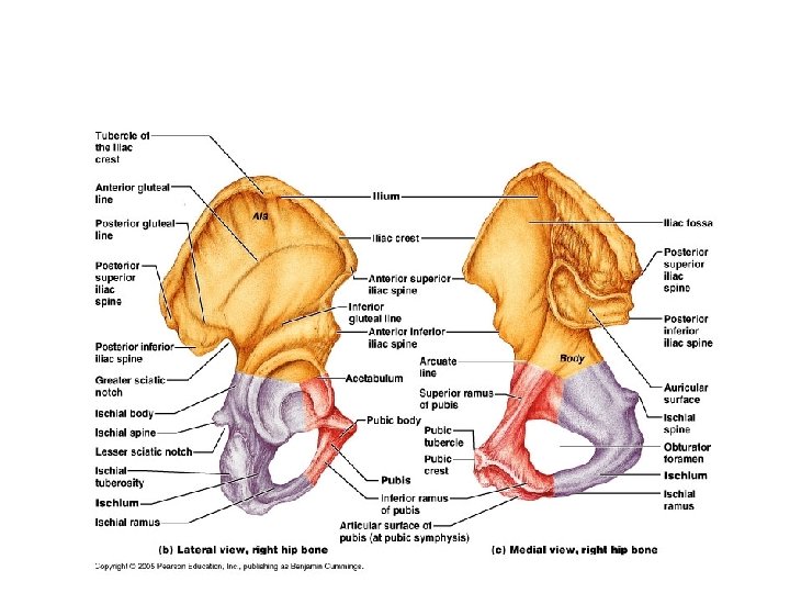
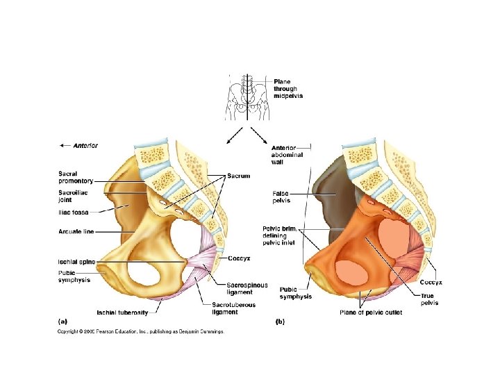
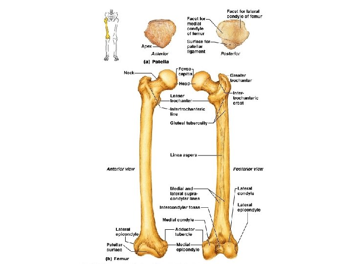
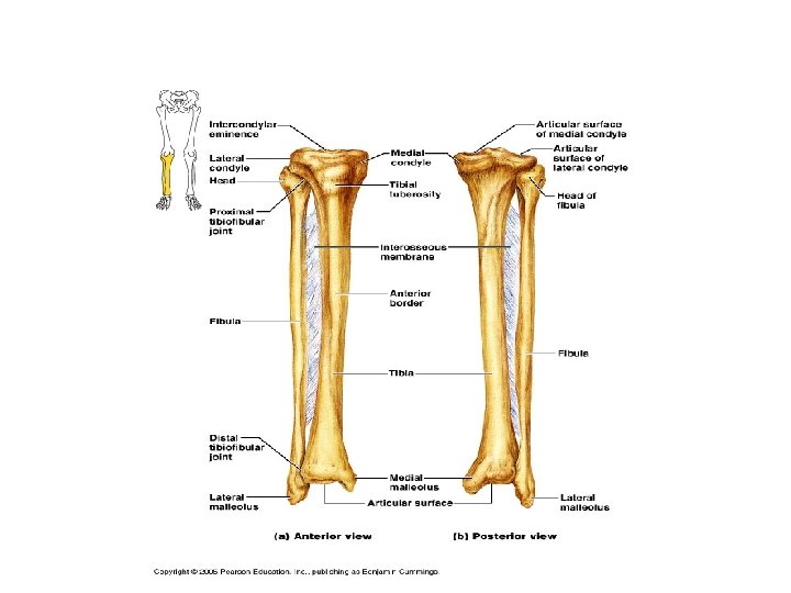
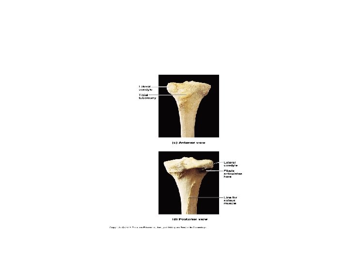
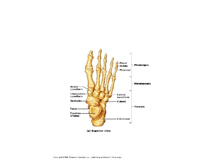
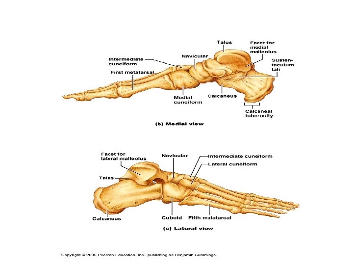
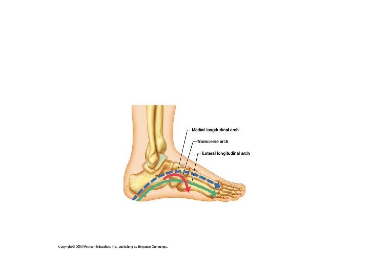
• VI. The pelvic girdle- consists of two coxal bones (os coxae) that attach to the axial skeleton by very strong ligaments, they bear the body’s weight and have deep joint sockets for the lower limbs. The two coxal bones are made up of three fused bones. • A. Ilium- large lateral flaring bones • B. Ischium-located posterior inferior to ilium, also known as the sit bones • C. Pubis-anterior bones of the pelvis • D. True and false pelvis- false pelvis refers to the greater pelvis that is superior to pelvic brim, forms part of the abdomen, and contains abdominal organs. The true pelvis is inferior to pelvic brim and holds pelvic organs. • E. Pelvic structure and childbearing- womens’ pelvic girdles are designed for child bearing. They have a true pelvis that is broad and shallow, bones are lighter and thinner, the acetabula are smaller and further apart and the pubic arch is more rounded. • VII. The lower limbs- they carry the entire weight of the body, it includes the thigh (femur) the leg (tibia and fibia) and the foot (tarsals, metatarsals, and phalanges).
Disorders of the appendicular skeleton • Hip dysplasia- common birth defect in which the acetabulum is incompletely formed or the ligatments are lose allowing the femur to slip out of its socket. The treatment involves properly positioning the head of the femur so cause the acetabulum to deepen. • Club foot-a congenital disorder that is caused by heredity or malpositioning of the fetus in the womb. The soles of the feet point medially and the toes point inferiorly. It is treated with casts that reposition the foot or with surgery.
Growth of body and proportions of limbs to trunk-. After birth limbs grow faster than the trunk.