
DISC EDEMA Prof Vasudev Anand Rao CAUSES UNILATERAL

- Slides: 29
DISC EDEMA Prof. Vasudev Anand Rao
CAUSES • • • UNILATERAL Papillitis Anterior Ischemic optic neuropathy Neuroretinitis Papillophlebitis Ischemic CRVO Anterior compressive optic neuropathies (orbital tumors) Infiltrative optic neuropathies Ocular hypotony Foster-Kennedy syndrome • • BILATERAL Papilledema Hypertension Diabetic papillopathy Advanced Graves disease Cavernous sinus thrombosis Carotid cavernous fistula Leber hereditary optic neuropathy
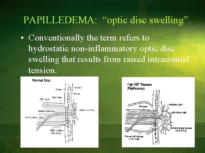
PAPILLEDEMA: “optic disc swelling” • Conventionally the term refers to hydrostatic non-inflammatory optic disc swelling that results from raised intracranial tension.
ETIOLOGY 1. Intracranial space occupying lesion § neoplasm (location of the tumor is more important than size) § abscess/inflammatory mass § hemorrhage/infarct § A-V malformation 2. Obstruction of ventricular system 3. Cerebral edema 4. Impaired CSF absorption by arachnoid villi: § Meningitis § Raised venous pressure § SAH/trauma § Communicating hydrocephalus 5. Severe systemic hypertension 6. Idiopathic (pseudo tumor cerebri): 7. Decreased size of cranial vault: § Craniosynostosis § Thickening of skull 8. Hypersecretion of choroids plexus tumor
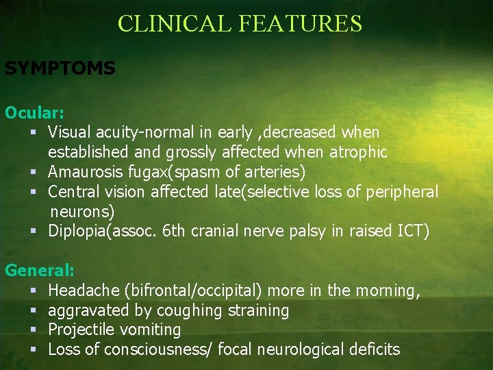
CLINICAL FEATURES SYMPTOMS Ocular: § Visual acuity-normal in early , decreased when established and grossly affected when atrophic § Amaurosis fugax(spasm of arteries) § Central vision affected late(selective loss of peripheral neurons) § Diplopia(assoc. 6 th cranial nerve palsy in raised ICT) General: § Headache (bifrontal/occipital) more in the morning, § aggravated by coughing straining § Projectile vomiting § Loss of consciousness/ focal neurological deficits
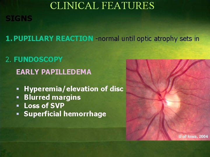
CLINICAL FEATURES SIGNS 1. PUPILLARY REACTION -normal until optic atrophy sets in 2. FUNDOSCOPY EARLY PAPILLEDEMA § § Hyperemia/elevation of disc Blurred margins Loss of SVP Superficial hemorrhage
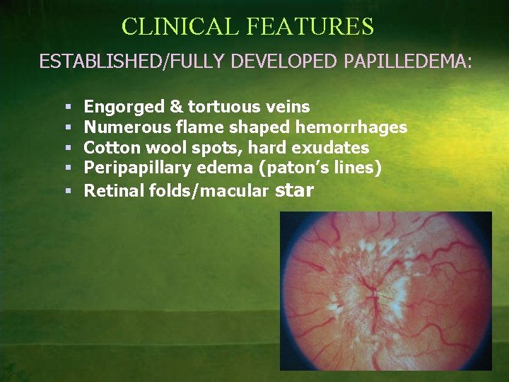
CLINICAL FEATURES ESTABLISHED/FULLY DEVELOPED PAPILLEDEMA: § § § Engorged & tortuous veins Numerous flame shaped hemorrhages Cotton wool spots, hard exudates Peripapillary edema (paton’s lines) Retinal folds/macular star
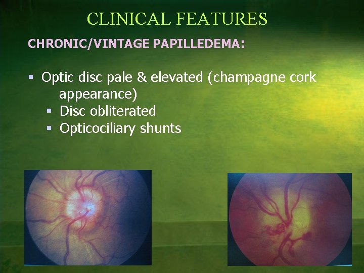
CLINICAL FEATURES CHRONIC/VINTAGE PAPILLEDEMA: § Optic disc pale & elevated (champagne cork appearance) § Disc obliterated § Opticociliary shunts
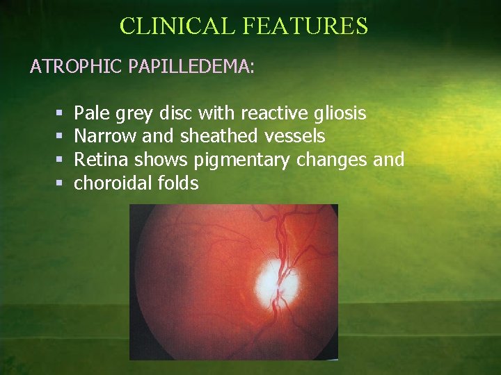
CLINICAL FEATURES ATROPHIC PAPILLEDEMA: § § Pale grey disc with reactive gliosis Narrow and sheathed vessels Retina shows pigmentary changes and choroidal folds
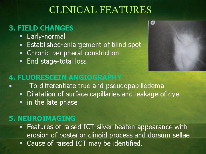
CLINICAL FEATURES 3. FIELD CHANGES § Early-normal § Established-enlargement of blind spot § Chronic-peripheral constriction § End stage-total loss 4. FLUORESCEIN ANGIOGRAPHY § To differentiate true and pseudopapilledema § Dilatation of surface capillaries and leakage of dye § in the late phase 5. NEUROIMAGING § Features of raised ICT-silver beaten appearance with erosion of posterior clinoid process and dorsum sellae § Cause of raised ICT may be identified.
UNILATERAL PAPILLEDEMA – Asymmetric – Foster Kennedy syndrome Seen in patients with frontal lobe/olfactory lobe tumors, meningiomas of olfactory groove/sphenoidal wing, characterized by optic atrophy on the side of the tumor caused by direct pressure on the nerve and papilledema on the opposite side because of raised ICT. – Prior optic atrophy, congenital abnormality in disc, high myopia
PSEUDOTUMOR CEREBRI Or Benign Intracranial hypertension Defined by 4 criteria 1. Increased intracranial pressure 2. Normal or small ventricles 3. No evidence of intracranial mass lesion 4. Normal CSF composition Usually idiopathic seen in young obese women
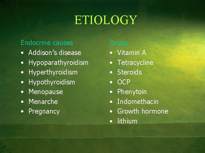
ETIOLOGY Endocrine causes • Addison’s disease • Hypoparathyroidism • Hyperthyroidism • Hypothyroidism • Menopause • Menarche • Pregnancy Drugs • Vitamin A • Tetracycline • Steroids • OCP • Phenytoin • Indomethacin • Growth hormone • lithium
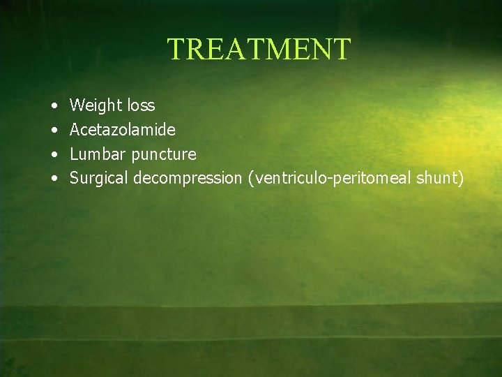
TREATMENT • • Weight loss Acetazolamide Lumbar puncture Surgical decompression (ventriculo-peritomeal shunt)
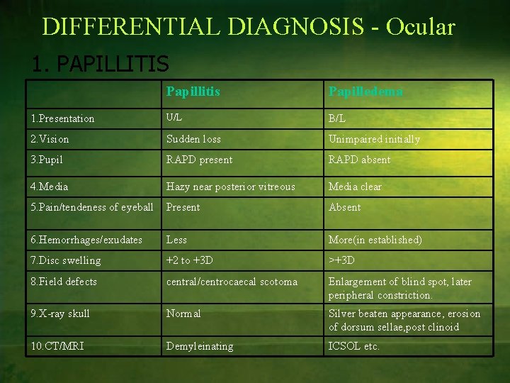
DIFFERENTIAL DIAGNOSIS - Ocular 1. PAPILLITIS Papillitis Papilledema 1. Presentation U/L B/L 2. Vision Sudden loss Unimpaired initially 3. Pupil RAPD present RAPD absent 4. Media Hazy near posterior vitreous Media clear 5. Pain/tendeness of eyeball Present Absent 6. Hemorrhages/exudates Less More(in established) 7. Disc swelling +2 to +3 D >+3 D 8. Field defects central/centrocaecal scotoma Enlargement of blind spot, later peripheral constriction. 9. X-ray skull Normal Silver beaten appearance, erosion of dorsum sellae, post clinoid 10. CT/MRI Demyleinating ICSOL etc.
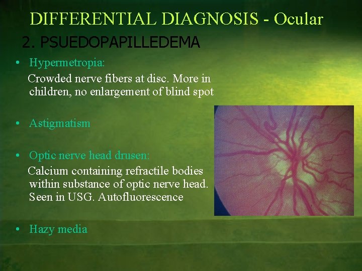
DIFFERENTIAL DIAGNOSIS - Ocular 2. PSUEDOPAPILLEDEMA • Hypermetropia: Crowded nerve fibers at disc. More in children, no enlargement of blind spot • Astigmatism • Optic nerve head drusen: Calcium containing refractile bodies within substance of optic nerve head. Seen in USG. Autofluorescence • Hazy media
DIFFERENTIAL DIAGNOSIS - Ocular 3. AION/LHON/TOXIC AMBLYOPIAS 4. OCULAR HYPOTONY Effusion from choroidal vessels 5. RAISED INTRAOCULAR PRESSURE: Obliteration of peripapillary vessels by raised IOP 6. CRVO
OPTIC NEURITIS : “Inflammation of the optic nerve” ETIOPATHOGENESIS 1. 2. 3. IDIOPATHIC DEMYELINATING (Always Retrobulbar) Isolated – a/w multiple sclerosis – neuromyelitis optica – schilder’s disease
• ETIOPATHOGENESIS INFECTIOUS AND PARAINFECTIOUS – LOCAL: • Orbital cellulites • Sinusitis • Teeth, tonsil • Meninges, brain or base of skull. – SYSTEMIC: • VIRAL-measles, mumps, rubella, chickenpox, herpes, CMV and EBV. • BACTERIAL-T. B, syphilis, cat scratch disease, lyme’s • FUNGAL-cryptococcosis, histoplasmosis • PARASITImalaria, pneumocystis, toxoplasma, toxocara, cystice rcosis – VACCINES: BCG, DPT, TT, Hep. B, variola and influenza
ETIOPATHOGENESIS 4. IMMUNE RELATED LOCAL Uveitis, sympathetic ophthalmitis. SYSTEMIC sarcoidosis, Wegener’s polyarteritis nodosa, SLE etc. 5. METABOLIC Anemia Diabetes Starvation 6. DRUGS AND TOXINS INH, ethambutol, etanercept, INFa, tobacco, alcohol, quinine.
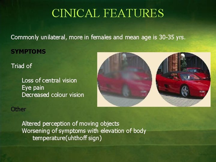
CINICAL FEATURES Commonly unilateral, more in females and mean age is 30 -35 yrs. SYMPTOMS Triad of Loss of central vision Eye pain Decreased colour vision Other Altered perception of moving objects Worsening of symptoms with elevation of body temperature(uhthoff sign)
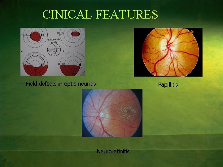
CINICAL FEATURES SIGNS • • • Decreased visual acuity Tenderness Marcus gunn pupil (RAPD) Decreased colour vision and contrast sensitivity Visual field defects: classically central/centrocaecal scotoma but other defects can also occur Fundus changes 1. Papillitis: edema, hyperemia, blurred margins, dilated tortuous vs, few exudates and vitreous haze 2. Retrobulbar neuritis: normal 3. Neuroretinitis: macular star with exudates • • VEP-Delayed latency and decreased amplitude FAG to differentiate from other causes-dilated and telangiectatic vs with leak from capillaries
CINICAL FEATURES Field defects in optic neuritis Papillitis Neuroretinitis
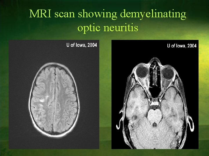
INVESTIGATIONS To determine cause for optic neuritis 1. Complete Hemogram 2. CRP, ESR, Mantoux 3. VDRL 4. Serology-ANA, Toxoplasma, Lymes 5. PNS X-ray, chest x ray(sarcoidosis) 6. X ray skull, CT 7. MRI(demyleinating plaques-2 or more predictive of deveplopment of MS) 8. Lumbar puncture-CSF pleocytosis and oligoclonal bands
MRI scan showing demyelinating optic neuritis
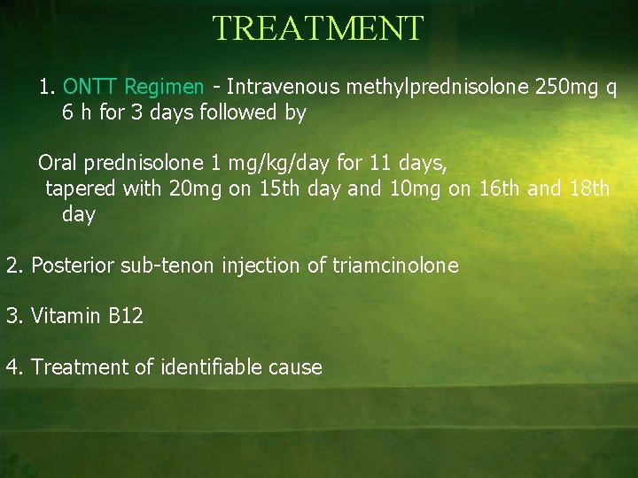
TREATMENT 1. ONTT Regimen - Intravenous methylprednisolone 250 mg q 6 h for 3 days followed by Oral prednisolone 1 mg/kg/day for 11 days, tapered with 20 mg on 15 th day and 10 mg on 16 th and 18 th day 2. Posterior sub-tenon injection of triamcinolone 3. Vitamin B 12 4. Treatment of identifiable cause
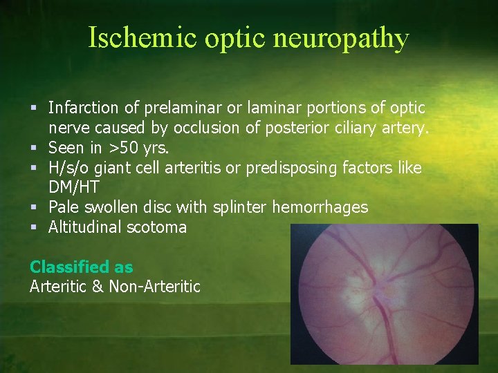
Ischemic optic neuropathy § Infarction of prelaminar or laminar portions of optic nerve caused by occlusion of posterior ciliary artery. § Seen in >50 yrs. § H/s/o giant cell arteritis or predisposing factors like DM/HT § Pale swollen disc with splinter hemorrhages § Altitudinal scotoma Classified as Arteritic & Non-Arteritic
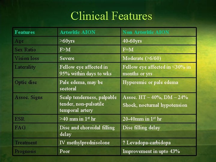
Clinical Features Arteritic AION Non Arteritic AION Age >60 yrs 40 -60 yrs Sex Ratio F>M F=M Vision loss Severe Moderate (>6/60) Laterality Fellow eye affected in 95% within days to wks Fellow eye affected in <30% in months or yrs Optic disc Pale edema, may be sectoral Hyperemic or pale edema Assoc. Signs Scalp tenderness, palpable tender, non-pulsatile temporal artery Assoc. HT – 40%, DM – 24% Shock, nocturnal hypotension ESR >40 mm in 1 st hr 20 -40 mm in 1 st hr FAG Disc and choroidal filling delay Disc filling delay Treatment IV methylprednisolone ? Levadopa-carbidopa Prognosis Poor Improvement in upto 43%
THANK YOU