
Can YOU Walk the EYE Doc Talk Dr





- Slides: 61
Can YOU Walk the EYE Doc Talk? ? Dr. Kris Kerestan krisgarbig@fuse. net Dr. Kerestan is a member of the Alcon Vision Care Speakers Alliance. Alcon, however, has no financial interest in this course or its content.
The Language of Eye Care Clinical Optometric Abbreviations Clinical Optometric Terminology Medication Rx Abbreviations
Clinical Optometric Abbreviations
AI Accommodative Insufficiency Inability to maintain focus on print at near Signs and Symptoms Blur N/D Eyestrain / Asthenopia Headache Difficulty reading Motion sickness Treatment VT – Success rate of close to 90% Near rx
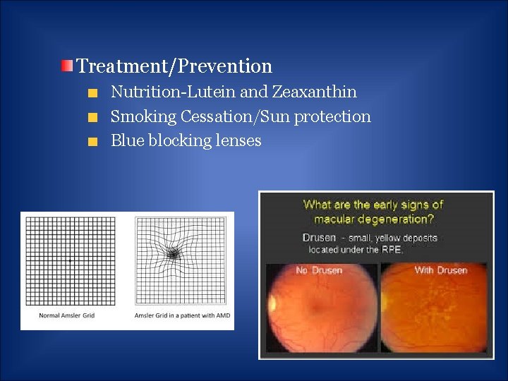
AMD Age-Related Macular Degeneration Acquired retinal disorder caused by atrophy and degeneration of the central retina Signs and Symptoms: Gradual onset of blurred vision Metamorphopsia Pigment degeneration Drusen
Treatment/Prevention Nutrition-Lutein and Zeaxanthin Smoking Cessation/Sun protection Blue blocking lenses
Amps Amplitudes of Accommodation Maximum increase in focusing power that an eye can achieve by switching from D to N Measurement via PD rule or Accomm. Rod Age dependent
ATR/WTR Against the Rulewhen the axis of the minus cylinder is orientated at X 90 q Adults With the Rule- when the axis of the minus cylinder is orientated at X 180 q Children
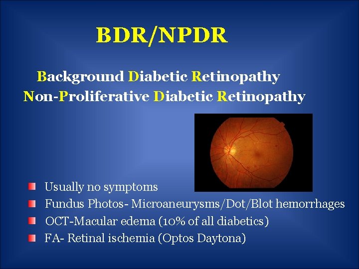
BDR/NPDR Background Diabetic Retinopathy Non-Proliferative Diabetic Retinopathy Usually no symptoms Fundus Photos- Microaneurysms/Dot/Blot hemorrhages OCT-Macular edema (10% of all diabetics) FA- Retinal ischemia (Optos Daytona)
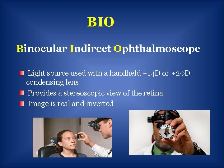
BIO Binocular Indirect Ophthalmoscope Light source used with a handheld +14 D or +20 D condensing lens. Provides a stereoscopic view of the retina. Image is real and inverted
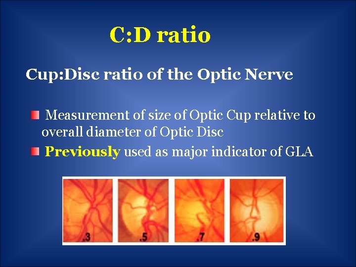
C: D ratio Cup: Disc ratio of the Optic Nerve Measurement of size of Optic Cup relative to overall diameter of Optic Disc Previously used as major indicator of GLA
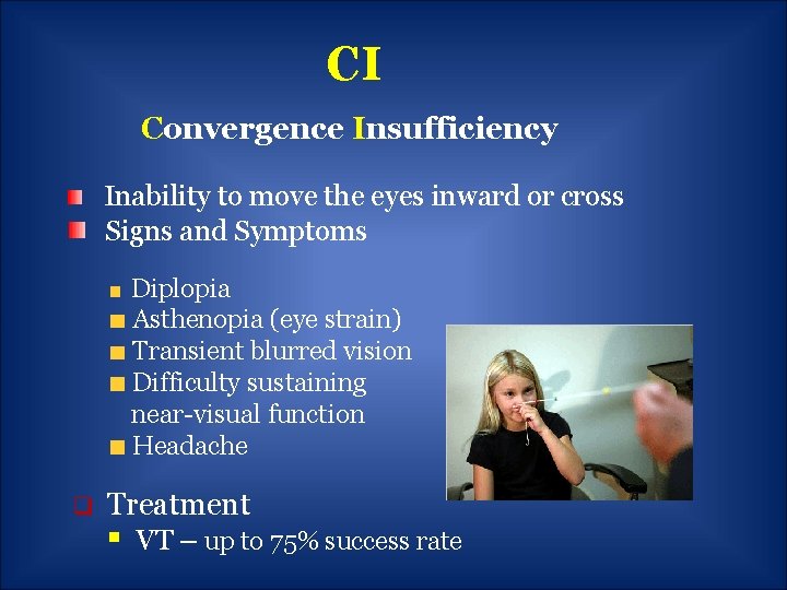
CI Convergence Insufficiency Inability to move the eyes inward or cross Signs and Symptoms Diplopia Asthenopia (eye strain) Transient blurred vision Difficulty sustaining near-visual function Headache q Treatment § VT – up to 75% success rate
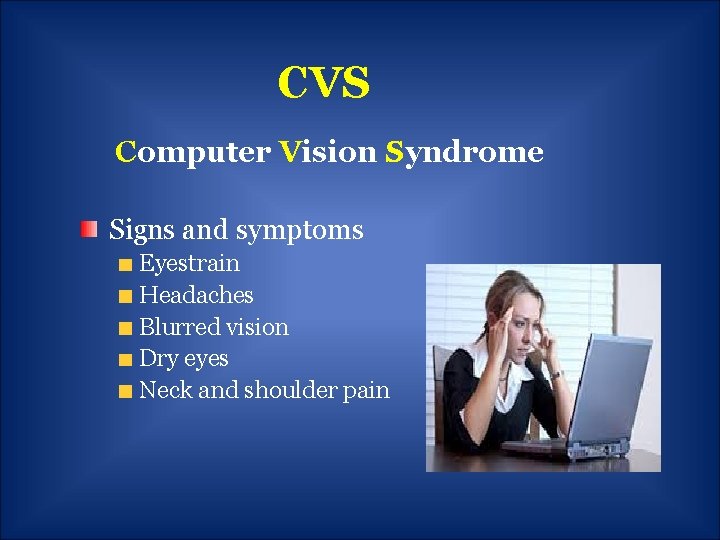
CVS Computer Vision Syndrome Signs and symptoms Eyestrain Headaches Blurred vision Dry eyes Neck and shoulder pain
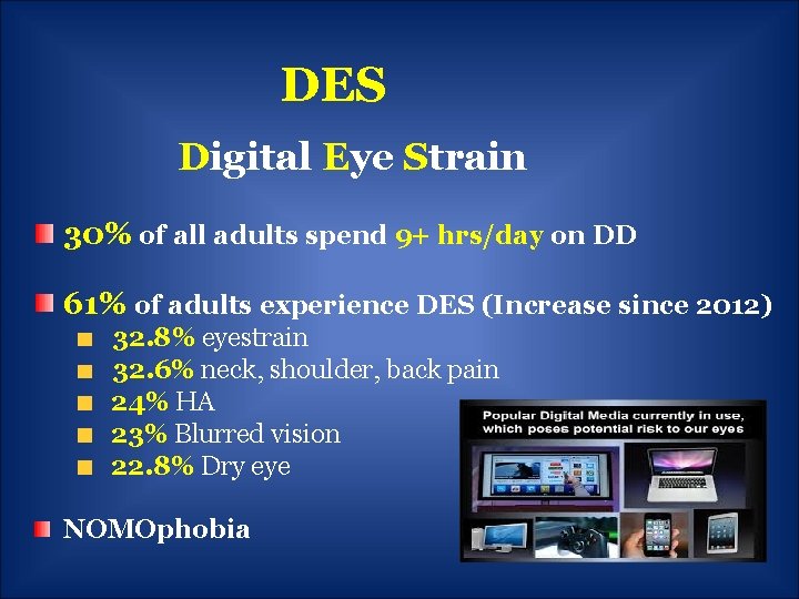
DES Digital Eye Strain 30% of all adults spend 9+ hrs/day on DD 61% of adults experience DES (Increase since 2012) 32. 8% eyestrain 32. 6% neck, shoulder, back pain 24% HA 23% Blurred vision 22. 8% Dry eye NOMOphobia
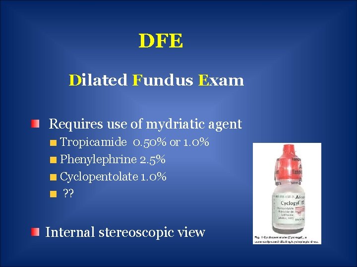
DFE Dilated Fundus Exam Requires use of mydriatic agent Tropicamide 0. 50% or 1. 0% Phenylephrine 2. 5% Cyclopentolate 1. 0% ? ? Internal stereoscopic view
D Diopter Unit of measurement of the optical power of a lens utilized to prescribe corrective lenses Plus (Convex-Magnifies an image) Minus (Concave-Minifies an image) Accommodation (Loss of focusing with age)
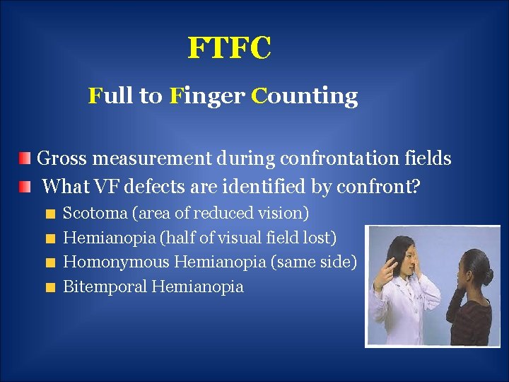
FTFC Full to Finger Counting Gross measurement during confrontation fields What VF defects are identified by confront? Scotoma (area of reduced vision) Hemianopia (half of visual field lost) Homonymous Hemianopia (same side) Bitemporal Hemianopia
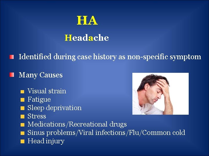
HA Headache Identified during case history as non-specific symptom Many Causes Visual strain Fatigue Sleep deprivation Stress Medications/Recreational drugs Sinus problems/Viral infections/Flu/Common cold Head injury
Hx Case History/History of Present Illness CC/RFV HPI ROS Medication Allergies PFSH FH
IOL Intra Ocular Lens Cataract Technology Multifocal- Restore/Tecnis Spherical Toric- Up to 3. 5 cyl Aspheric- Acry. Sof Blue Light filtering Myopic Technology -3 diopters to -16 diopters Less than - 2. 5 cyl. Verisyse Phakic IOL- Anterior chamber Visian Implantable Collamer Lens- Posterior Chamber Between 92 -95% achieve 20/20 -20/40 vision
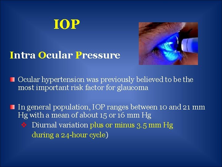
IOP Intra Ocular Pressure Ocular hypertension was previously believed to be the most important risk factor for glaucoma In general population, IOP ranges between 10 and 21 mm Hg with a mean of about 15 or 16 mm Hg v Diurnal variation plus or minus 3. 5 mm Hg during a 24 -hour cycle)
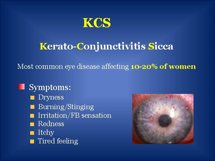
KCS Kerato-Conjunctivitis Sicca Most common eye disease affecting 10 -20% of women Symptoms: Dryness Burning/Stinging Irritation/FB sensation Redness Itchy Tired feeling
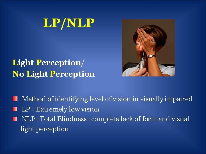
LP/NLP Light Perception/ No Light Perception Method of identifying level of vision in visually impaired LP= Extremely low vision NLP=Total Blindness=complete lack of form and visual light perception
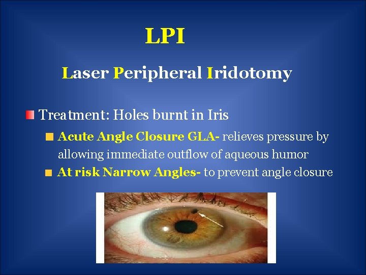
LPI Laser Peripheral Iridotomy Treatment: Holes burnt in Iris Acute Angle Closure GLA- relieves pressure by allowing immediate outflow of aqueous humor At risk Narrow Angles- to prevent angle closure
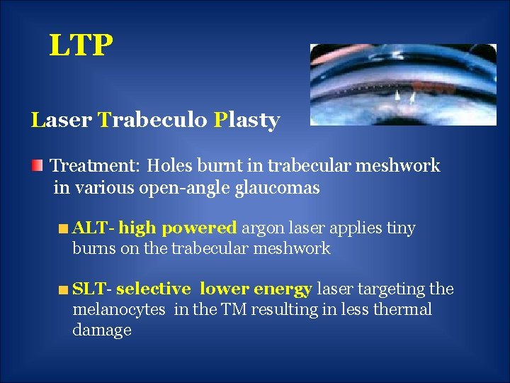
LTP Laser Trabeculo Plasty Treatment: Holes burnt in trabecular meshwork in various open-angle glaucomas ALT- high powered argon laser applies tiny burns on the trabecular meshwork SLT- selective lower energy laser targeting the melanocytes in the TM resulting in less thermal damage
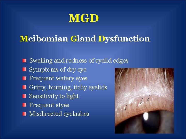
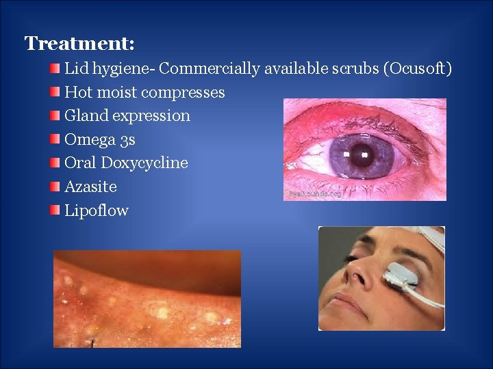
MGD Meibomian Gland Dysfunction Swelling and redness of eyelid edges Symptoms of dry eye Frequent watery eyes Gritty, burning, itchy eyelids Sensitivity to light Frequent styes Misdirected eyelashes
Treatment: Lid hygiene- Commercially available scrubs (Ocusoft) Hot moist compresses Gland expression Omega 3 s Oral Doxycycline Azasite Lipoflow
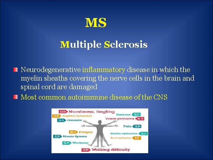
MS Multiple Sclerosis Neurodegenerative inflammatory disease in which the myelin sheaths covering the nerve cells in the brain and spinal cord are damaged Most common autoimmune disease of the CNS
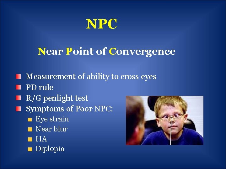
NPC Near Point of Convergence Measurement of ability to cross eyes PD rule R/G penlight test Symptoms of Poor NPC: Eye strain Near blur HA Diplopia
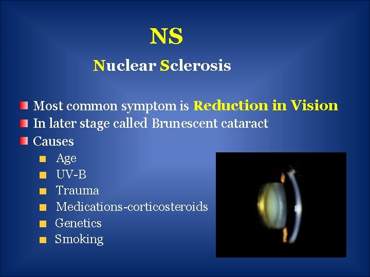
NS Nuclear Sclerosis Most common symptom is Reduction in Vision In later stage called Brunescent cataract Causes Age UV-B Trauma Medications-corticosteroids Genetics Smoking
OD/OS/OU Oculus Dexter Oculus Sinister Oculi Uterque
PRK vs. Lasik Photo Refractive Keratectomy Laser Assisted in situ Keratomileusis Increased inflammation and slower recovery More postoperative discomfort, irritation, pain Cornea's structural integrity is less altered No difference in ocular dryness Can correct between − 1. 00 to − 12. 00 D. of Myopia
POAG Primary Open Angle Glaucoma 2 nd leading cause of Blindness World Wide after Cataracts 90% of all glaucoma cases in the United States Painless/No symptoms-Only 50% know they have it!! Gradual progressive visual field loss Increased cup: disc May have normal or high IOP (50%-67% of all POAG) Thinning of retinal fiber layers
Causes Physiology § Decreased drainage through TM § Increased production in CB Ethnicity - African descent 3 X Genetics – 2 to 4 X if family hx. Medication use- Steroids Ocular disease-BDR, Uveitis Trauma Caffeine? Systemic hyertension ? (UK study showed 29% incidence)
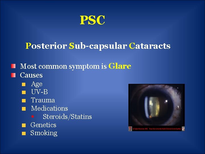
PSC Posterior Sub-capsular Cataracts Most common symptom is Glare Causes Age UV-B Trauma Medications § Steroids/Statins Genetics Smoking
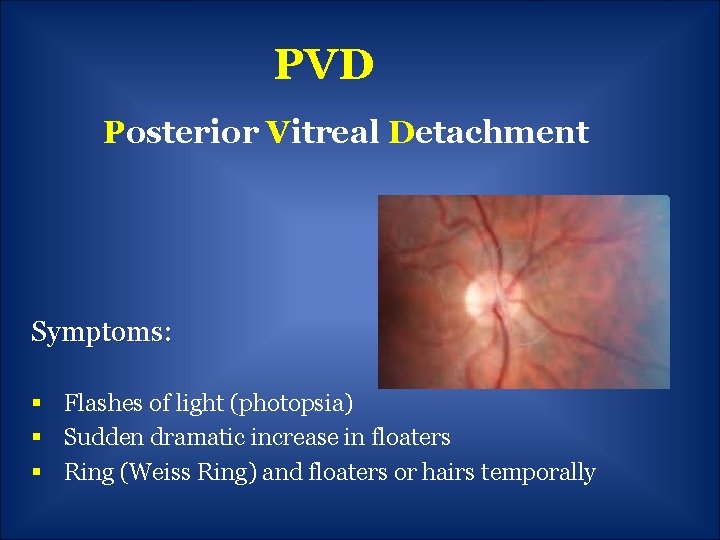
PVD Posterior Vitreal Detachment Symptoms: § Flashes of light (photopsia) § Sudden dramatic increase in floaters § Ring (Weiss Ring) and floaters or hairs temporally
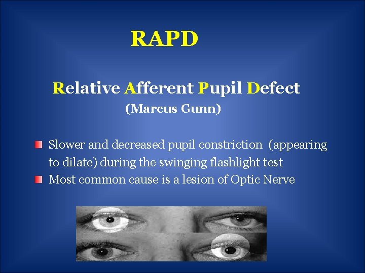
RAPD Relative Afferent Pupil Defect (Marcus Gunn) Slower and decreased pupil constriction (appearing to dilate) during the swinging flashlight test Most common cause is a lesion of Optic Nerve
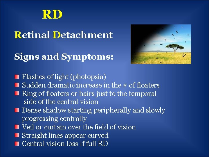
RD Retinal Detachment Signs and Symptoms: Flashes of light (photopsia) Sudden dramatic increase in the # of floaters Ring of floaters or hairs just to the temporal side of the central vision Dense shadow starting peripherally and slowly progressing centrally Veil or curtain over the field of vision Straight lines appear curved Central vision loss if full RD
RP Retinitis Pigmentosa Signs and Symptoms Night blindness or nyctalopia Tunnel vision Peripheral vision loss Aversion to glare Slow dark to light adaptation time Blurring of vision Poor color separation Extreme tiredness
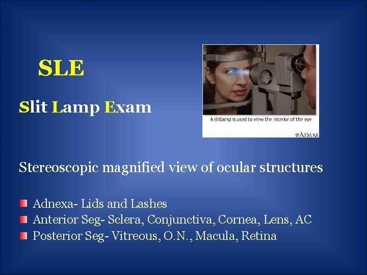
SLE Slit Lamp Exam Stereoscopic magnified view of ocular structures Adnexa- Lids and Lashes Anterior Seg- Sclera, Conjunctiva, Cornea, Lens, AC Posterior Seg- Vitreous, O. N. , Macula, Retina
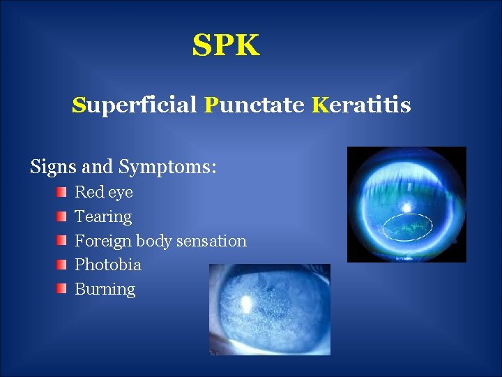
SPK Superficial Punctate Keratitis Signs and Symptoms: Red eye Tearing Foreign body sensation Photobia Burning
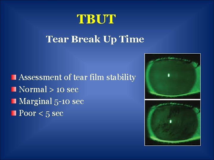
TBUT Tear Break Up Time Assessment of tear film stability Normal > 10 sec Marginal 5 -10 sec Poor < 5 sec
Clinical Terminology A through Z
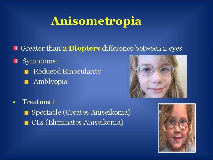
Anisometropia Greater than 2 Diopters difference between 2 eyes Symptoms: Reduced Binocularity Amblyopia • Treatment: Spectacle (Creates Aniseikonia) CLs (Eliminates Aniseikonia)
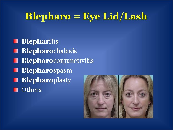
Blepharo = Eye Lid/Lash Blepharitis Blepharochalasis Blepharoconjunctivitis Blepharospasm Blepharoplasty Others
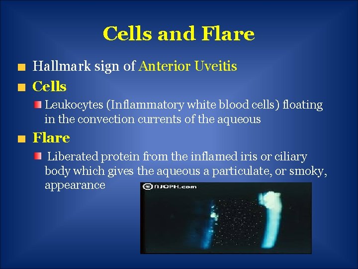
Cells and Flare Hallmark sign of Anterior Uveitis Cells Leukocytes (Inflammatory white blood cells) floating in the convection currents of the aqueous Flare Liberated protein from the inflamed iris or ciliary body which gives the aqueous a particulate, or smoky, appearance
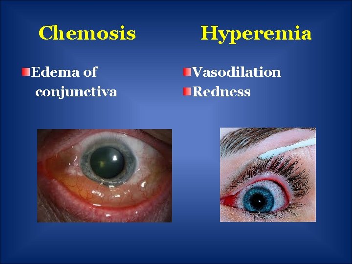
Chemosis Edema of conjunctiva Hyperemia Vasodilation Redness
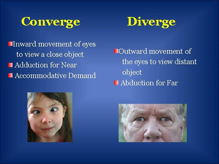
Converge Inward movement of eyes to view a close object Adduction for Near Accommodative Demand Diverge Outward movement of the eyes to view distant object Abduction for Far
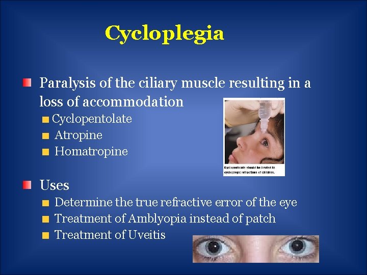
Cycloplegia Paralysis of the ciliary muscle resulting in a loss of accommodation Cyclopentolate Atropine Homatropine Uses Determine the true refractive error of the eye Treatment of Amblyopia instead of patch Treatment of Uveitis
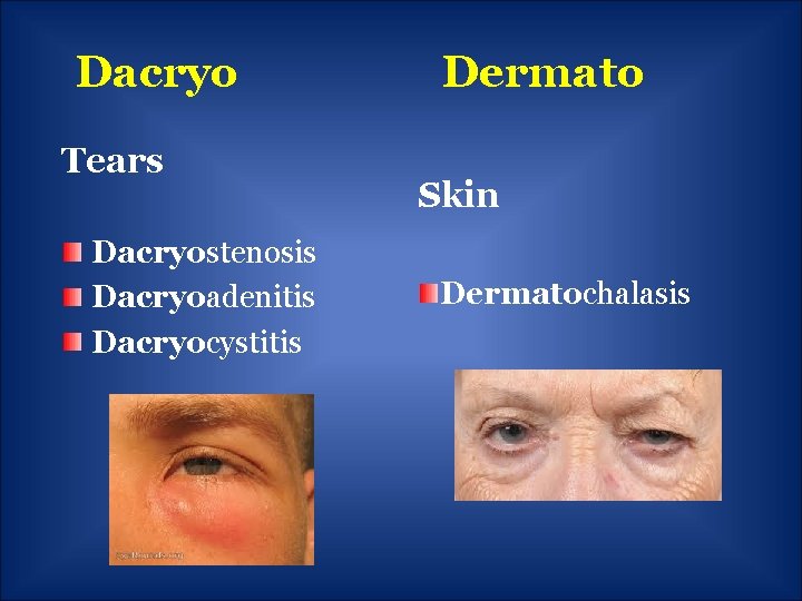
Dacryo Tears Dacryostenosis Dacryoadenitis Dacryocystitis Dermato Skin Dermatochalasis
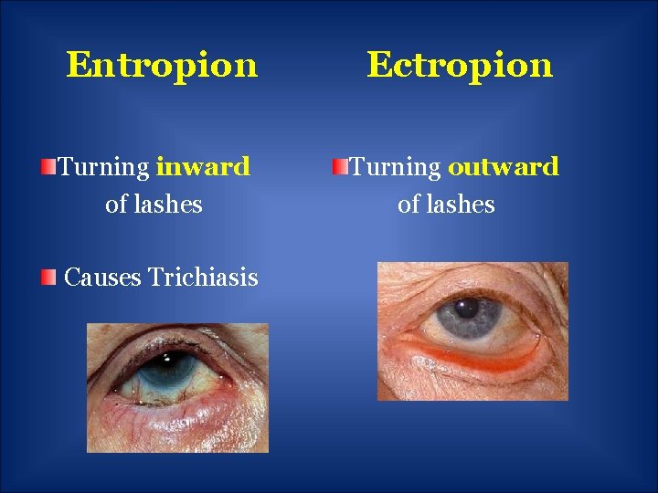
Entropion Ectropion Turning inward of lashes Turning outward of lashes Causes Trichiasis
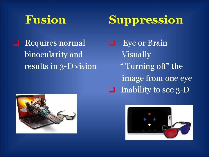
Fusion q Requires normal binocularity and results in 3 -D vision Suppression q Eye or Brain Visually “ Turning off” the image from one eye q Inability to see 3 -D
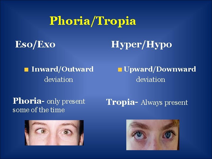
Phoria/Tropia Eso/Exo Inward/Outward deviation Hyper/Hypo Upward/Downward deviation Phoria- only present some of the time Tropia- Always present
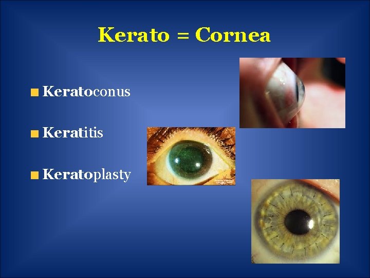
Kerato = Cornea Keratoconus Keratitis Keratoplasty
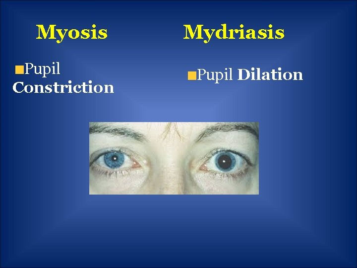
Myosis Pupil Constriction Mydriasis Pupil Dilation
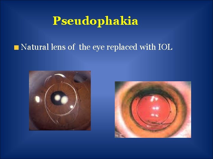
Pseudophakia Natural lens of the eye replaced with IOL
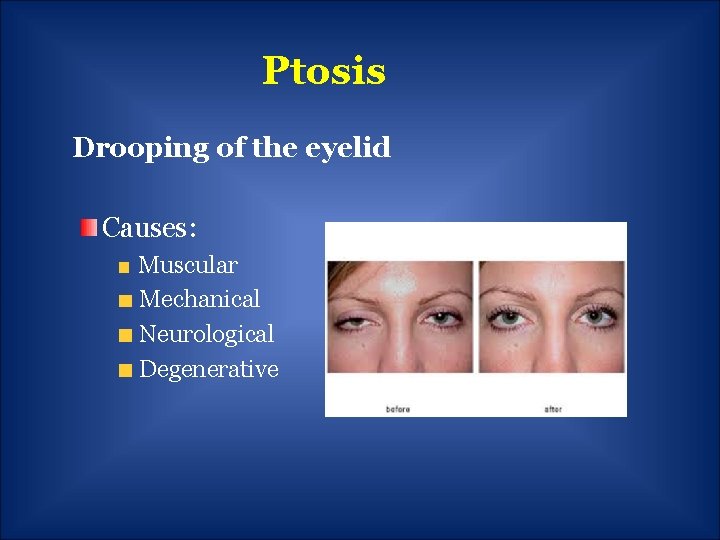
Ptosis Drooping of the eyelid Causes: Muscular Mechanical Neurological Degenerative
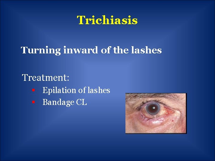
Trichiasis Turning inward of the lashes Treatment: § Epilation of lashes § Bandage CL
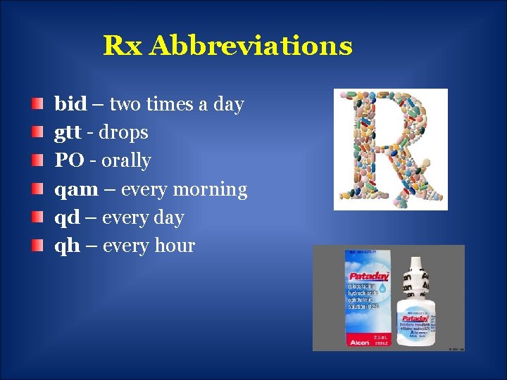
Rx Abbreviations bid – two times a day gtt - drops PO - orally qam – every morning qd – every day qh – every hour
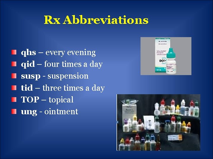
Rx Abbreviations qhs – every evening qid – four times a day susp - suspension tid – three times a day TOP – topical ung - ointment
Thank You!! It’s been a pleasure to present to your group today! Any questions? ? Kris Kerestan Garbig, OD krisgarbig@fuse. net