By Prof Saeed Abuel Makarem By the 4

By Prof. Saeed Abuel Makarem

• By the 4 th week, as the pharyngeal arches develop, they are supplied by arteries from the aortic sac. • Aortic sac formed from fusion of the 2 primitive aortas. • The aortic arches terminate in the dorsal aorta of the ipsilateral side. • Aortic arches are not all present in the same time • By 8 th week the primordial aortic arch pattern is transformed into the final fetal arterial arrangement. Aortic Arch Derivatives

• Initially there are 6 pairs of aortic arches. • Three aortic arches disappear (1 st, 2 nd & 5 th) • Three aortic arches persist ( 3 rd, 4 th & 6 th). • The fate of 4 th arch differs in the right & the left sides. • Also the fate of the dorsal segments of the sixth arch differs in right & left sides.

• The 1 st aortic arch disappears, only small part of it remains forming the maxillary artery. • The 2 nd aortic arch disappears, only small part of it remains forming stapedial artery.

• Proximal part of the 3 rd aortic arch forms the common carotid artery, while its distal part forms the internal carotid artery • The remaining part of internal carotid artery is formed by cranial part of dorsal aorta.

• 4 th arch has different fate on right & left sides • Right: Forms the stem of the right subclavian artery. • Left: Forms part of the arch of the aorta. • NB. Completion of right subclavian artery is formed by right dorsal aorta & right 7 th intersegmental artery. • Lt Subclavian artery develops from left 7 th intersegmental artery. • Proximal part of aortic arch develops from aortic sac.

• 5 th aortic arch is rudimentary and disappears early. • 6 th arch (pulmonary arch) • Each 6 th arch divides into proximal & dorsal parts. • The proximal part in each side gives the pulmonary artery. • Distal part in right side disappears. • Distal part in left side gives ductus arteriosus.

• So, the recurrent laryngeal nerve differs in both sides. • In Left side it turns around the ductus arteriosus (ligamentum arteriosum) in thorax. • In the right side it turns around the subclavian artery so it is found in the neck.
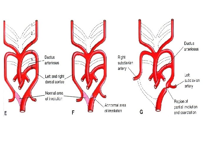

Coarcitation of the aorta • It means narrowing of aorta below the origin of the left subclavian artery. 2 types: I- Postductal. The duct will be closed forming ligamentum arteriosum,

Coarcitation of the aorta So circulation to the lower limb will maintained by collateral? Anastomosis around scapula. X- ray chest will show notching of the inferior border of the ribs from enlarging intercostal arteries.
")
• II- Pre-ductal type: • So the ductus arteriosus will remain open (patent) to maintain the circulation to the lower part of the body.

• Abnormal persistence of entire right dorsal aorta • Abnormal persistence of dorsal part of the right 6 th aortic arch. • Normal involution of the distal part of the left dorsal aorta

Double Aortic Arches.

• Right aortic arch with retroesophageal component • Notice that the right aortic arch and ligamentum arteriosum form a ring that may compresses the trachea and esophagus.

• Abnormal right subclavian artery. • It arises from the aorta and passes behind trachea and esophagus.
- Slides: 16