GOOD MORNING EXTENSOR COMPARTMENT OF FOREARM Dr QUDSIA


 Origin. Lateral epicondyle of humerus • Insertiondorsal surface of")


- Slides: 34
GOOD MORNING
EXTENSOR COMPARTMENT OF FOREARM Dr. QUDSIA SULTANA
Extensor compartment of forearm Muscles. Occur in two layers • Superficial layer • Deep layer Deep fascia. Extensor retinaculum. Principal artery. Posterior interosseous artery. Nerve. Posterior interosseous nerve
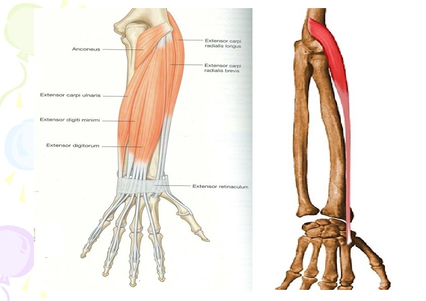
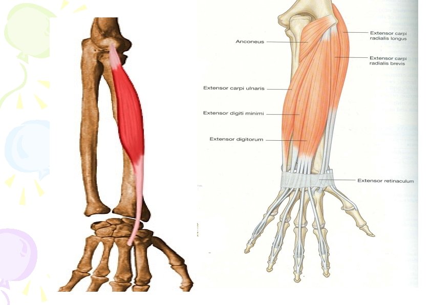
Superficial layer • Brachioradialis • Extensor carpi radialis longus • Extensor carpi radialis brevis • Extensor digitorum • Extensor digiti minimi • Extensor carpi ulnaris • Anconeus
Superficial muscles posterior compartment of foreman Anconeus Extends the elbow joint Brachioradialis Flexes the elbow joint Extensor carpi radialis longus Extends and abducts the wrist Extensor carpi radialis brevis Extends and abducts the wrist Extensor digitorum Extends wrist joint Extensor digiti minimi 5 Extensor carpi Extends the finger Extends and
Deep layer • Supinator • Abductor pollicis longus • Extensor pollicis brevis • Extensor indicis
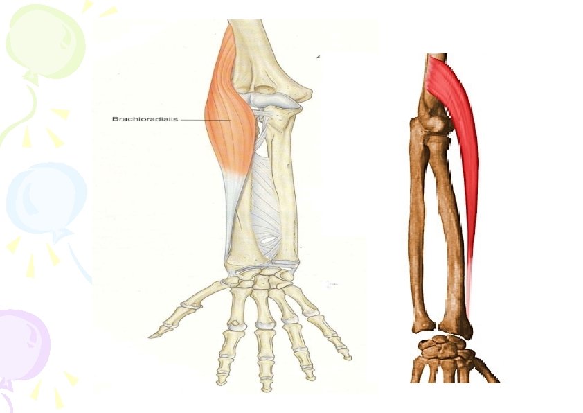
Brachioradialis • Origin- upper 2/3 rd of lateral supracondylar ridge of humerus & lateral intermuscular septum • Insertion- Base of styloid process of radius • Action- Flexes arm in midprone position • Supinates the fully pronated forearm & pronates the fully supinated forearm Nerve- Radial nerve •
• EXTENSOR CARPI RADIALIS LONGUS Origin. Lower third of supracondylar ridge of humerus & lateral intermuscular septum • Insertion. Lateral side of dorsal surface of base of the second metacarpal bone • Action. Extends hand with extensor carpi ulnaris abduction of hand with flexor carpi radialis • Nerve- Radial nerve
EXTENSOR CARPI RADIALIS BREVIS • Origin. Lateral epicondyle of humerus, radial collateral ligament of elbow joint • Insertiondorsal surface of second and third metacarpal bones. • Action • Extends hand with flexor carpi ulnaris • Abduction of with flexor carpi radialis. • Nerve • Deep branch of radial nerve(Post interosseous nerve)
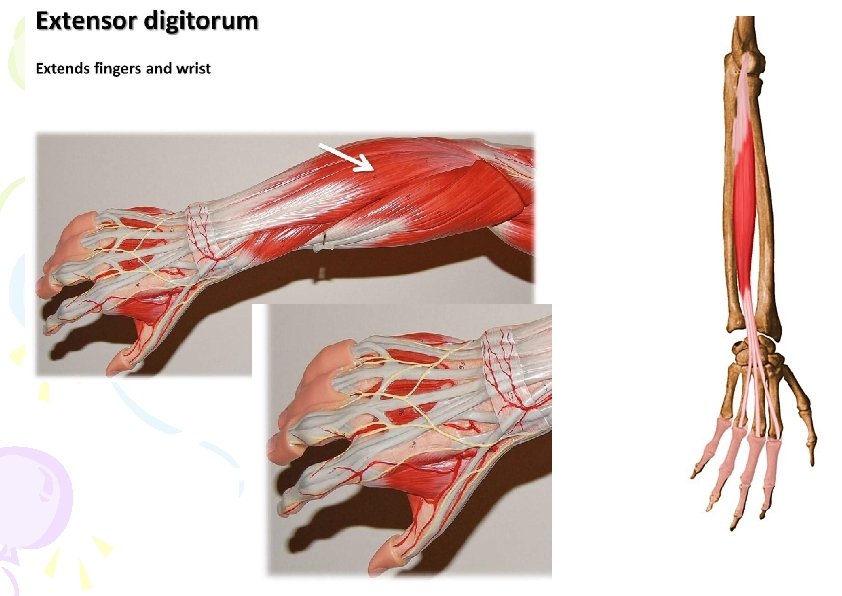
• EXTENSOR DIGITORUM (COMMUNIS) Origin. Lateral epicondyle of humerus • Insertiondorsal surface of middle phalanges of 2 -5 fingers and forms dorsal digital expansion
• Action • Extends the fingers of hand at metecarpophalyngeal joint and interphalyngeal joint. • Nerve- Deep branch of radial nerve(Post interosseous nerve)
Dorsal digital expansion
EXTENSOR DIGITI MINIMI Origin. Common tendon attached to lateral epicondyle of humerus Insertion. Dorsal digital expansion of the fifth finger Action. Extends Metacarpal joint of the fifth finger Nerve. Deep branch of radial nerve
EXTENSOR CARPI ULNARIS Origin • lateral epicondyle of humerus. • Posterior border of ulna. Insertion – dorsal surface of base of fifth metacarpal bone Action. Extends hand with Flexor carpi radialis longus and brevis adduction of hand with flexor carpi ulnaris Nerve- Deep branch of radial nerve
ANCONEUS Origin. Posterior part of lateral epicondyle of humerus Insertion. Lateral surface of the olecranon process & upper 1/4 th posterior surface of ulna Action. Week Extensor of elbow, moves ulna laterally during pronation. Nerve. Radial nerve
Origin of deep muscles of back of forearm
SUPINATOR • ORIGIN: • SUPERFICIAL STRATALATERAL EPICONDYLE OF HUMERUS, ANNULAR LIGAMENT • DEEP STRATA-SUPINATOR CREST OF ULNA • INSERTION: • UPPER 1/3 OF THE LAT SURFACE OF THE RADIUS ABOVE ANT OBLIQUE LINE. • NS: DEEP BRANCH OF RADIAL NERVE • ACTION: • SUPINATION
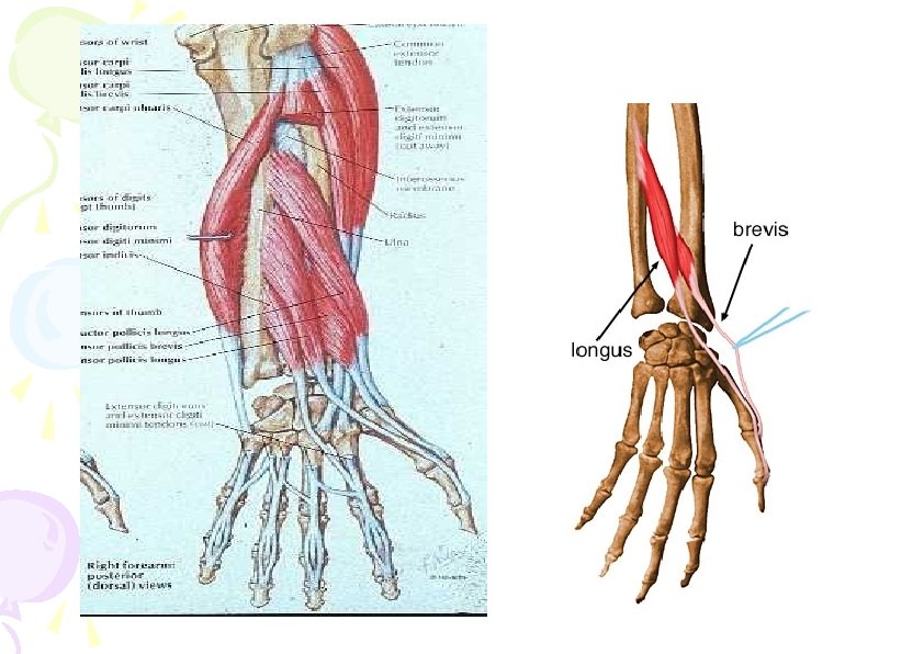
ABDUCTOR POLLICIS LONGUS Origin. Posterior surface of shafts of radius, ulna & interosseous membrane Insertion. Dorsal surface of base of first metacarpal bone. Actionabducts and extends thumb, abducts wrist Nerve- Deep branch of radial nerve
EXTENSOR POLLICIS BREVIS Origin • Dorsal surface of Radiusbelow the abductor pollicis longus, • adjacent part of interosseous membrane Insertion. Base of proximal phalanx of thumb Action. Extends metacarpophalyngeal joint of the thumb Nerve. Deep branch of radial nerve
EXTENSOR POLLICIS LONGUS Origin. Middle third of dorsal surface of ulna below the Abductor pollicis longus & interosseous membrane Insertion. Base of distal phalanx of thumb Action – Extends distal phalanx of the thumb Nerve. Deep branch of radial nerve
EXTENSOR INDICIS Origin • Posterior surface of ulna below the extensor pollicis longus • adjacent part of interosseous membrane Insertion. Extensor expansion of index finger Action. Extends index finger Nerve. Deep branch of radial nerve
Arteries & veins • Posterior interosseous artery • Anterior interosseous artery in lower part.
Posterior interosseous artery • Originate in anterior compartment from common interosseous artery • Passes dorsally over the proximal margin of interosseous membrane into posterior compartment • Gives out recurrent interosseous artery • Terminates by joining the dorsal carpal arch of wrist
Radial nerve • Most of the muscles are supplied by deep branch of radial nerve (posterior interosseous nerve) • Brachioradialis, anconeus & Extensor Carpi Radialis Longus are supplied by radial nerve directly in arm.
Extensor retinaculum • Thickened band of fascia • Runs across the back of wrist Attachment • laterally to lower part of anterior border of radius • Medially it is attached to triquetral & pisiform
Anatomical snuff box
Applied anatomy • Injury to radial nerve in forearm leads to wrist drop
Thank you • Thank you
Supinator Origin • Lateral epicondyle of humerus. • Radial collateral ligament. • Annular ligament • Supinator crest of ulna. Insertion – • Dorsal and lateral surface of shaft of upper 1/3 rd of ulna. Action – • Supinates forearm Nerve supply • Deep branch of radial nerve.