Good morning Deep Cervical Fascia DR QUDSIA SULTANA






- Slides: 59
Good morning
Deep Cervical Fascia DR. QUDSIA SULTANA
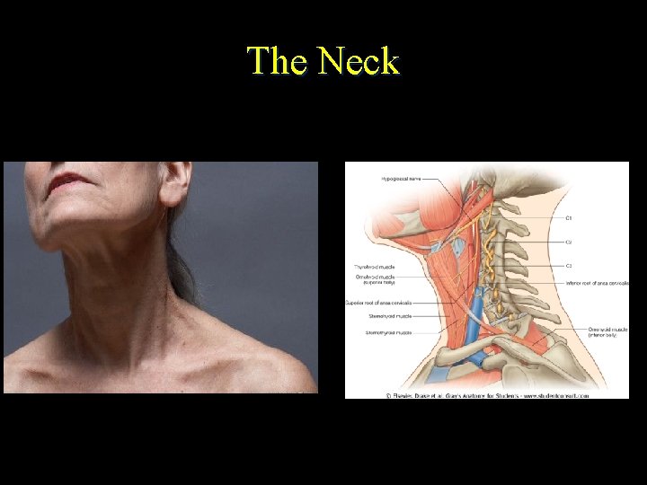
The Neck
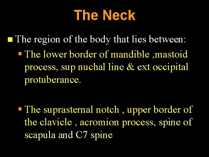
The Neck n The region of the body that lies between: § The lower border of mandible , mastoid process, sup nuchal line & ext occipital protuberance. § The suprasternal notch , upper border of the clavicle , acromion process, spine of scapula and C 7 spine
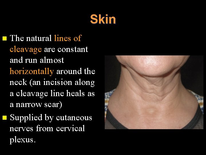
Skin The natural lines of cleavage are constant and run almost horizontally around the neck (an incision along a cleavage line heals as a narrow scar) n Supplied by cutaneous nerves from cervical plexus. n
Superficial fascia
Platysma
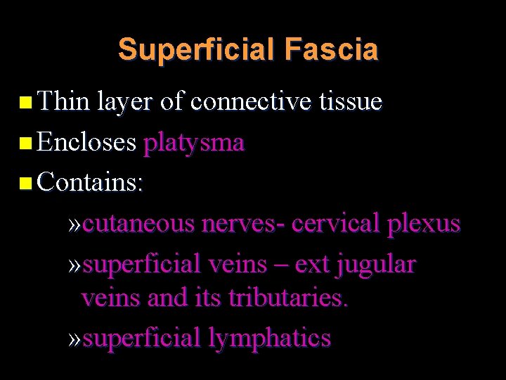
Superficial Fascia n Thin layer of connective tissue n Encloses platysma n Contains: » cutaneous nerves- cervical plexus » superficial veins – ext jugular veins and its tributaries. » superficial lymphatics
Deep cervical fascia n n Fascia colli Surrounds the neck like a collar
Deep Cervical Fascia Deep fascia around the neck is known as deep cervical fascia or fascia colli as it forms the collar around the neck n It lies deep to platysma muscle in the interval between muscles, vessels & organs of the cervical region n And it gives various extensions or lamina around various structures n
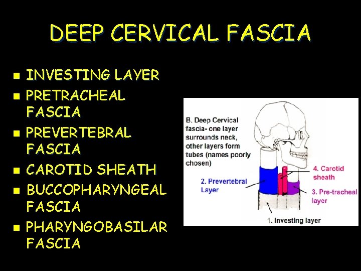
DEEP CERVICAL FASCIA n n n INVESTING LAYER PRETRACHEAL FASCIA PREVERTEBRAL FASCIA CAROTID SHEATH BUCCOPHARYNGEAL FASCIA PHARYNGOBASILAR FASCIA
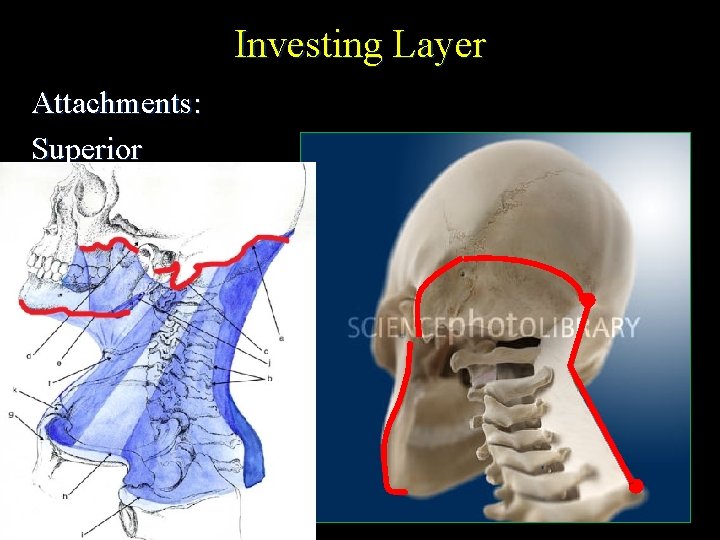
Investing Layer Attachments: Superior
Superiorly n External occipital protuberance, superior nuchal line n Mastoid process n Cartilagenous part of external ear n Lower margin of zygomatic arch n Lower border of mandible
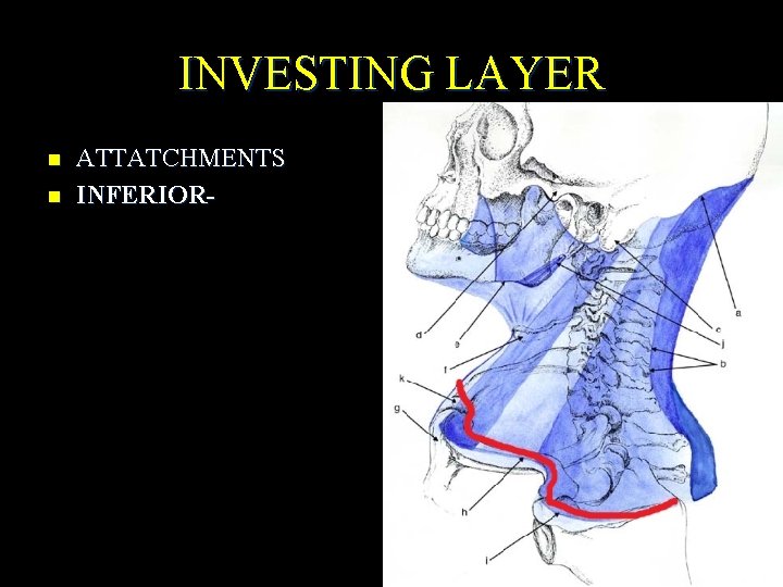
INVESTING LAYER n n ATTATCHMENTS INFERIOR-
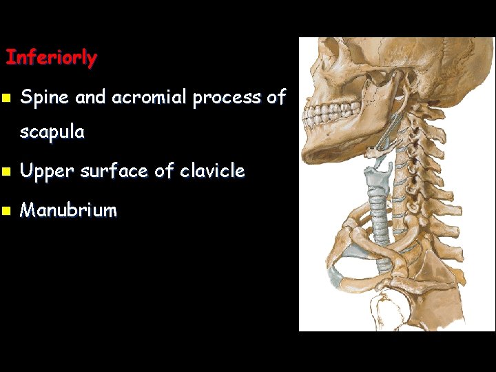
Inferiorly n Spine and acromial process of scapula n Upper surface of clavicle n Manubrium
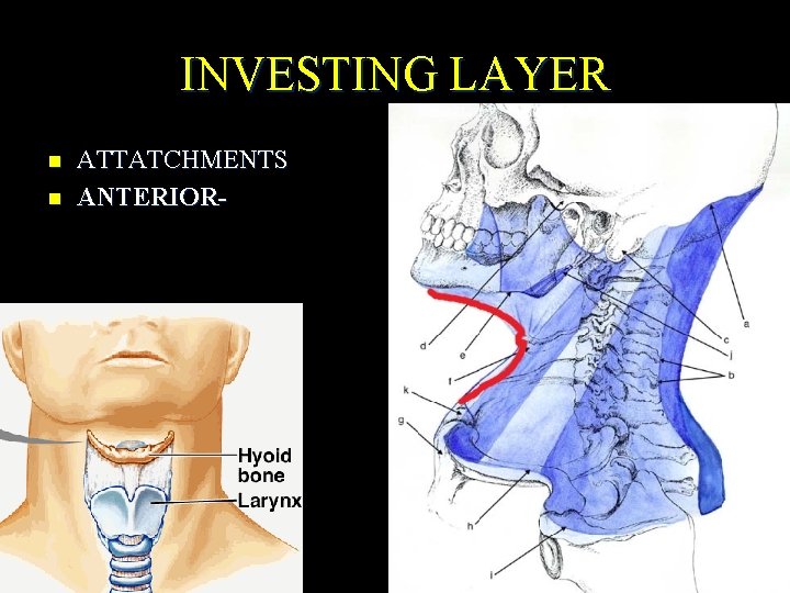
INVESTING LAYER n n ATTATCHMENTS ANTERIOR-
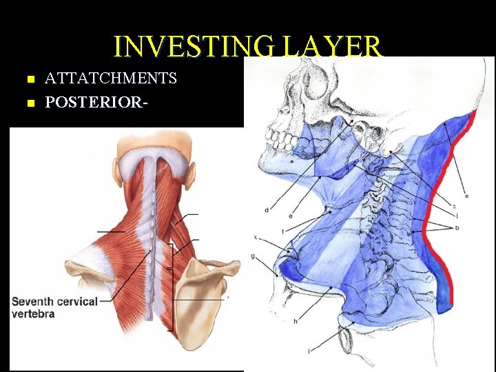
INVESTING LAYER n n ATTATCHMENTS POSTERIOR-
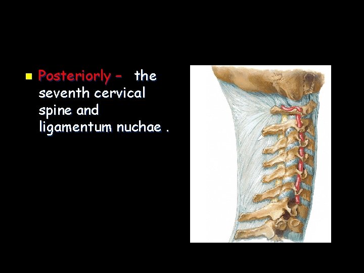
n Posteriorly – the seventh cervical spine and ligamentum nuchae.
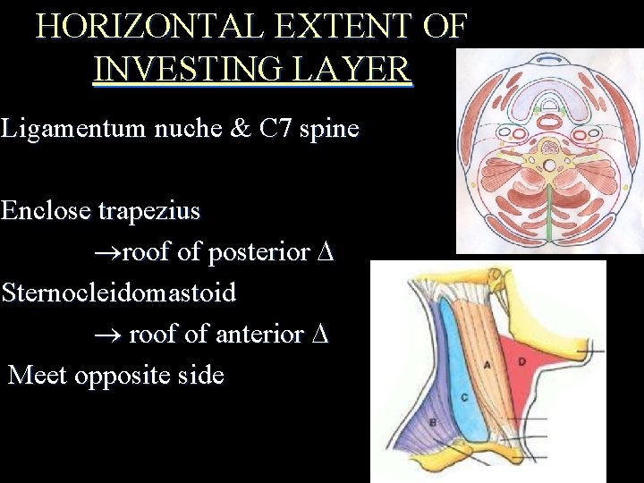
HORIZONTAL EXTENT OF INVESTING LAYER Ligamentum nuche & C 7 spine Enclose trapezius roof of posterior Sternocleidomastoid roof of anterior Meet opposite side
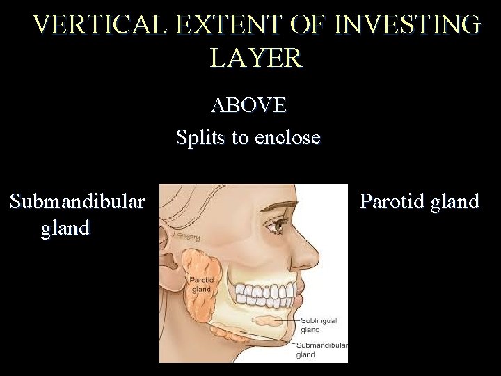
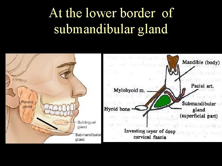
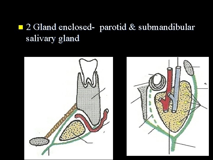
VERTICAL EXTENT OF INVESTING LAYER ABOVE Splits to enclose Submandibular gland Parotid gland
At the lower border of submandibular gland
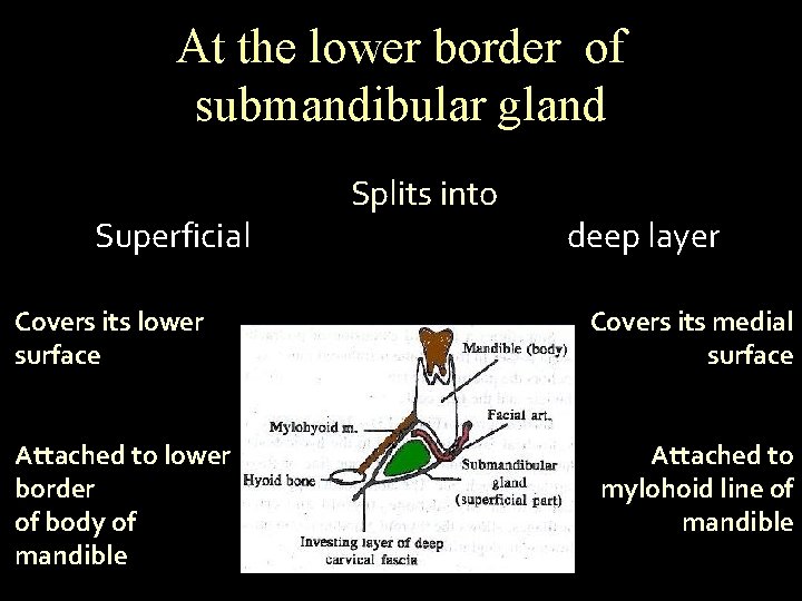
At the lower border of submandibular gland Superficial Covers its lower surface Attached to lower border of body of mandible Splits into deep layer Covers its medial surface Attached to mylohoid line of mandible
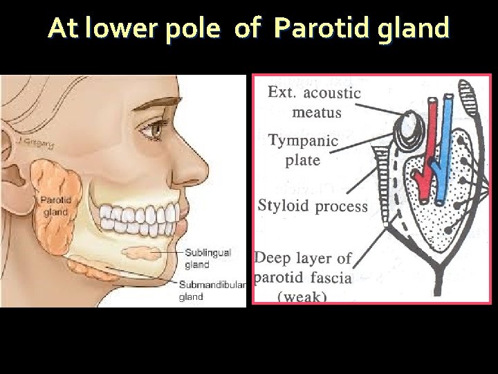
At lower pole of Parotid gland
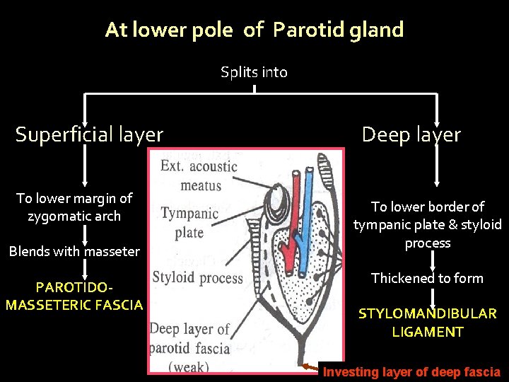
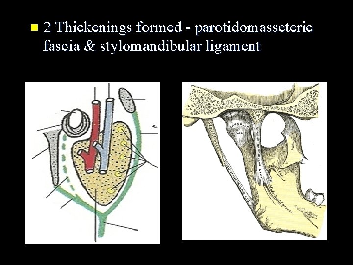
At lower pole of Parotid gland Splits into Superficial layer To lower margin of zygomatic arch Blends with masseter PAROTIDOMASSETERIC FASCIA Deep layer To lower border of tympanic plate & styloid process Thickened to form STYLOMANDIBULAR LIGAMENT Investing layer of deep fascia
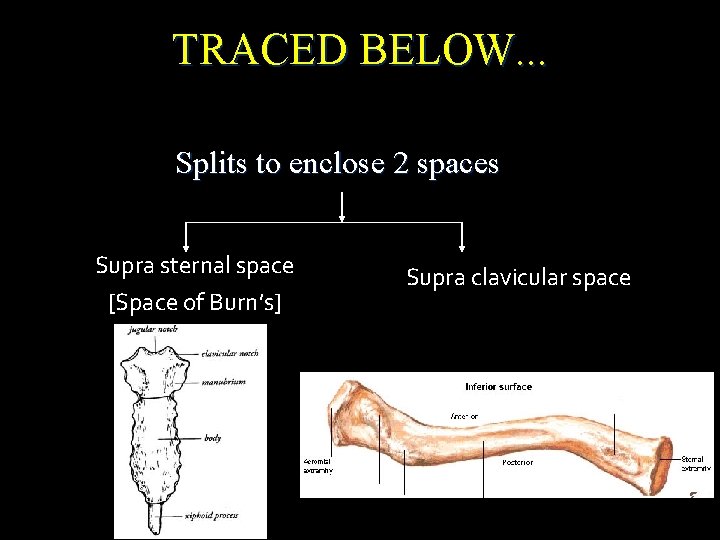
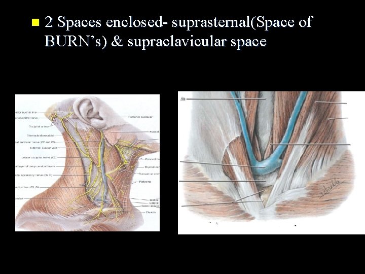
TRACED BELOW. . . Splits to enclose 2 spaces Supra sternal space [Space of Burn’s] Supra clavicular space
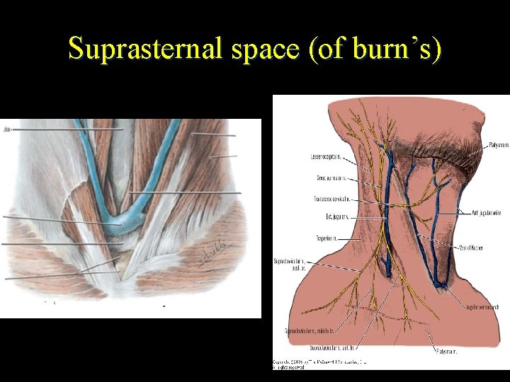
Suprasternal space (of burn’s)
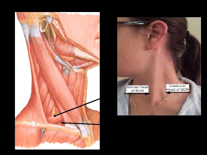
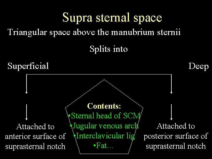
Supra sternal space Triangular space above the manubrium sternii Splits into Superficial Deep Contents: • Sternal head of SCM • Jugular venous arch Attached to anterior surface of • Interclavicular lig posterior surface of • Fat… suprasternal notch
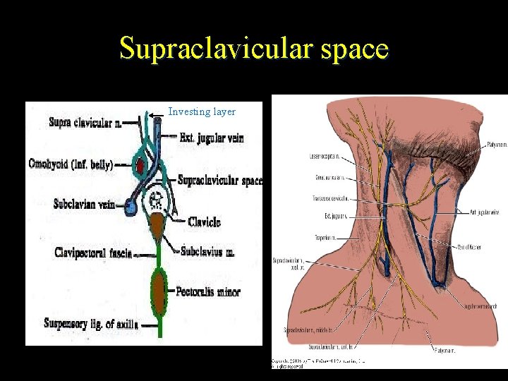
Supraclavicular space Investing layer
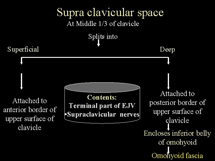
Supra clavicular space At Middle 1/3 of clavicle Splits into Superficial Attached to anterior border of upper surface of clavicle Deep Contents: Terminal part of EJV • Supraclavicular nerves Attached to posterior border of upper surface of clavicle Encloses inferior belly of omohyoid Omohyoid fascia
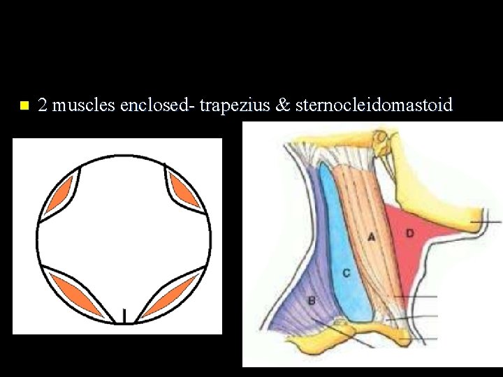
n 2 muscles enclosed- trapezius & sternocleidomastoid
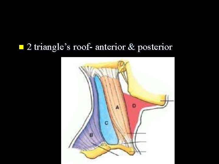
n 2 triangle’s roof- anterior & posterior
n 2 Gland enclosed- parotid & submandibular salivary gland
n 2 Spaces enclosed- suprasternal(Space of BURN’s) & supraclavicular space
n 2 Thickenings formed - parotidomasseteric fascia & stylomandibular ligament
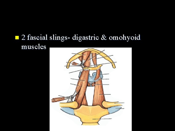
n 2 fascial slings- digastric & omohyoid muscles
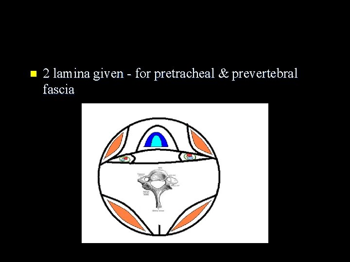
n 2 lamina given - for pretracheal & prevertebral fascia
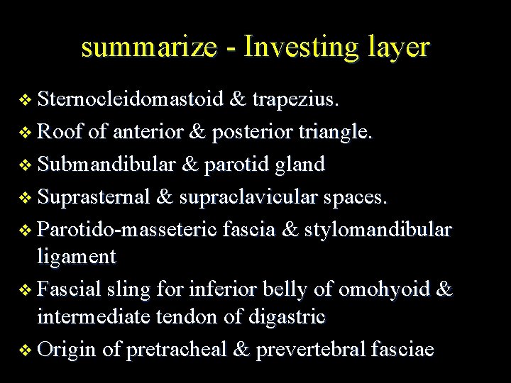
summarize - Investing layer v Sternocleidomastoid & trapezius. v Roof of anterior & posterior triangle. v Submandibular & parotid gland v Suprasternal & supraclavicular spaces. v Parotido-masseteric fascia & stylomandibular ligament v Fascial sling for inferior belly of omohyoid & intermediate tendon of digastric v Origin of pretracheal & prevertebral fasciae
Applied anatomy n n n surrounds muscles, blood vessels, and nerves, binding those structures together Limit the spread of infections natural cleavage planes for tissues, direct spread of pathology (fluid/growth) Provide slipperiness – allows the structures to move & pass over one another Ext jugular vein if cut remains retractedattachment to cervical fascia-air embolism
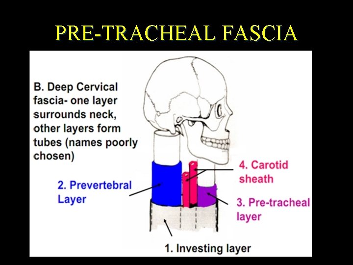
PRE-TRACHEAL FASCIA
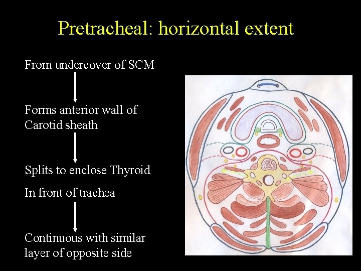
Pretracheal: horizontal extent From undercover of SCM Forms anterior wall of Carotid sheath Splits to enclose Thyroid In front of trachea Continuous with similar layer of opposite side
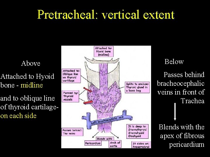
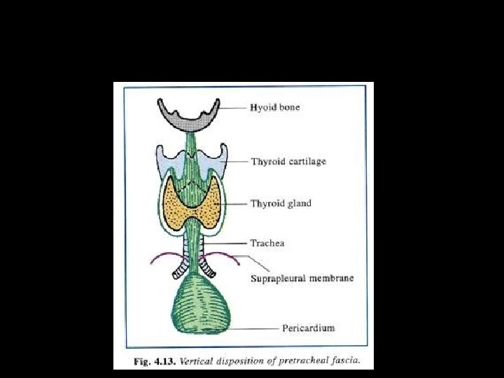
Pretracheal: vertical extent Above Attached to Hyoid bone - midline and to oblique line of thyroid cartilageon each side Below Passes behind bracheocephalic veins in front of Trachea Blends with the apex of fibrous pericardium
Applied anatomy Ligament of berry
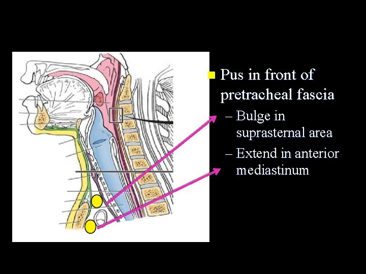
n Pus in front of pretracheal fascia – Bulge in suprasternal area – Extend in anterior mediastinum
Carotid sheath
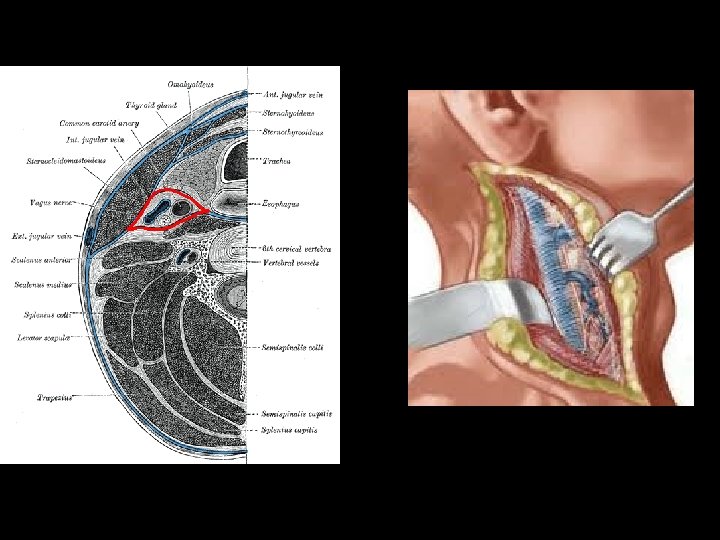
CAROTID SHEATH IT IS A FASCIAL SHEATH SITUATED DEEP TO STERNOCLEIDOMASTOID MUSCLE ON EACH OF THE FRONT OF THE NECK FORMATION • ANTERIOR WALL- BY PRETRACHEAL LAYER OF DEEP CERVICAL FASCIA Ø POSTERIOR WALL- BY PREVERTEBRAL LAYER OF DEEP CERVICAL FASCIA CONTENTS Ø INTERNAL JUGULAR VEIN LATERALLY, COMMON CAROTID ARTERY ( IN THE LOWER PART) & INTERNAL CAROTID ARTERY ( IN THE UPPER PART) MEDIALLY, VAGUS NERVE IN B/W THEM IN A POSTERIOR PLANE RELATIONS Ø Ø Ø ANTERIORLY- ANSA CERVICALIS POSTERIORLY – SYMPATHETIC TRUNK
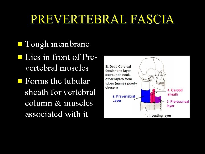
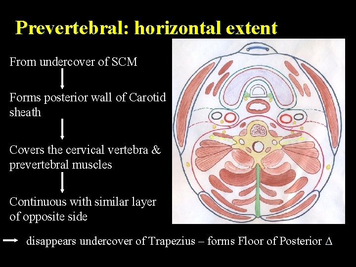
PREVERTEBRAL FASCIA Tough membrane n Lies in front of Prevertebral muscles n Forms the tubular sheath for vertebral column & muscles associated with it n
Prevertebral: horizontal extent From undercover of SCM Forms posterior wall of Carotid sheath Covers the cervical vertebra & prevertebral muscles Continuous with similar layer of opposite side disappears undercover of Trapezius – forms Floor of Posterior
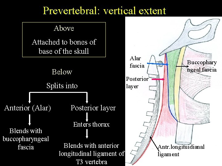
Prevertebral: vertical extent Above Attached to bones of base of the skull Below Alar fascia C 2 Splits into Buccophary ngeal fascia Posterior C 3 layer C 4 Anterior (Alar) Blends with buccopharyngeal fascia Posterior layer C 5 C 6 Enters thorax C 7 T 1 Blends with anterior longitudinal ligament of T 2 T 3 vertebra T 3 Antr. longituidianal ligament
Special feature Covers cervical & brachial plexus nerves n 4 superficial branches of cervical plexus pierce the fascia n Between Sc. Anterior & medius - Axillary sheath around cords of Br. plexus and Subclavian artery n Pus from an abscess posterior to the prevertebral layer may extend laterally posterior to SCM n
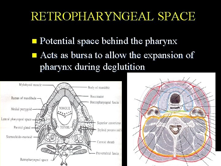
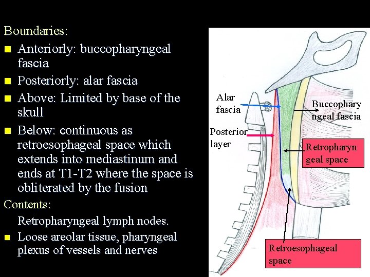
RETROPHARYNGEAL SPACE Potential space behind the pharynx n Acts as bursa to allow the expansion of pharynx during deglutition n Retro pharyngeal space
Boundaries: n Anteriorly: buccopharyngeal fascia n Posteriorly: alar fascia n Above: Limited by base of the skull n Below: continuous as retroesophageal space which extends into mediastinum and ends at T 1 -T 2 where the space is obliterated by the fusion Contents: Retropharyngeal lymph nodes. n Loose areolar tissue, pharyngeal plexus of vessels and nerves Alar fascia Posterior layer Buccophary ngeal fascia Retropharyn geal space Retroesophageal space
Clinical significance: n Route through which the infections of mouth & throat reach the mediastinum n Infection can break through the posterior wall of space and can enter danger space n Fatal haemorrhage could result from an extension of retropharyngeal abscess to the deep vessels of neck. n Retropharyngeal abscess –dysphagia, dysarthria.
Imp questions Investing layer of deep cervical fascia? n Pre tracheal fascia. n Retropharyngeal space. n
Thank you