Skin and Fascia Dr Rashmi Malhotra Associate Professor


- waterproofs & protects skin, nails, hair, stratum corneum • Melanocytes")


, histiocytes, endothelial cells, perivascular macrophages and")


 hair and skin. Sebum An")
 of the sebaceous duct.")


- Slides: 64
Skin and Fascia Dr. Rashmi Malhotra Associate Professor
OBJECTIVES Describe layers of skin. Enlist the functions of skin. Define appendages of skin. Define Fascia. Ø Differentiate between Superficial and Deep Fascia. Ø Applied Anatomy Ø Ø
Tissues of the body The tissue: is a group of cells which perform a specific function There are four basic tissues: 1. Epithelium 2. Connective tissue 3. Muscular tissue 4. Nervous tissue
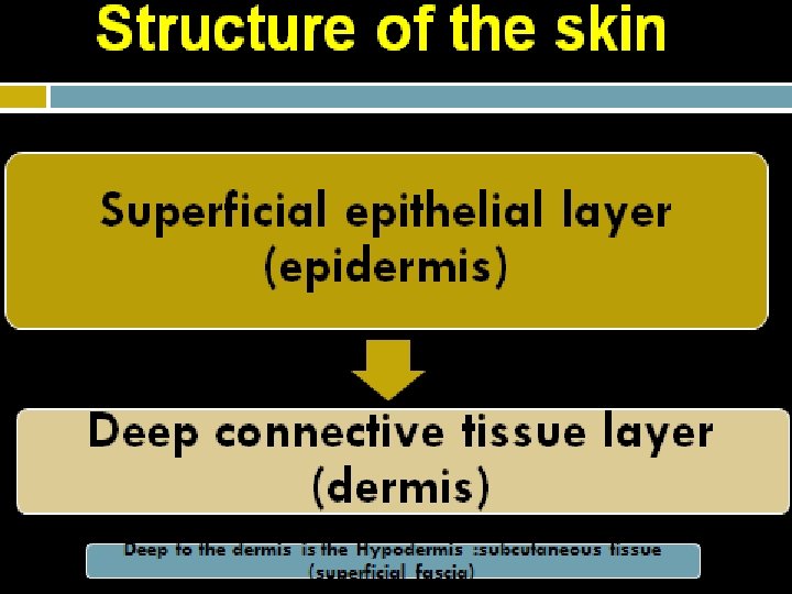
Skin
IMPORTANCE OF SKIN q Normal skin is a very complex organ �The skin is one of the largest organs of the body: 76 oo sq cm
FUNCTIONS OF THE SKIN � 1 -Protection � continuous and covers the body as well as protects the deep tissues ��abrasion, invasion, water loss, UV protection � 2 -Vitamin D synthesis ��epidermal keratinocytes when exposed to UV light ��helps maintain health of skeleton by increasing absorption of Ca 2+
� 3 -Sensation ��receptors for heat, cold, touch, pressure, vibration and pain � 4 - Thermoregulation ��thermo receptors and sweat glands ��hypothalamus controls cutaneous arteries and sweat glands to retain or dissipate heat
5 - Excretion through the secretion of sweat. � 6 - Psychological and social functions ��appearance and social acceptance ��facial expression and nonverbal communication
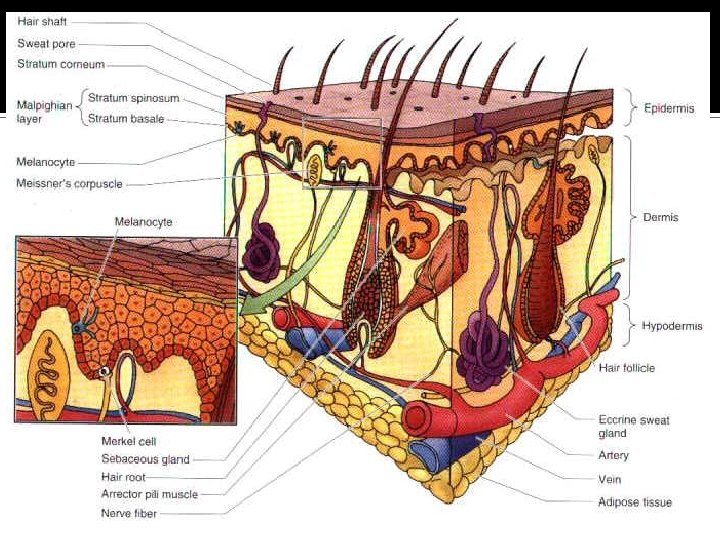
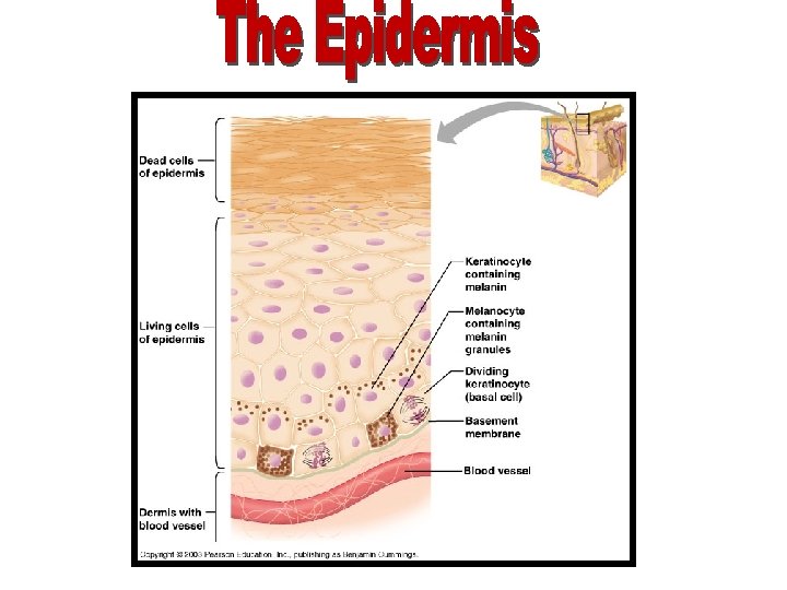
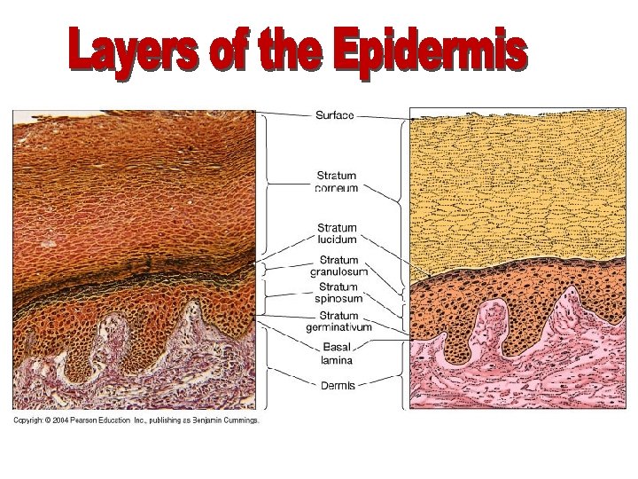
q Keratinized stratified squamous epithelium devoid of blood vessels Connective tissue containing (bl. v. lymph v. , sensory nerve endings, smooth m, hair follicles, sweat and sebaceous glands) In its deep part the collagen bundles are arranged in parallel rows
• Keratinocytes (90%)- waterproofs & protects skin, nails, hair, stratum corneum • Melanocytes (8%)- produce melanin • Merkel Cells- slow mechanoreceptors • Langerhans’ Cells- immunological defense
• Stratum Corneum • Stratum Lucidum • Stratum Granulosum • Stratum Spinosum • Stratum Basale(Germinativum)
Epidermis Thickness is increased: The epidermis is generally thin except in : • The palms of the hand. • The soles of the feet. Why? To protect these parts and withstand friction, wear and tear that occurs in these regions.
Dermis
papillary dermis reticular dermis
a. Cellular Fibroblasts (synthesize collagen, elastin, and reticulin), histiocytes, endothelial cells, perivascular macrophages and dendritic cells, mast cells, smooth muscle, and cells of peripheral nerves b. Fibrous Collagen & reticulin - provide tensile strength Elastic fibers- provide for restoration of shape after a deformation c. Ground substance glycosaminoglycans: hyaluronic acid, chondroitin sulfate, and dermatan sulfate.
Hypodermis This layer contains adipose tissue and serves to attach the dermis to its underlying tissues.
Lines of cleavage • The collagen fibers, arranged in parallel rows, called: Lines of cleavage (langer’s lines): • The direction of the rows of collagen fibers in the dermis: It runs • Longitudinally in the limbs. • Circumferentially in the neck and the trunk.
Lines of cleavage These lines are important to determine the direction for an incision (cut) during a surgery to avoid obvious scars.
• A surgical incision along or between these lines causes the minimum disruption of collagen so that the wound heals with a small scar. • Conversely, an incision made across the rows of collagen makes a disruption resulting in the massive production of fresh collagen and the formation of a broad scar.
Skin creases Folded skin over the joints. Skin is thin and is firmly adherent to underlying structures.
Some variations in human skin color Sub-Saharan African, Indian, Southern European, Northwest European
Skin Color Ø Due to Melanin, a pigment in the epidermis and Carotene, Ø Melanin is synthesized in cells called Melanocytes (found in basal layer). Ø Number of Melanocytes is essentially the same in all races. Ø The differences in skin color is due to the amount of pigment the melanocytes produce.
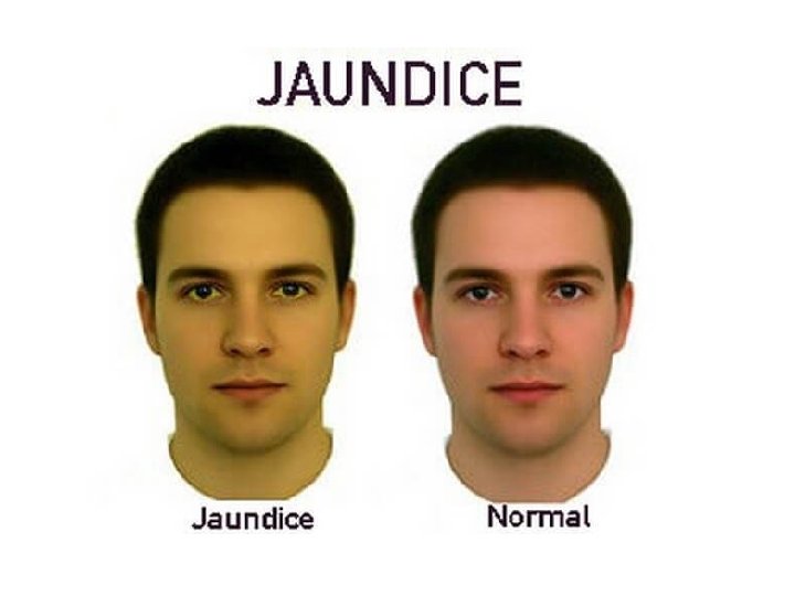
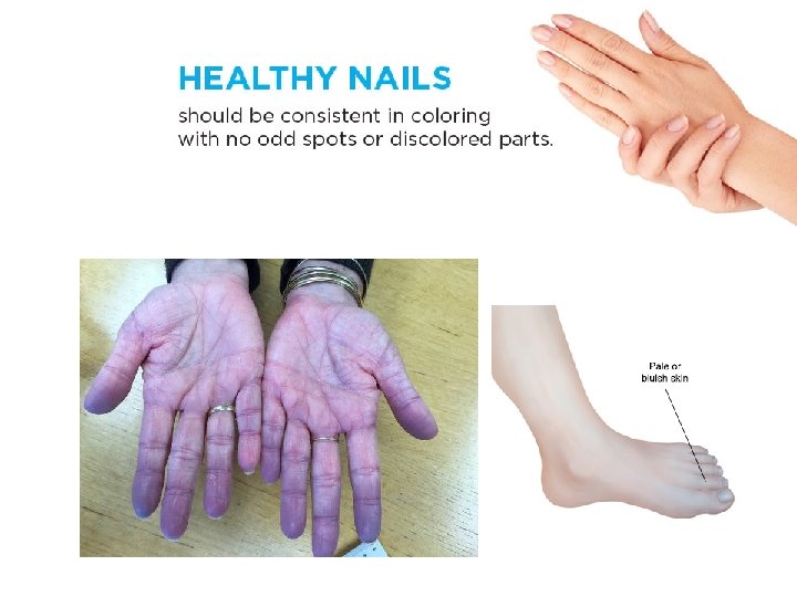
• Cyanotic • Jaundice • Erythema • Pallor
• Pigmentation levels usually increase with age. - exception: premature graying • Normal pigmentation may be altered by genetic defects or by acquired diseases. -Hyperpigmentation- age spots -Hypopigmentation- vitiligo, albinism
External agents can also alter skin color. • lightening agents • carotene • dyes • Some internal compounds--such as the byproducts of hemoglobin metabolism--may color the skin.
SKIN IN ANAEMIA
CHICKEN POX
SKIN ERUPTIONS
ACNE
Skin infections Pathogenic organisms can enter to the tissue through : • Nail Folds • Hair Follicles • Sebaceous Glands Staphylococcus: A type of bacteria that causes skin infections.
Ring worm
Malignant melanoma • 2% of all cancers Risks: 1. Skin type 2. Sun exposure 3. Family history 4. Age 5. Immunological status Normal mole Melanoma • A= asymmetry • B= border • C= color • D= diameter
Skin Cancers
The appendages of the skin • Nails • Hairs • Sebaceous glands • Sweat glands
Nails A nail is a flat horny plate on the dorsal surface of tips of the fingers and toes It has: Root: proximal edge (part embedded in skin) body: exposed part & has a free distal edge Nail fold: folds of skin surround and overlap the nail • Nail bed is very vascular causing pink color of the nail • The germinative zone lies beneath the root& is responsible for growth of nail
Hair Cover whole surface of the body except some areas as lips, palms, soles, some genital areas
Hair follicles: invaginations of the epidermis into the dermis, the hair grows out of these follicles (hair shaft). Hair bulb: the expanded extremity of the follicle, concaved at the end (located deep in the dermis). Hair papilla: a vascular connective tissue that occupies the concavity of the bulb.
Arrector Pilli muscle • A band of smooth muscle connects the undersurface of the follicle to the superficial part of the dermis. • It is innervated by sympathetic nerve fibers. • It is involuntary.
Arrector Pilli muscle Functions: • Its contraction causes the hair to move into a more vertical position. • It compresses the sebaceous gland causes it to extrude sebum.
Sebaceous glands Function It secrets sebum to oil (lubricate) hair and skin. Sebum An oily material that keeps the flexibility of the hair and oils the epidermis around the mouth of the follicle.
Sebaceous cyst It occurs because of the obstruction (blocking) of the sebaceous duct.
Sweat glands • long tubular glands with deep coiled part. • All over the body except red margins of lips, nail beds, glans penis and clitoris. • The most deeply penetrated structure.
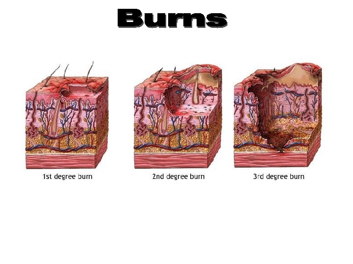
Skin burns Superficial Heals rapidly from the edges, . Heals quickly. Doesn’t need a skin graft. Deep Heals slowly from the edges. Usually needs skin grafting.
Clinical notes Graft is transferring tissue from one site to another. Skin graft is needed when the skin is damaged ( usually by deep burning )
Clinical notes Skin Graft Split thickness grafting Transferring epidermis only Full thickness grafting Transferring both epidermis and dermis.
Fascia
Fascia Collection of connective tissue Superficial fascia Deep fascia
Superficial fascia
Deep Fascia
Superficial fascia: • • Loose, mixture of adipose and loose areolar tissues. It unites the skin to the underlying structures. It is dense in some places as scalp, palm of hand sole of foot and contains collagen bundles It is thin in the eyelids, auricle, scrotum (devoid of adipose tissue). Functions: • • • Facilitates movement of skin over underlying structures. Passage for cutaneous vessels, nerves. Protects the body against heat loss.
Superficial fascia
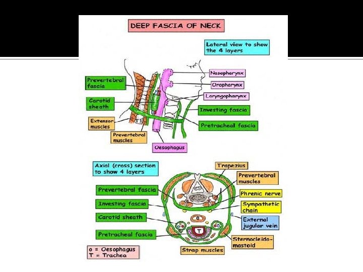
Deep fascia It is more dense than superficial fascia Collagenous bundles are more compact and more regularly arranged It is usually present in the form of membranes
Examples of deep fascia A. Intermuscular septa lie between muscles dividing the limb into compartments
Examples of deep fascia B. Investing fascia • • Covers the surfaces of muscles In the neck: it forms well-defined layers, bounds fascial spaces so limits spread of infection or determine the path of infection In the abdomen: it is thin In the limbs: forms a definite sheath around the muscles
Examples of deep fascia C. Retinacula Localized thickening of deep fascia around joints, hold the tendons in place, prevent bowstringing of tendons
Thank you