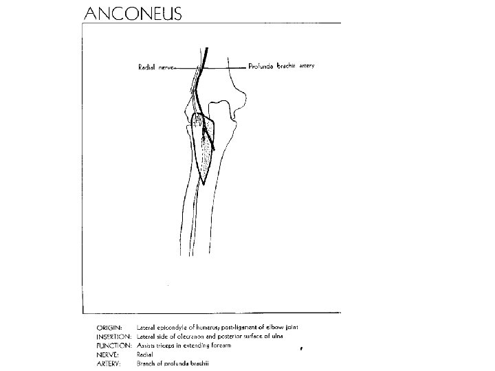
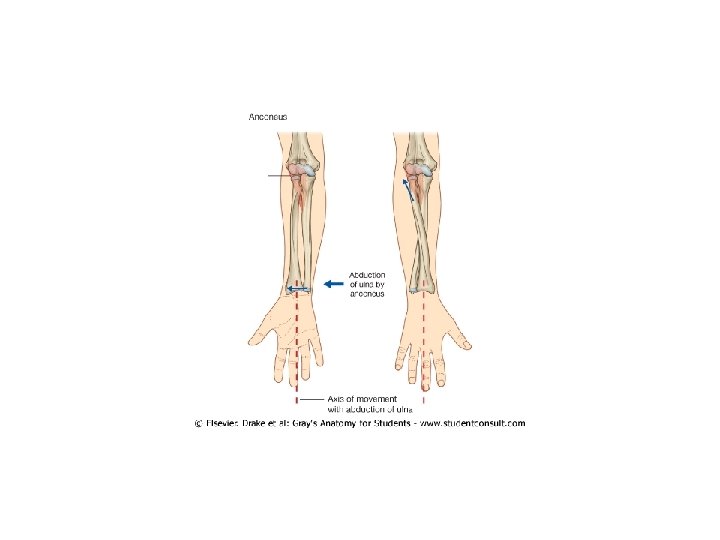
KOL KASLARI Devam M articularis cubiti M anconeus
")
KOL KASLARI (Devam)

M. articularis cubiti
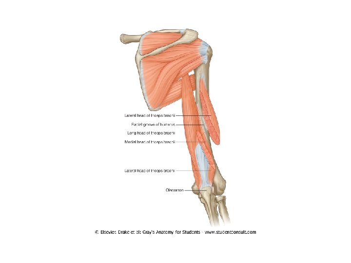
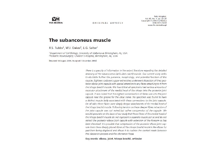
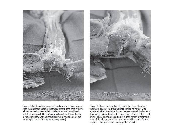

M. anconeus


ÖNKOL ve ELİN ARKA YÜZ KASLARI 11 Mart 2010 Saat: 10: 30 -12: 15

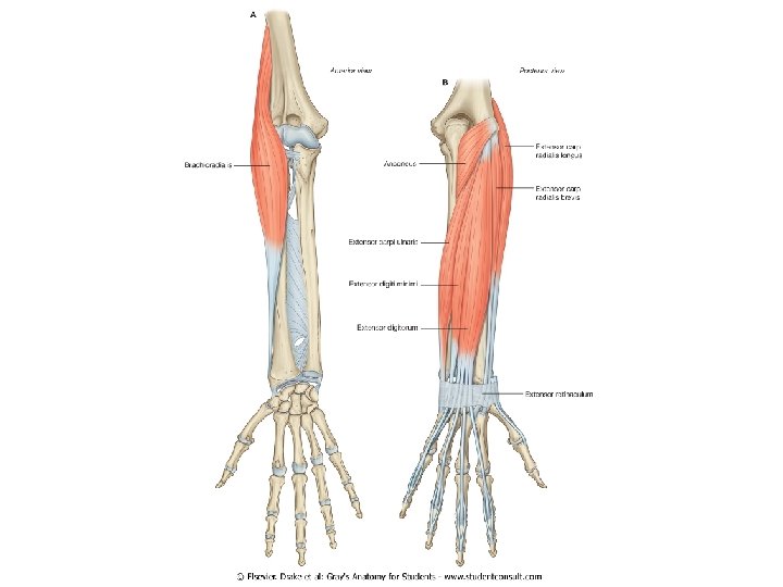
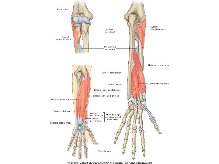
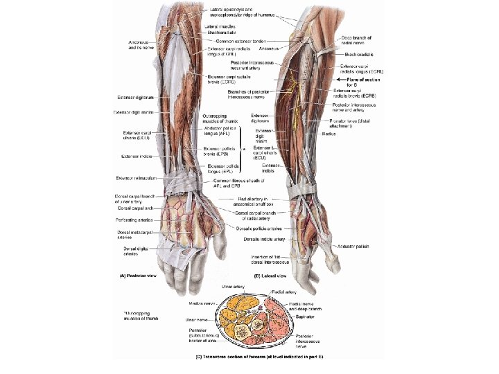
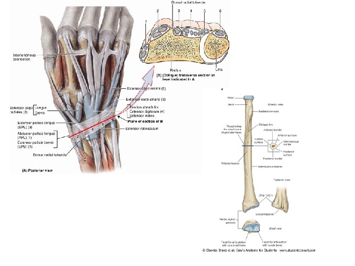
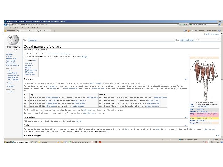
YÜZEYEL GRUP KASLAR M. brachioradialis M. extensor carpi radialis longus M. extensor carpi radialis brevis M. extensor digitorum M. extensor digiti minimi M. extensor carpi ulnaris DERİN GRUP KASLAR M. supinator M. extensor indicis M. abductor pollicis longus M. extensor pollicis brevis Baş parmağa gidecek şekilde yüzeyelleşirler

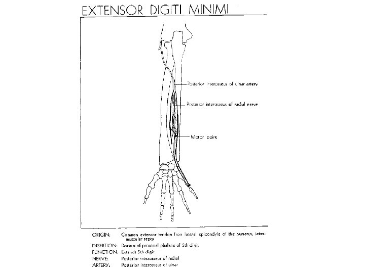
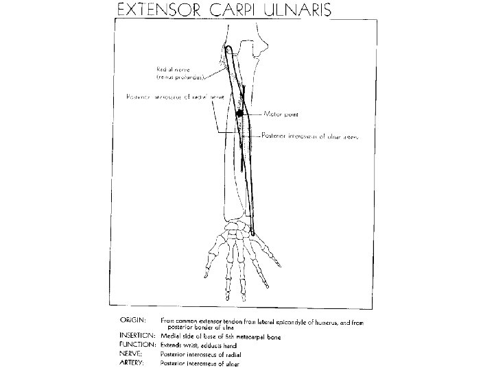
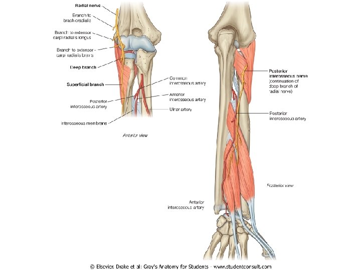
Kol, önkol ve elin arka yüz kaslarının tamamı n. radialis tarafından innerve edilir

Önkol’un arka yüz kaslarından M. supinator ve M. brachioradialis hariç diğer kaslar elbileğini çağrazlar

YÜZEYEL GRUP KASLAR

M. brachioradialis
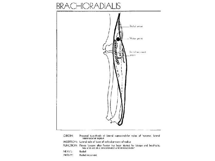


M. brachioradialis, fossa cubiti’nin lateral kenarını oluşturur


M. brachioradialis, extensor kompartmanda bulunmasına rağmen önkola fleksiyon yaptırır

M. extensor carpi radialis longus
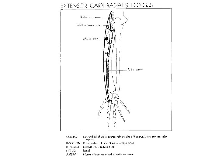

Tendonu, abductor pollicis longus ve extansor pollicis brevis tarafından çaprazlanır

M. extensor carpi radialis brevis


M. brachioradialis, extensor kompartmanda bulunmasına rağmen önkola fleksiyon yaptırır

M. extensor digitorum
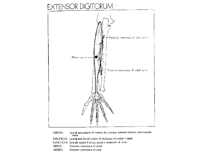

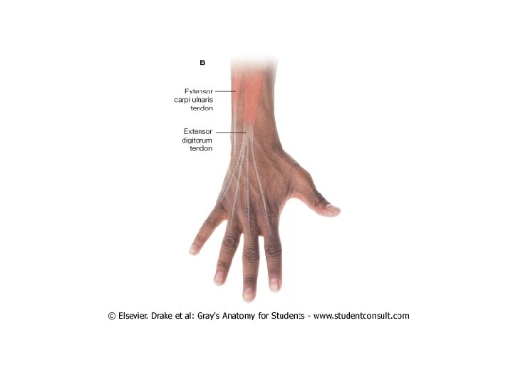

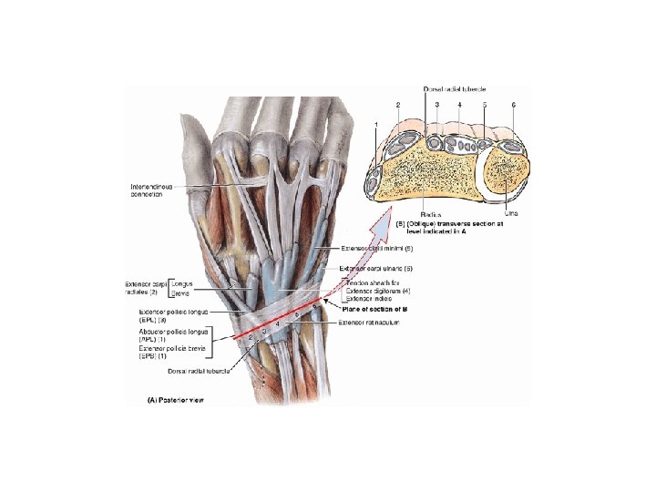
Dört tendonu, retinaculum extensorum’un altından ortak bir vagina synovialis ile sarılı olarak geçer

Connexus intertendinea

M. extensor digiti minimi


M. extensor carpi ulnaris


DERİN GRUP KASLAR


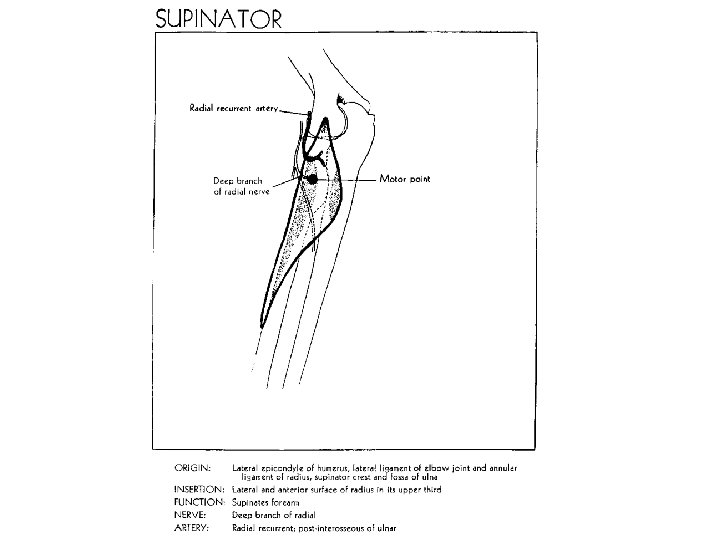
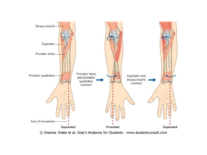
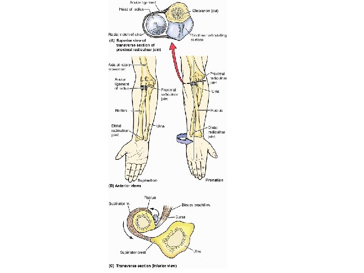
M. supinator

")
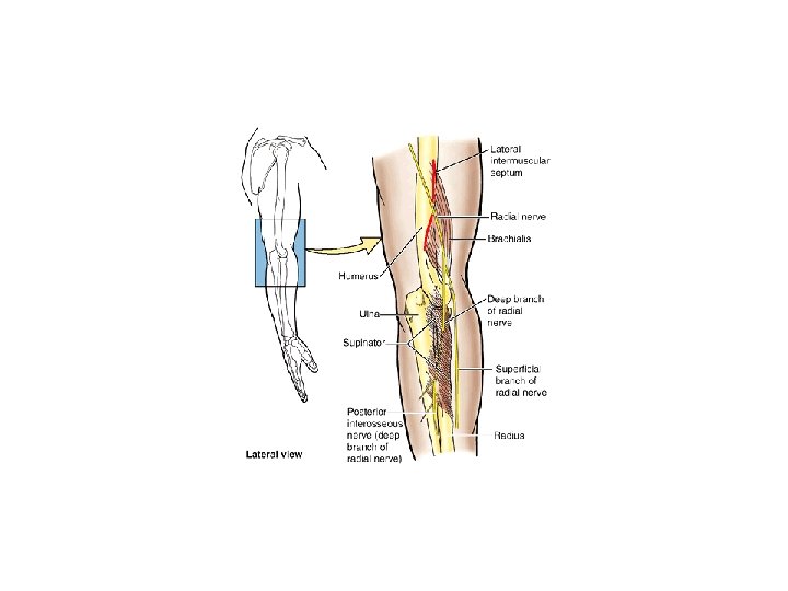
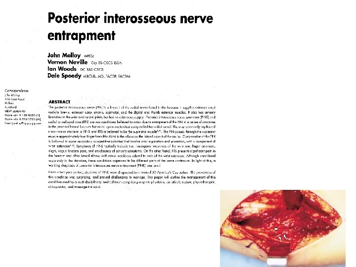
Supinator Syndrome (Posterior Interosseous Nerve Palsy)
 tarafından delinir")
M. supinator, N. radialis’in r. profundus’u (N. interosseus posterior) tarafından delinir

M. extensor indicis
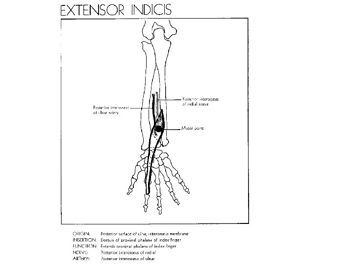


İşaret parmağının iki ekstensor kası vardır M. extensor digitorum M. extensor indicis

M. abductor pollicis longus
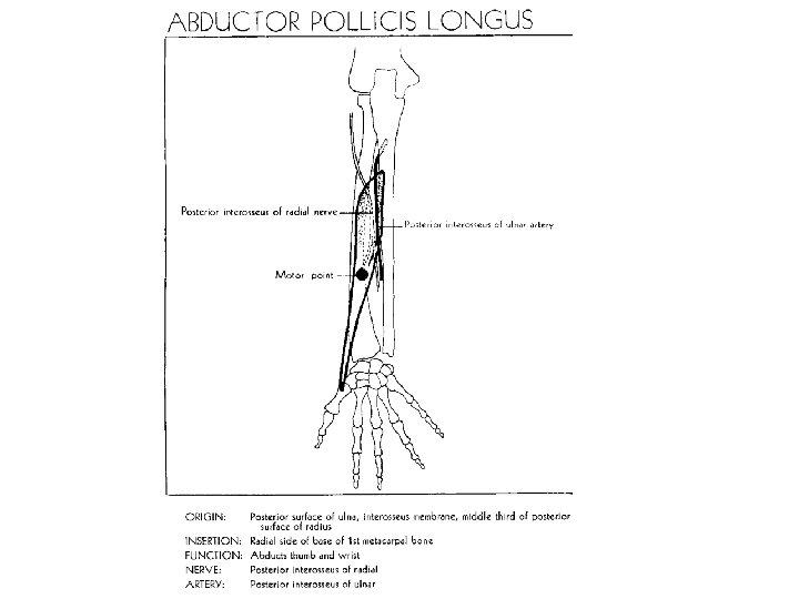


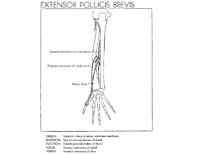
M. abductor pollicis longus ve M. extensor pollicis brevis’in tendonları ortak bir vagina synovialis ile sarılır ve 1. kanaldan geçer


M. extensor pollicis brevis
")
Fritz de Quervain (1868– 1940)
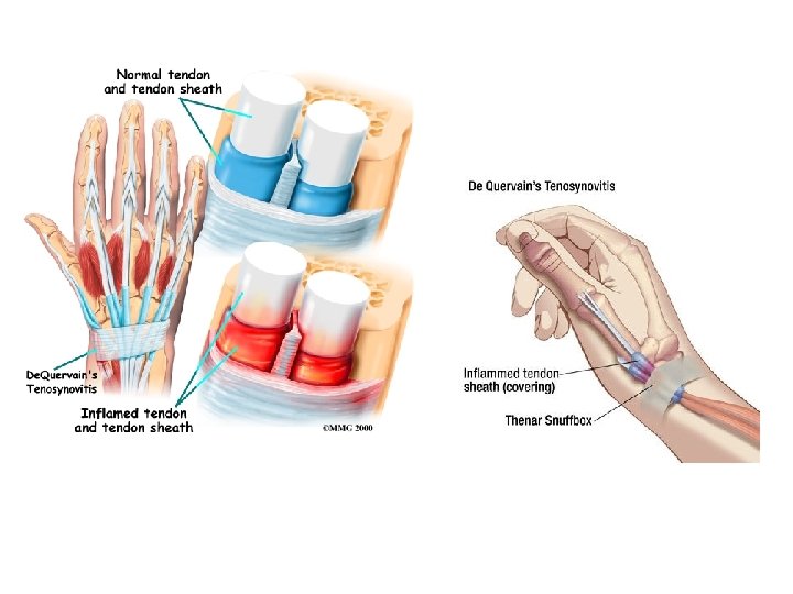

The Finkelstein test draws the tendons of the first dorsal compartment distally and causes sharp, local pain when tendon entrapment has occurred and inflammation is present In de Quervain tenosynovitis, the first dorsal compartment is thickened, raising the skin and creating a prominence at the radial styloid


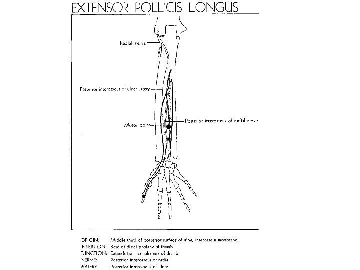
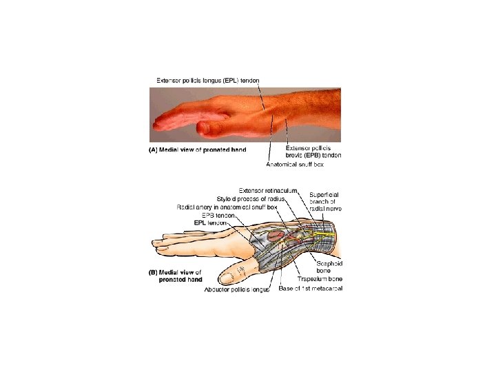
M. extensor pollicis longus

")
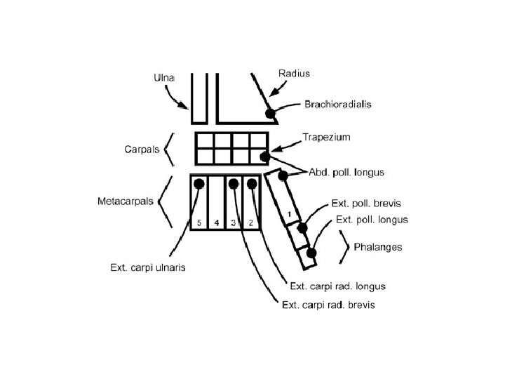
Compartment 1 2 3 4 5 6 Tendon Associated Pathology Extensor pollicis brevis (EPB) Abductor pollicis longus (APL) De Quervain's tenosynovitis Extensor carpi radialis longus (ECRL) Extensor carpi radialis brevis (ECRB) Intersection syndrome Extensor pollicis longus (EPL) Drummer's wrist, traumatic rupture with distal radius fx Extensor indicis proprius (EIP) Extensor digitorum communis (EDC) Extensor tenosynovitis Extensor digiti minimi (EDM) Vaughn-Jackson Syndrome Extensor carpi ulnaris (ECU) Snapping ECU
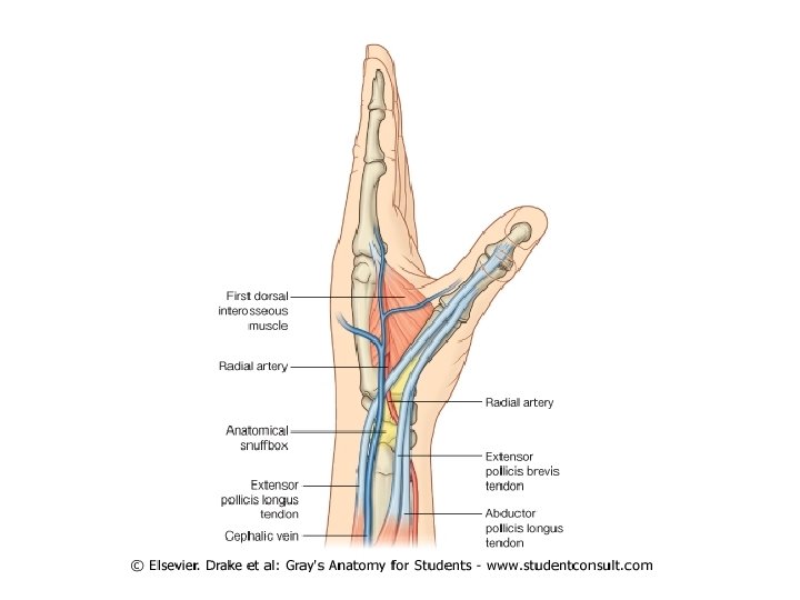
: -İçte M. extensor pollicis longus, -dışta M. abductor pollicis longus")
Fovea radialis (enfiye çukuru): -İçte M. extensor pollicis longus, -dışta M. abductor pollicis longus ve M. extensor pollicis brevis krişleri, -aşağıda başparmağın dorsal yüzü arasındaki çukurluktur. -Baş parmağı ekstensiyon durumuna getirirsek, bu çukurda deri çökerek çukur belirginleşir. -Bu çukurdan A. radialis geçer.
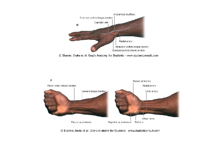

Nabız oluğu: -M. brachioradialis ile M. flexor carpi radialis’in krişleri arasında bulunan oluk İçinde a. radialis bulunur.

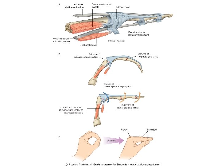
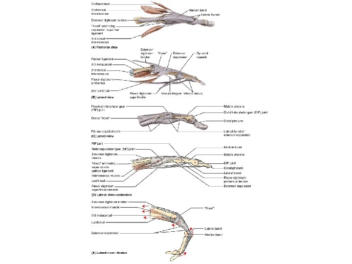
ELİN ARKA YÜZ KASLARI

Mm. interossei dorsales

bipennat diziliş gösterirler
 Lateral supraepicondylar ridge of humerus Lateral")
Superficial layer Brachioradialis Extensor carpi radialis longus (ECRL) Lateral supraepicondylar ridge of humerus Lateral surface of distal end of Radial nerve (C 5, C 6, C 7) radius proximal to styloid process Dorsal aspect of base of 2 nd Radial nerve (C 6, C 7) metacarpal Extensor carpi radialis brevis (ECRB) Lateral epicondyle of humerus (common extensor origin) Doral aspect of base of 3 rd metacarpal Extensor digitorum Extensor expansions of medial Posterior interosseous nerve (C 7, C 8), four fingers continuation of deep branch of radial nerve Extensor digiti minimi (EDM) Extensor expansion of 5 th finger Extensor carpi ulnaris (ECU) Deep layer Supinator Extensor indicis Proximal two thirds of supraepicondylar ridge of humerus Extend abduct hand at the wrist joint; ECRL active during fist clenching Deep branch of radial nerve (C 7, C 8) Lateral epicondyle of humerus; Dorsal aspect of base of 5 th posterior border of ulna via a shared metacarpal aponeurosis Lateral epicondyle of Lateral posterior, and anterior surfaces of humerus; radial proximal third of radius collateral and anular ligaments; supinator fossa; crest of ulna Posterior surface of Extensor expansion of 2 nd finger distal third of ulna and interosseous membrane Relatively week flexion of forearm, maximal when forearm is in midpronated position Extends medial four fingers primarily at metacarpophalangeal joints, secondarily at interphalangeal joints Extends 5 th finger primarily at metacarpophalangeal joint, secondarily at interphalangeal joint Extends and adducts hand at wrist joint (also active during fist clenching) Deep branch of radial nerve (C 7, C 8) Supinates forearm; rotates radius to turn palm anteriorlyor superiorly (if elbow is flexed) Posterior interosseous nerve (C 7, C 8), continuation of deep branch of radial nerve Extends 2 nd finger (enabling its independent extension); helps extend hand at wrist Outcropping muscles of deep layer Abductor pollicis longus (APL) Posterior surface of Base of 1 st metacarpal Posterior interosseous nerve (C 7, C 8), Abducts thumb and extends it at proximal halves of continuation of deep branch of radial nerve carpometacarpal joint ulna, radius, and interosseous membrane Extensor pollicis longus (EPL) Posterior surface of Dorsal aspect of base of distal phalanx of Extends distal phalanx of thumb at middle third of ulna thumb interphalangeal joint; extends and interosseous metacarpophalangeal and carpometacarpal membrane joints Extensor pollicis brevis (EPB) Posterior surface of Dorsal aspect of base of proximal phalanx of Extends proximal phalanx of thumb at distal third of radius thumb metacarpophalangeal joint; extends and interosseous carpometacarpal joint membrane a. The spinal cord segmental innervation is indicated (e. g. , “C 7, C 8� means that the nerves supplying the extensor carpi radialis brevis are derived from the seventh and eighth cervical segments of the spinal cord). Numbers in boldface (C 7) indicate the main segmental innervation. Damage to one or more of the listed spinal cord segments or to the motor nerve roots arising from them results in paralysis of the muscles concerned.

- Slides: 96