
Good morning BLOOD SUPPLY of HEART Dr Qudsia

Good morning
BLOOD SUPPLY of HEART Dr. Qudsia Sultana
üCORONARY ARTERIES üCORONARY SINUS AND TRIBUTARIES
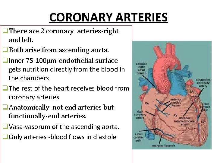
CORONARY ARTERIES q. There are 2 coronary arteries-right and left. q. Both arise from ascending aorta. q. Inner 75 -100µm-endothelial surface gets nutrition directly from the blood in the chambers. q. The rest of the heart receives blood from coronary arteries. q. Anatomically not end arteries but functionally-end arteries. q. Vasa-vasorum of the ascending aorta. q. Only arteries -blood flows in diastole
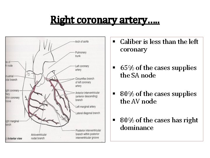
Right coronary artery…. . § Caliber is less than the left coronary § 65% of the cases supplies the SA node § 80% of the cases supplies the AV node § 80% of the cases has right dominance
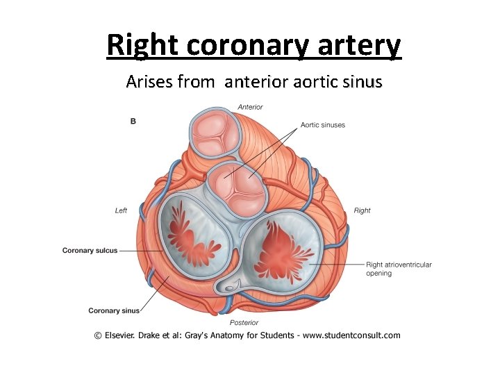
Right coronary artery Arises from anterior aortic sinus

Course of right coronary artery……. • Appears b/w right auricle & pulmonary trunk--- right ant. Coronary sulcus. • Descends—junction of the right and inf border of the heart—right post. Coronary sulcus on the diaphragmatic surface • Reaches crux & ends little left to crux & terminates by anastomosing –circumflex branch of left coronary artery.
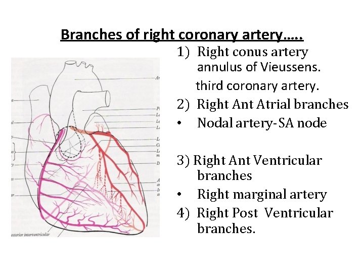
Branches of right coronary artery…. . 1) Right conus artery annulus of Vieussens. third coronary artery. 2) Right Ant Atrial branches • Nodal artery-SA node 3) Right Ant Ventricular branches • Right marginal artery 4) Right Post Ventricular branches.
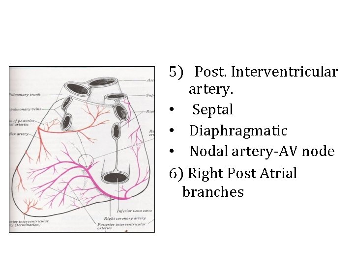
5) Post. Interventricular artery. • Septal • Diaphragmatic • Nodal artery-AV node 6) Right Post Atrial branches
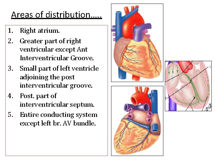
Areas of distribution…. . 1. Right atrium. 2. Greater part of right ventricular except Ant Interventricular Groove. 3. Small part of left ventricle adjoining the post interventricular groove. 4. Post. part of interventricular septum. 5. Entire conducting system except left br. AV bundle.
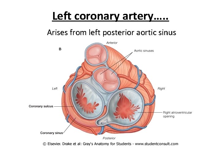
Left coronary artery…. . Arises from left posterior aortic sinus
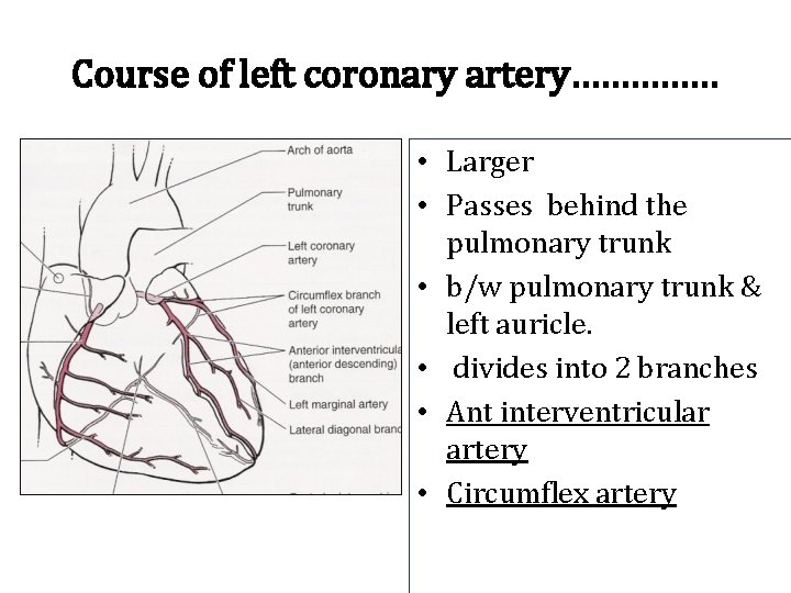
Course of left coronary artery…………… • Larger • Passes behind the pulmonary trunk • b/w pulmonary trunk & left auricle. • divides into 2 branches • Ant interventricular artery • Circumflex artery

1. Anterior interventricular artery…… Left anterior descending (LAD) artery…. . Descends in anterior interventricular groove…. Terminates on inferior surface- by anastomosing –post interventricular br of right coronary artery in the post interventricular groove

Branches…. . 1. Left conus artery(annulus of Vieussens) 2. Right ant Ventricular branches adjoining the ant Interventricular groove 3. Left ant. Ventricular branches – left diagonal artery. 4. Septal branches
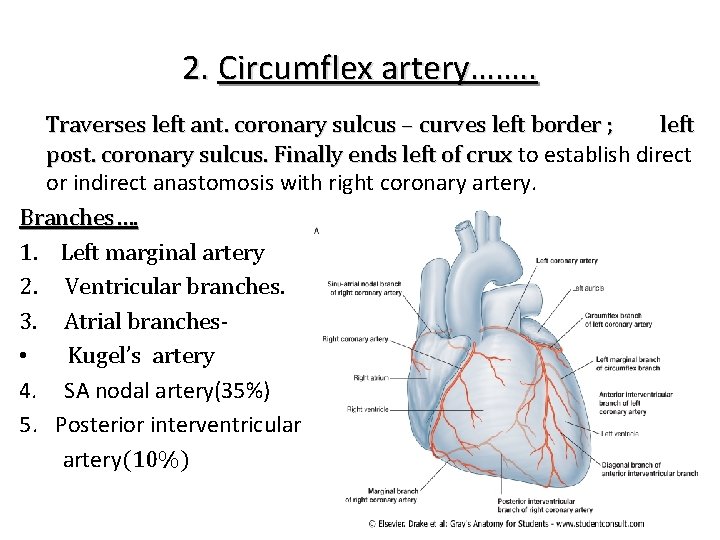
2. Circumflex artery……. . Traverses left ant. coronary sulcus – curves left border ; left post. coronary sulcus. Finally ends left of crux to establish direct or indirect anastomosis with right coronary artery. Branches…. 1. Left marginal artery 2. Ventricular branches. 3. Atrial branches • Kugel’s artery 4. SA nodal artery(35%) 5. Posterior interventricular artery(10%)
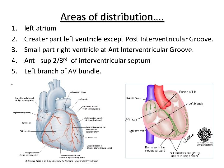
1. 2. 3. 4. 5. Areas of distribution…. left atrium Greater part left ventricle except Post Interventricular Groove. Small part right ventricle at Ant Interventricular Groove. Ant –sup 2/3 rd of interventricular septum Left branch of AV bundle.
Variations • LCA- arise-Pulmonary trunk cyanosis-ventricular failure • LCA-anterior aortic sinus longer course-compressed-sudden cardiac death
Cardiac Predominance… • Right coronary artery-post interventricular branch- Right cardiac dominance. (majority) • Left coronary artery- post interventricular branch- left cardiac dominance (10%) commonly affected by coronary diseasesentire left ventricle and ventricular septumunder its control. • Rarely-post interventricular artery-from both – coronaries-least affected by coronary diseases.
Myocardial circulation • Coronaries anastomose with each other in myocardium. • Arterio venous anastomosis • Arterio sinusoidal anastomosis Collateral circulation • • Vasa vasorum-Aorta ad pulmonary arteries. Internal thoracic arteries. The bronchial arteries. The phrenic arteries.
Clinical……. Angina pectoris Incomplete obstruction of coronary artery due to spasmodic contraction results decrease blood supply to heart –insufficient oxygen-severe pain in chest region & also in left arm. Myocardial infarction Complete obstruction coronary artery due to thrombosis results in death of myocardium - heart attack.
INVESTIGATIONS: ECG CARDIAC ENZYMES e. g. CK, LDH, TROPONIN etc. ECHOCARDIOGRAPHY TREADMILL EXERCISE TEST THALLIUM STRESS TEST CORONARY ANGIOGRAPHY 21
TREATMENT: – CORONARY DILATORS E. g. NITRATES – BETA-BLOCKERS – ANGIOPLASTY (DILATE AREA OF CONSTRICTION) – STENT – BYPASS SURGERY 22
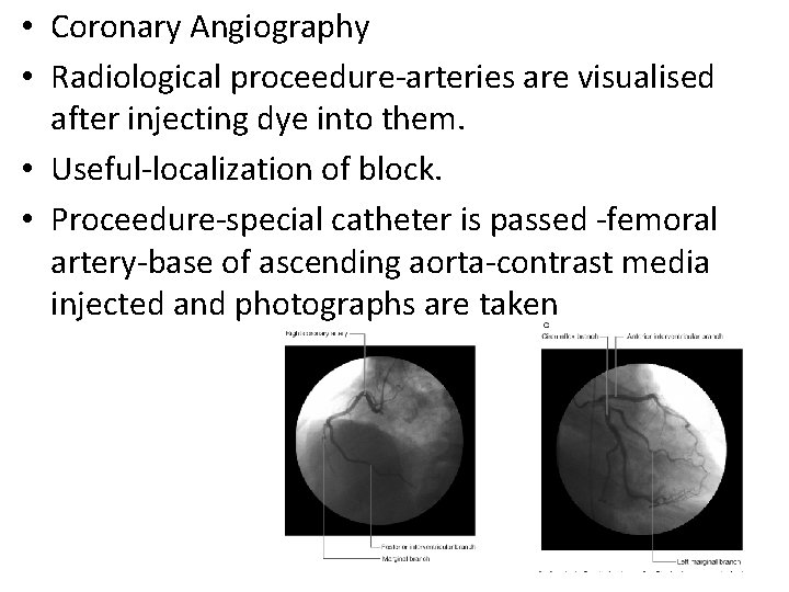
• Coronary Angiography • Radiological proceedure-arteries are visualised after injecting dye into them. • Useful-localization of block. • Proceedure-special catheter is passed -femoral artery-base of ascending aorta-contrast media injected and photographs are taken
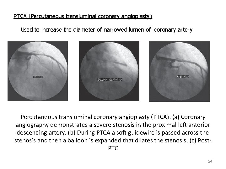
PTCA (Percutaneous transluminal coronary angioplasty) Used to increase the diameter of narrowed lumen of coronary artery Percutaneous transluminal coronary angioplasty (PTCA). (a) Coronary angiography demonstrates a severe stenosis in the proximal left anterior descending artery. (b) During PTCA a soft guidewire is passed across the stenosis and then a balloon is expanded that dilates the stenosis. (c) Post. PTC 24
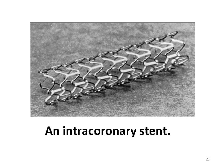
An intracoronary stent. 25
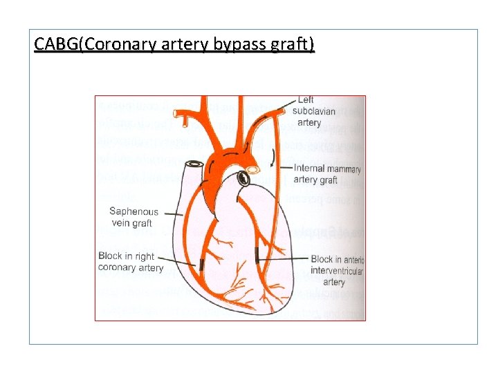
CABG(Coronary artery bypass graft)
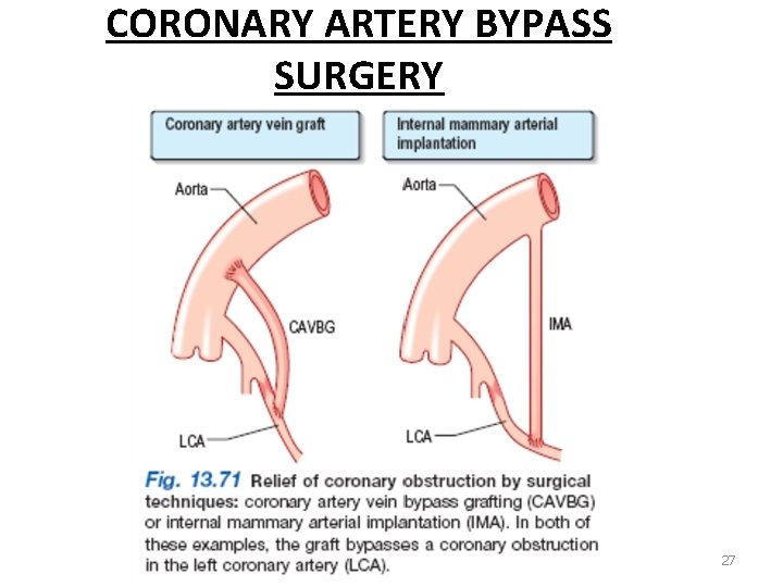
CORONARY ARTERY BYPASS SURGERY 27
Venous drainage…. Venous blood from drains into right atrium through a. Coronary sinus b. Anterior cardiac veins c. Venae cordis minimae (thebesian veins)
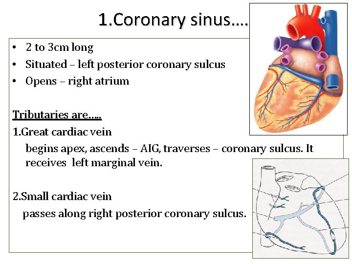
1. Coronary sinus…. . • 2 to 3 cm long • Situated – left posterior coronary sulcus • Opens – right atrium Tributaries are…. . 1. Great cardiac vein begins apex, ascends – AIG, traverses – coronary sulcus. It receives left marginal vein. 2. Small cardiac vein passes along right posterior coronary sulcus.
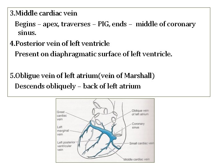
3. Middle cardiac vein Begins – apex, traverses – PIG, ends – middle of coronary sinus. 4. Posterior vein of left ventricle Present on diaphragmatic surface of left ventricle. 5. Obligue vein of left atrium(vein of Marshall) Descends obliquely – back of left atrium
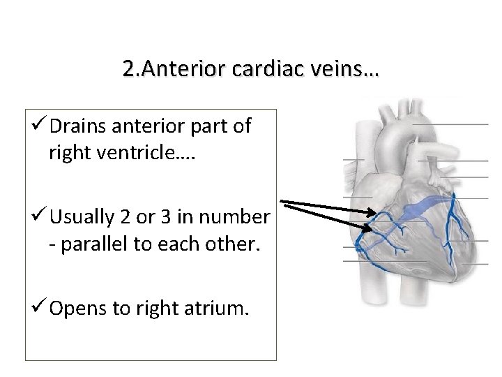
2. Anterior cardiac veins… ü Drains anterior part of right ventricle…. ü Usually 2 or 3 in number - parallel to each other. ü Opens to right atrium.
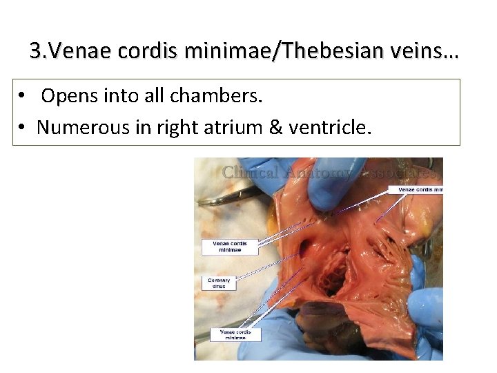
3. Venae cordis minimae/Thebesian veins… • Opens into all chambers. • Numerous in right atrium & ventricle.
Question • Blood supply of heart? • Arterial and venous drainage.

Thanks for listening. .
- Slides: 34