Good morning Caecum and Appendix Dr Qudsia Sultana


 Retro-caecal or Retrocolic(120")


- Slides: 34
Good morning
Caecum and Appendix Dr. Qudsia Sultana
The Caecum
General features Asymmetrical cul-desac n Commencement of large intestine n Furnished with taeniae coli n Lies in the right iliac fossa Size n Length- 6 cm n Breadth- 7. 5 cm n
Types Depending on the shape FINE Normal
Position n Normally right iliac fossa Occupies a triangular area Due to faulty rotation of midgut may occupy any other abnormal locations.
Abnormal locations n n n Subhepatic Right lumbar Pouch of Douglas Left iliac fossa Umbilical region
Peritoneal recesses n n n Superior ileocal recess Inferior ileocal recess Retroceacal recess
Relations n n ü ü ü Peritoneal- all sides covered by peritoneum General. Above- continuous with ascending colon Below- rests on inguinal ligament Anteriorly-anterior abdominal wall, Greater omentum, coils of intestine.
Relations ü Posteriorly n Right Ilio-psoas n Lateral femoral cutaneous nerve n Femoral nerve n Genito-femoral nerve n Retro-caecal recess of peritoneum
Medially n Terminal part of ileum. n Vermiform appendix suspended by mesoappendix. n Inferior ileocaecal recess.
Blood supply Arterial supply n Anterior and posterior caecal branches of ileocolic artery Venous drainage n Ileocolic vein and thence into superior mesenteric vein
Lymphatic drainage n Ileocolic lymphnodessup mesentric group of preaortic lymphnodes. Nerve supply Sympathetic nervesn Superior mesentric plexus (T 10 -T 11) Parasympatheticn Vagus nerves.
Internal features n Ileo-caecal orifice n Measures 2. 5 cm n Situated in the posteromedial wall of Caecum n Guarded by valve having upper and lower lip n Lips meet and continue as mucous folds known as frenula. n Prevents regurgitation.
Appendicular orifice n n n Small circular opening 2 cm below and behind the ileocaecal orifice Guarded by valve of Gerlach
Investigations
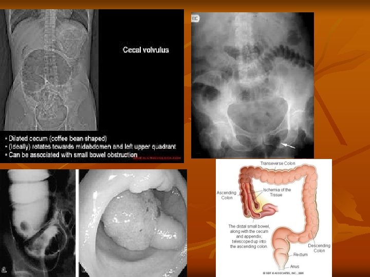
Applied anatomy n n Acts as a guide in operation of intestinal obstruction. Caecum-bloated-large intestinal odstruction Caecum-empty-small intestinal obstruction Intussusception- telescopical invagination of terminal part of ileum into the caecum and ascending colon.
The vermiform Appendix/Abdominal Tonsil
General features n n Worm like narrow tubular diverticulum Arises from posteromedial wall of the Caecum, 2 cm below the ileo-caecal junction Suspended by a peritoneal fold – mesoappendix It is a vestigial organ?
n n n Devoid of taenia coli, sacculations, appendices epiploicae 2 -20 cm in length. ( average -9 cm) The body of appendixkinked on itselfmesoappendix shortcoiled like worm. VERMIFORM
Surface anatomy n n n Base of appendix Mc. Burney’s pointmaximum tendernessinflammation of appendix Spinoumbilical line
Presenting parts n n Base Body Tip Mesoappendix
n n n BASEposteromedial wall, 2 cm below ileocaecal junction. Taenia of caecum converge at baseidentification of appendix
n n BODY – narrow, tubular and contains a canal-opens -caecum-gaurded mucosal fold “valve of Gerlach” TIPLeast vascular-various positions
Types n n n n Sub-caecal or parcolic (110 Clock ) Retro-caecal or Retrocolic(120 Clock ) Splenic type(20 clock) n Pre-ileal n Post-ileal Promonteric type(30 clock) Pelvic type(40 clock) Mid-inguinal type(60 clock) Ectopic
Blood supply n n Appendicular artery, a branch of inferior division of ileocolic artery End artery Tip least vascular Veins drain into Superior mesentric vein
LYMPHATIC DRAINAGE Ileo colic nodes Superior mesenteric nodes q NERVE SUPPLY Sympathetic T 10 – Superior mesenteric plexus Parasympathetic-vagus q
Applied anatomy n n Inflammation of appendix known as appendicitis Murphy's triad-Pain, vomiting, temperature. Tenderness –Mc Burneys point Pain first felt in umbilical region, then settles in right iliac fossa(Rowlings sign)
Predisposing factors 1. Blind tube 2. End artery 3. Hiatus muscularis 4. Lymphatic follicles— Abdominal Tonsil
n n n Hiatus musculare-muscles of wall are deficient -gap-infection–extend-peritoneum. Abdominal Tonsil-submucous coat-numerous lymphatic follicles. Appendicular lumen –obstructed-fecolithprecipitate attack of appendicitis.
ANAMOLIES n Undescended caecum and appendix n Agenesis of appendix Left sided appendix � Double appendix �
DEVELOPMENT OF CAECUM AND APPENDIX CAECAL BUD diverticulum arising from post arterial segment of primitive midgut.
Thank you