Perineum Dr Qudsia Sultana Questions Colles fascia n


 n Sacrum n Coccyx n Joined")


 Mons pubis(F). n POSTERIORLY: Buttocks n ON EACH SIDE: The")


- Slides: 60
Perineum Dr. Qudsia Sultana
Questions Colle’s fascia n Perineal body n Superficial perineal pouch n Perineal membrane n Deep perineal pouch n Urogenital diaphragm n
Pelvic Cavity Enclosed by bony, ligamentous and muscular wall n Contains the urinary bladder, ureters, uterus, genital organs, rectum, blood vessels, lymphatics and nerves n
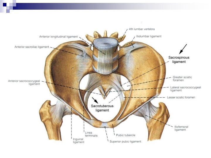
Bony Pelvis Innominate bone (Ilium, ischium and pubis) n Sacrum n Coccyx n Joined anteriorly by pubic symphysis n Posteriorly by sacro-iliac joint n
Bony Pelvis
Superior Apeture
Inferior aperture
Greater Pelvis/false pelvis / pelvis major n n Location of some abdominal viscera (ileum and sigmoid colon) Bounded by abdominal wall anteriorly, the iliac fossa posteriolaterally and L 5 S 1 vertebrae posteriorly
Lesser Pelvis/true pelvis/ pelvis minor. Location of pelvic viscera – the urinary bladder and reproductive organs such as the uterus and ovaries n Bounded by the pelvic surfaces of the hip bones, sacrum, and coccyx n Limited inferiorly by the musculofascial pelvic diaphragm n
Pelvic Floor Formed by the funnel shaped pelvic diaphragm – consists of the levator ani and coccygeus muscles and their fascia n Stretches between the pubis anteriorly and the coccyx posteriorly and from one lateral pelvic wall to the other n
Pelvic Floor - Male
Pelvic Floor - Female
Perineum
Introduction ü ü ü Perineum is the region at the lower end of the trunk in the interval between two thighs. The external genitalia are located in the perineum. Perineum forms the lower division of pelvis below the pelvic diaphragm & fills the pelvic outlet.
Introduction
Contents of the perineum
SUPERFICIAL BOUNDARIES ANTERIORLY: Scrotum(M) Mons pubis(F). n POSTERIORLY: Buttocks n ON EACH SIDE: The upper part of the medial side of the thigh. n
DEEP BOUNDARIES OF PERINEUM Deep boundaries of perineum are the same as those of pelvic outlet.
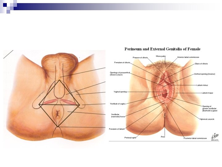
DIVISIONS OF PERINEUM A transverse line joining the anterior parts of ischial tuberosities divides the perineum into 2 triangular areas. 1. An anterior urogenital triangle. 2. Posterior anal triangle.
Subdivisions
ANAL TRIANGLE It consists of n IN THE MIDLINE Perineal body Termination of anal canal Anococcygeal raphe n ON EACH SIDE A fascial lined wedge shaped space, the ischioanal fossa. q PUDENDAL CANAL is a fascial tunnel in the lateral wall of ischioanal fossa. It contains the neurovascular bundle of perineum.
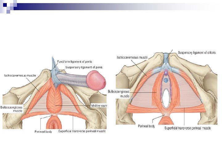
MUSCLES CONVERGING IN PERINEAL BODY PAIRED v Bulbospongiosus. v Superficial Transverse Perenei. v Deep Transverse Perenei. v Levator ani. UNPAIRED v External anal sphincter. v Fibres of longitudinal muscle coat of anal canal.
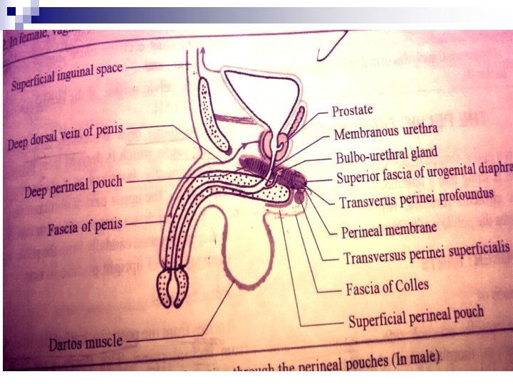
UROGENITAL TRIANGLE The Urogenital triangle • Skin • Fatty layer of superficial fascia. • Membranous layer of superficial fascia(COLLES’ FASCIA). • Superficial perineal pouch. • Perineal membrane. • Deep perineal pouch. • Urogenital diaphragm & its fasciae.
UROGENITAL TRIANGLE Skin n Male – midline medial raphe of the scrotum n Female – midline cleft (vestibule)-urethra and vagina n CUTANEOUS INNERVATIONn n n DORSAL NERVE PENIS/CLITORIS ILIOINGUINAL NERVE GENITAL BR. OF GENITOFEMORAL NERVE PERINEAL BR. OF POST. CUTANEOUS NERVE POSTERIOR SCROTAL /LABIAL NERVE
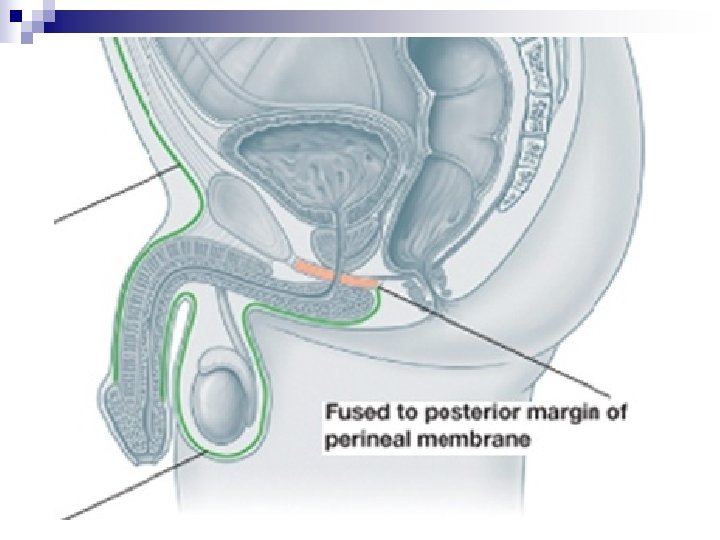
SUPERFICIAL FASCIA Superficial fascia is made up of 2 layers as in the lower part of anterior abdominal wall. 1. A superficial fatty layer 2. A deep membranous layer (colles’ fascia) n On each side colles’ fascia is attached to the ischiopubic rami. n
n n Anteriorly – dartos muscle of scrotum, fascia of penis and extends as fascia of scarpa of anterior abdominal wall. Posteriorly –fuses with the perineal membrane around superficial transverse perenei muscle.
COLLES’ FASCIAAPPLIED ANATOMY n n Since it is continuous up the anterior abdominal wall , fluids or infected material that accumulates in the superficial pouch can track out of the perineum & onto the lower abdominal wall. This material will not track into the anal triangle or the thigh because the fascia fuses with the deep fasciae at their borders.
SUPERFICIAL PERINEAL POUCH n It is an interfascial space stretching across the pubic arch & is situated superficial to the perineal membrane.
BOUNDARIES SUPERFICIAL: colle’s fascia. n DEEP: perineal membrane. n ON EACH SIDE: ischiopubic rami. n
n n POSTERIORLY: closed by the fusion of perineal membrane with colles’ fascia. ANTERIORLY: open & continuous with the spaces of scrotum, penis & anterior abdominal wall.
CONTENTS OF SUPERFICIAL PERINEAL POUCH n • • ROOT OF THE PENIS made up of 2 corpora cavernosa 1 corpus spongiosum (traversed by urethra). BODY OF CLITORIS made up of 2 corpora cavernosa. 2 BULBS OF VESTIBULE, 1 on each side of vaginal & urethral orifice.
MUSCLES IN THE SUPERFICIAL PERINEAL POUCH ISCHIOCAVERNOSUS covering the corpus cavernosum of penis/clitoris. n BULBOSPONGIOSUS covering the corpus spongiosum/bulb of the vestibule. n SUPERFICIAL TRANSVERSUS PERENEI. n
SUPERFICIAL POUCH- VESSELS n 1. 2. 3. 4. 3 branches of perineal artery 2 Posterior scrotal/labial arteries. Transverse perineal arteries. 4 branches Artery to the bulb of penis/clitoris. Dorsal artery of penis/clitoris. Deep artery of penis/clitoris. Urethral artery.
SUPERFICIAL POUCH-NERVES 3 branches from the perineal nerve • 2 Posterior scrotal/ labial n of internal pudendal. • Long perineal n. from posterior cutaneous nerve of thigh. n
SUPERFICIAL POUCHOTHER CONTENTS MALE n Spongy urethra n Ducts of bulbourethral glands FEMALE n Urethra n Vagina n Greater vestibular glands
Perineal membrane
Perineal membrane
PERINEAL MEMBRANE It is a strong sheet of fascia extending across the pubic arch. n It is triangular in shape with apex directed anteriorly. n It separates the superficial perineal pouch from the deep pouch. n In males it is tough due to the attachment of root of the penis & associated perineal muscles. n
PERINEAL MEMBRANEATTACHMENTS n n n LATERALLY: periosteum of ischiopubic rami. ANTERIORLY: thickened to form transverse perineal ligament. POSTERIORLY: fused to the deep parts of perineal body & is continuous with the fascia over deep transverse perinei.
STRUCTURES PIERCING PERINEAL MEMBRANE NEAR THE APEX: Dorsal A of penis/clitoris. IN THE MIDDLE: deep A of penis/clitoris. A to the bulb of penis on each side of urethra(M). AT THE BASE: 2 posterior scrotal/ labial vessels & nerves on each side. IN THE MIDLINE: Urethra Duct of bulbourethral glands & vagina behind urethra(F).
PERINEAL MEMBRANE 3 structures pass in the small triangular gap between arcuate pubic ligament & transverse perineal ligament. n A deep dorsal vein of penis/ clitoris (midline). n Dorsal nerve of penis/clitoris on each side.
Structures piercing perineal membrane
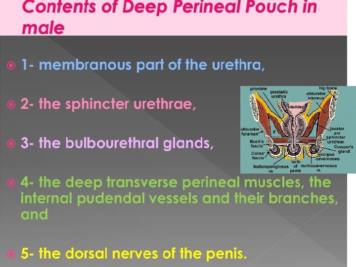
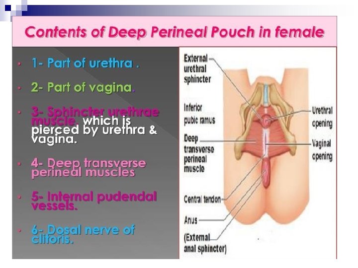
Deep perineal pouch
Deep perineal pouch Superficial- perineal membrane Deep- superior fascia of urogenital diaphragm Each side – Ischiopubic rami Posteriorly – closed by superior fascia of urogenital diaphragm perineal membrane Anteriorly – closed
Contents –muscles of deep perineal pouch
UROGENITAL DIAPHRAGM
Applied Anatomy n Episiotomy
UROGENITAL DIAPHRAGM It is a musculo-fascial partition across the pubic arch separating the pelvic cavity from the anterior part of the pelvic outlet. Consists of 2 muscles: 1. sphincter urethrae & 2. deep transverse perinei. n 2 fasciae 1. Superior fascia 2. Inferior fascia/ perineal membrane. n
UROGENITAL DIAPHRAGM Both the muscles are supplied by muscular branches of perineal nerve. n It is pierced by I. Urethra. II. Vagina(F). n
RELATIONS SUPERIORLY: n Anterior fibres of both levator ani muscles. n Anterior recess of ischorectal fossa. n apex of prostate (M). n Neck of bladder(F). INFERIORLY: n contents of superficial perineal pouch.
RELATIONS n ANTERIORLY: transverse perineal ligament. n POSTERIORLY: ischiorectal fossa.
ACTIONS OF UROGENITAL DIAPHRAGM It supports the prostate & bladder. n It constricts the vagina(F). n It fixes the perineal body. n Sphincter urethrae muscle exerts voluntary control of micturition & expels the last drops of urine after bladder stops contracting. n [IN FEMALES UROGENITAL DIAPHRAGM IS LESS DEFINED DUE TO THE PRESENCE OF VAGINA & IS PREDOMINANTLY FIBROUS. HENCE REFERRED AS TRIANGULAR LIGAMENT. ]
THANK YOU