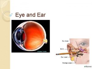
Eye and Ear Histology Orientation Images Eyelid structures


- Slides: 31

Eye and Ear Histology Orientation Images

Eyelid structures - from Netter Orbicularis oculi muscle Superior tarsus Inferior tarsus Levator palpebrae superioris muscle Lacrimal glands Orbital part Palpebral part Lacrimal ducts

Eyelids: Netter pl. 76

UCSF #173: human eyelid Lev ato r Lacrimal gland pal peb rae . S. Ta rsal m uscle Orbicularis oris m. Superior tarsus Apocrine gland s. m land Tarsal g Palpebral conjunctiva

Three Tunics of the Eye

Arteries and Veins of Orbit Ciliary arteries Optic nerve Ophthalmic artery Central retinal artery and vein Internal carotid artery

Cornea Anterior chamber Iris Posterior chamber UCSF #164 Ciliary body and processes Vitreous body Hyaloid canal Sclera Choroid Retina Optic nerve Central retinal artery & vein # Eye-2

Cornea Stroma Corneal epithelium Bowman’s membrane Corneal stroma Descemet’s membrane Corneal endothelium

Lens Zonule (of Zinn) Lens capsule Lens epithelium Lens fibers

Lens fiber formation at the equator region Zonule equator

A: Anterior chamber P: Posterior chamber A P

Canal of Schlemm Cornea Trabecular meshwork Anterior chamber Iris Posterior chamber

A: Trabecular meshwork B: Canal of Schlemm B A

Ciliary body and ciliary processes Pigmented and non-pigmented ciliary epithelial cells Ciliary processes Ciliary body

Sphincter (constrictor) pupillae (smooth muscle) Eye-2 Dilator pupillae (myoepithelial cells)

Retina Inner limiting membrane Optic nerve fibers Ganglion cell layer Inner plexiform layer Inner nuclear layer Outer plexiform layer Outer nuclear layer Outer limiting membrane Rods and cones Retinal pigment epithelium Choroid Bruch’s membrane Choriocapillary layer Sclera

EYE-2 Ora serrata EYE-1 Fovea centralis

Eye-2: Optic papilla (disc) Optic nerve Central retinal artery & vein

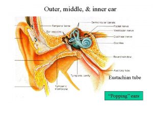
Ear Outer, middle and inner ear

Semicircular ducts Endolymphatic sac and duct Cochlear duct Saccule Utricle

Ear-2 Netter pl. 90 Cochlear duct (basal turn) Utricle Ampulla(e) Saccule Cochlear nerve Vestibular ganglion and nerve CN VIII: vestibulocochlear nerve Utricle ampulla

# Ear-4 Cochlear nerve Spira l (co Cochlear nerve chlea r) ga n glion

Ear-2 Vestibulocochlear Nerve (CN VIII) Cochlear Vestibular nerve Vestibular ganglion Cochlear Nerve Vestibular nerve Vestibulocochlear Nerve (CNS VIII) Vestibular ganglion

EAR-2 Macula Crista ampullaris Otolithic membrane with otoconia (otoliths) Cupula Ear-4

Ear-3 and other ear slides Stapes Saccule Cochlear duct Saccule ?

Cochlear Nerve and spiral ganglia Helicotrema Ear - 4 Base Spiral ganglion Cochlear Nerve

Organ of Corti in Scala Media ? Scala vestibuli ? Scala media ? Scala tympani

Scala vestibuli Vestibular membrane Stria vascularis Scala media (cochlear duct) Tectorial m. Basilar membrane Scala tympani

Organ of Corti Outer hair cells Inner hair cell Inner tunnel O. phalangeal cells Inner and outer pillar cells I. phalangeal cell

Perilymph and endolymph filled spaces in cochlea Blue: perilymph Yellow: endolymph

Helicotrema ? EAR-3 Low pitch sound (longer basilar membrane) Cochlear duct ? Osseous spiral lamina High pitch sound (shorter basilar membrane)
 Kimberly cockerham
 Floppy eyelid syndrome
 Camel 3 eyelids
 An animal with ears like leaves name
 Animals who have ears but we cannot see
 Cochlear nerve
 Nose teeth & hair
 Ethnocentric orientation adalah
 Homologous
 Bird's eye view and worm's eye view
 Taste anatomy
 Mácula lútea
 Accessory structure of the eye
 Lesson 7.1 internal structures of the eye
 Accessory structures of eye
 Hammurabi mesopotamia
 Hammurabi code eye for an eye
 Anemia eyes vs normal
 Code of hammurabi activity
 An eye for an eye a tooth for a tooth sister act
 Explain the image
 An eye for an eye meaning
 Behold he is coming
 Virtual vs real image
 Do f
Do f Https://tw.images.search.yahoo.com/images/view
 How to save images on google images
 Https://tw.images.search.yahoo.com/images/view
 If you don't like my story write your own things fall apart
 Stoma granulation tissue
 Tunica media
 Esophageal cardiac junction histology

