HISTOLOGY Epithelium I What is Histology Histology is


 and basal (inferior) surfaces • Cells have")


 (Silver painting)")

 (Böbrek Bowman kapsülü)")
")
")
")
")
")
")
 TEM’de görünüşü.")


")
.")
")
")
")

")
")
 (H&E) (x")

 http: //pathology.")
 cells with oval")


")

")
 (Periyodik asit-Schiff, PAS, boyaması). Epitelin bazal laminası (Ok)")
 Ekzokrin bez")


 http: //www. city. ac. uk/optometry/Biolabs/Tissue/")
")


")

")

")


")


")
")
")

")
")
 (x 350)")
 (H & E)")
 http: //www. city. ac. uk/optometry/Biolabs/Tissue/")

")
 http: //www. city. ac.")
 http: //www. city. ac.")

 of the conjunctiva covering")
 Bağ dokusu")

")

")
")

- Slides: 108
HISTOLOGY Epithelium I
What is Histology ? • Histology is the scientific study of the fine detail of biological cells and tissues that have been carefully prepared using "histological techniques” by microscopes • It is a discipline that is essential for the understanding and advancement of biology, medicine, veterinary medicine and many subdisciplines within those scientific subjects.
• The word "histology" comes from the combination of the Greek words , meaning "tissue", and which is sometimes translated as "-logia" and is a suffix generally used to denote the study of a subject or the branch of knowledge of a discipline
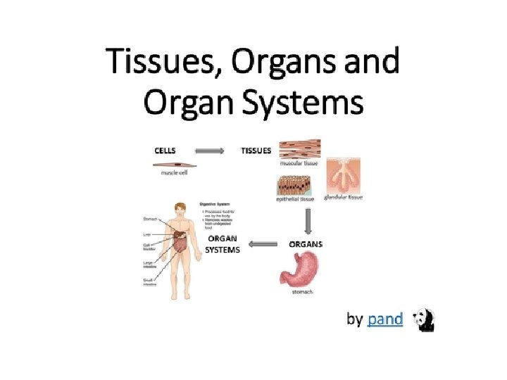
Tissue • Human’s body consists of billions of cells. • In biology, tissue is a cellular organizational level between cells and a complete organ. • A tissue is a collection of cells and noncellular structures, which have the similar origin, structure and carry out a specific function • Organs are formed by groups of tissues • Most organs are complex groups of different tissue types. • An organism is composed of organs that are grouped together and functionally integrated.
• Cells are bound together with varying amounts of intercellular substance to form tissues and organs • Single-celled livings do not have tissue. • Simple multi-cells, such as sponges, do not have tissue, they have cells which are slightly differentiated
• Tissues differs from the – ectoderm, – endoderm and – mesoderm layers of embryo • Differentiation of the tissues from these embryo layers is called histogenesis. • The transformation of an undifferentiated cell population into an organ during development is called organogenesis
• The cells that make up the tissues are surrounded by the extracellular matrix secreted by these cells. • The composition of ECM material differs from one tissue to another. • The ECM can be liquid, semi-solid or solid. • Each tissue has its own unique cells.
Three primitive germ layers give rise to 4 primary tissues There are 4 types of tissue: – 1. Epithelial – 2. Connective – 3. Muscle – 4. Nervous Similarities between tissue types: – 1. All contain cells – 2. Cells that make up tissues have similar functions
Connective tissue • Connective tissues are fibrous tissues. • They are made up of cells separated by non-living material, which is called an extracellular matrix. liquid or rigid – For example, blood contains plasma as its matrix and bone's matrix is rigid. • Connective tissue gives shape to organs and holds them in place.
• Blood, bone, cartilage, adipose tissues are examples of connective tissues. • One method of classifying connective tissues is to divide them into three types: – fibrous connective tissue – skeletal connective tissue – fluid connective tissue
Muscle tissue • Muscle cells form the active contractile tissue of the body known as muscle tissue. • Muscle tissue functions to produce force and cause motion, either locomotion or movement within internal organs. • Muscle tissue is separated into three distinct categories: – smooth muscle, found in the inner linings of organs – skeletal muscle, typically attached to bones, which generate gross movement – cardiac muscle, found in the heart where it contracts to pump blood throughout an organism.
Nervous tissue • Cells comprising the central nervous system and peripheral nervous system are classified as nervous (or neural) tissue. • In the central nervous system, neural tissues form the brain and spinal cord. • In the peripheral nervous system, neural tissues forms the cranial nerves and spinal nerves, inclusive of the motor neurons.
Epithelial tissue • The epithelial tissues are formed by cells that cover the organ surfaces such as – the surface of skin, – the airways, – the reproductive tract, and the – inner lining of the digestive tract. • The cells comprising an epithelial layer are linked via semi-permeable, tight junctions, this tissue provides a barrier between the external environment and the organ it covers.
• Epithelial tissue helps to protect organs from microorganisms, injury, and fluid loss. • In addition to this protective function, epithelial tissue may also be specialized to function in – secretion, – excretion and – absorption.
Origin and Distribution of Epithelium Consists of all layers of the embryo Ectoderm - epidermis of skin and epithelium of cornea together covers the entire surface of the body; sebaceous and ECTODERM mammary glands Endoderm - alimentary tract, liver, pancreas, gastric glands, intestinal glands • Endocrine glands - lose connection with surface Mesoderm MESODERM • male and female reproductive tracts • Endothelium - lining of blood vessels • Mesothelium - lining serous cavities ENDODERM
General Properties of Epithelial Tissue • It is common in organism, • Covers all surfaces of the body from inside to outside and lines organs/cavity walls • It forms secretory glands, • The cells are abundant and tightly together, ECM is very limited
• Exhibits polarity • Apical (superior) and basal (inferior) surfaces • Cells have special crossovers for special connections and communication • Supported by connective tissue • Cells are attached to a ‘basement’ membrane • The dead cells are replaced by new ones. *Ability to regenerate
Functions of epithelial tissue Ø Absorption, Ø Secretion Ø Transport, Ø Excretion Ø Protection, Ø Contraction, Ø Sensory intake etc
Types • Covering Epithelium • Glandular Epithelium Neuroepithelium cells Myoepiphelium cells
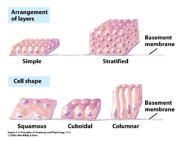
Classifications & Naming of Epithelia • First name of tissue indicates number of layers – Simple – one layer of cells – Stratified – more than one layer of cells
Classification & Naming of Epithelia • Last name of tissue describes shape of cells – Squamous – cells wider than tall (plate or “scale” like) – Cuboidal – cells are as wide as tall, as in cubes – Columnar – cells are taller than they are wide, like columns
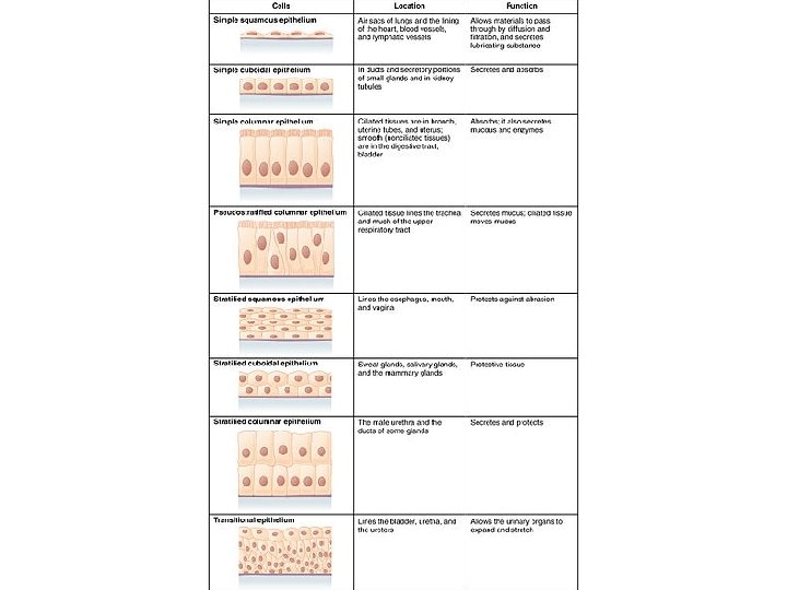
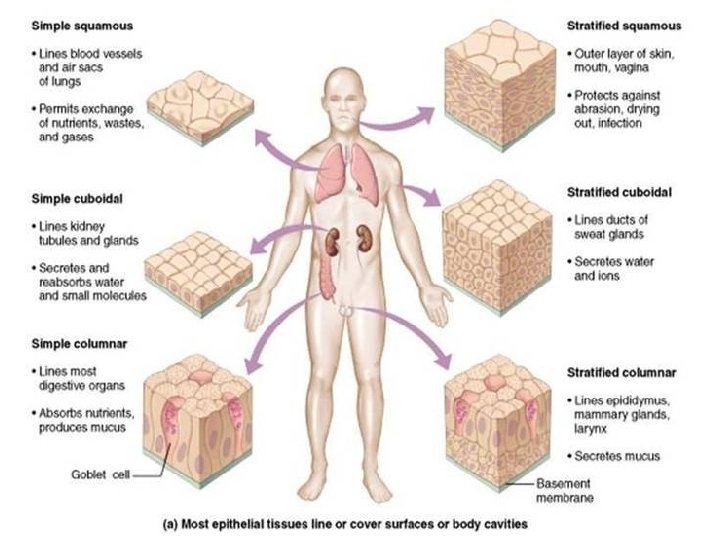
CLASSIFICATION OF EPITHELIA NUMBER OF LAYERS – SIMPLE - ONE LAYER – STRATIFIED - MORE THAN ONE LAYER SHAPE OF SUPERFICIAL CELLS – SQUAMOUS - FLAT – CUBOIDAL - CUBE – COLUMNAR – COLUMN OTHER – PSEUDOSTRATIFIED - NOT TRULY STRATIFIED – TRANSITIONAL - URINARY TRACT
Simple Squamous Epithelium • Description – single layer of flat cells with disc-shaped nuclei – are very thin and sensitive, easily damaged • Special types – Endothelium (inner covering) • slick lining of hollow organs – Mesothelium (middle covering) • Lines peritoneal, pleural, and pericardial cavities • Covers visceral organs of those cavities
Simple Squamous Epithelium • Function – Passage of materials by passive diffusion and filtration – Secretes lubricating substances in serosae • Location – Renal corpuscles – Alveoli of lungs – Lining of heart, blood and lymphatic vessels – Lining of ventral body cavity (serosae)
Simple squamous epithelium from the surface (Mesothelium) (Silver painting)
Bowman kapsülü Tek tabakalı yassı epitel Tek tabakalı kübik epitel Çıkan dalgalı kanal (Distal tüpçük) İnen dalgalı kanal (Proksimal tüpçük) Toplayıcı kanal Tek tabakalı yassı epitel Böbrek Nefron şeması Henle ilmeği
Glomerulus yumağı Tek tabakalı yassı epitel (Ok ucunda) (Böbrek Bowman kapsülü)
Tek tabakalı yassı epitel (Böbrek Bowman kapsülü)
Tek tabakalı yassı epitel (Böbrek Bowman kapsülü)
Tek tabakalı yassı epitel (Böbrek Bowman kapsülü)
Glomerulus yumağı Tek tabakalı yassı epitel (Böbrek Bowman kapsülü)
Tek tabakalı yassı epitel (Böbrek Bowman kapsülü’nde)
Tek tabakalı yassı epitel (Böbrek, Henle ilmeğinin ince kısmı)
Ç Bağ dokusu Kollagen teller Mezotelyumdaki tek tabakalı yassı epitelin (Ok) TEM’de görünüşü.
Simple Cuboidal Epithelium • Description – single layer of cube-like cells with large, spherical central nuclei • Function – secretion and absorption • Location – kidney tubules, secretory portions of small glands, ovary & thyroid follicles
Tek tabakalı kübik epitel
Tek tabakalı yassı epitel ve tek tabakalı kübik epitel, Böbrek enine kesit.
Tek tabakalı kübik epitel (Enine ve boyuna kesit)
Bazal lamina Tek tabakalı kübik epitel, boyuna kesit (Böbrek).
Tek tabakalı kübik epitel (İnsan böbreği)
Tek tabakalı kübik epitel (x 400)
Tek tabakalı kübik epitel (Böbrek kanallarında)
Tek tabakalı kübik epitel, enine kesit.
Tek tabakalı kübik epitel (Tiroit Bezi)
Tek tabakalı kübik epitel (Koroid pleksus)
Kübik Epitel Bağ dokusu Asinar bez Tek tabakalı kübik epitel (Pankreas kanalı) (H&E) (x 350)
Pankreatik kanalı çevreleyen kübik epitelin enine kesiti (TEM, x 3400
Tek tabakalı kübik epitel, Ovaryum ( x 132, H & E ) http: //pathology. mc. duke. edu/ research/
Simple Columnar Epithelium • Description – single layer of column-shaped (rectangular) cells with oval nuclei • Some bear cilia at their apical surface • May contain goblet cells • Function – Absorption; secretion of mucus, enzymes, and other substances – Ciliated type propels mucus or reproductive cells by ciliary action
Simple Columnar Epithelium • Location – Non-ciliated form • Lines digestive tract, gallbladder, ducts of some glands – Ciliated form • Lines small bronchi, uterine tubes, uterus
Goblet h. . Tek tabakalı silindirik epitel ve Goblet hücreleri
Tek tabakalı silindirik epitel ve Goblet hücreleri (İnce bağırsak)
Mikrovillüslü Silindirik epitel hücreleri G İnce bağırsak
Tek tabakalı silindirik epitel ve Goblet hücreleri (Jejenum)
Tek tabakalı silindirik epitel (Böbrek tüpçükleri) (Periyodik asit-Schiff, PAS, boyaması). Epitelin bazal laminası (Ok) ve fırça kenar (BB) yapısı.
Tek tabakalı silindirik epitel (Bez kanalında) Ekzokrin bez
Tek tabakalı silindirik epitel, safra kesesi. http: //www. city. ac. uk/optometry/Biolabs/Tissue
Tek tabakalı silli silindirik epitel
Siller Tek tabakalı silli silindirik epitel (Ovidukt kesit) http: //www. city. ac. uk/optometry/Biolabs/Tissue/
Tek tabakalı silli silindirik epitel ve bağ dokusu (İnsan ovidukt ampullası)
Pseudostratified Columnar Epithelium • Description – – All cells originate at basement membrane Only tall cells reach the apical surface May contain goblet cells and bear cilia Nuclei lie at varying heights within cells • Gives false impression of stratification • Function – secretion of mucus; propulsion of mucus by cilia
Pseudostratified Columnar Epithelium • Locations – Non-ciliated type • Ducts of male reproductive tubes • Ducts of large glands – Ciliated variety • Lines trachea and most of upper respiratory tract
Siller Epitel Yalancı tabakalı silli silindirik epitel, Trake.
Siller Yalancı tabakalı silli silindirik epitel (Trake)
Siller Goblet hücreleri Yalancı tabakalı silli silindirik epitel hücreleri ve Goblet hücreleri
Lamina propria Yalancı tabakalı silli silindirik epitel (Maymun, gırtlak)
Yalancı tabakalı silli silindirik epitel ve goblet hücreleri. Sillerin bazal cisimcik kısımlarının hücrenin üst yüzeyinde oluşturduğu eozinofilik bir çizgi (Ok ucu) ve siller (H&E; x 460)
Yalancı tabakalı silli silindirik epitel (Trake)
Sil Trakede yalancı tabakalı silli silindirik epitelin TEM’de görünüşü. Hücrelerin apikal yüzeyinde sil ve mikrovillüsler (x 1. 600)
Mikrovillüsler Siller Hücrelerin sınırı Trakenin lümene bakan yüzünde yalancı tabakalı silli silindirik epitelin silleri ve mikrovillüsleri. Mikrovillüslerin 1 mikronluk boyuna karşı sillerin 8 -10 mikron boyu kolaylıkla fark edilir. x 1. 450
Yalancı tabakalı silli silindirik epitel Siller
Siller G Yalancı tabakalı silli silindirik epitel ve Goblet hücreleri
Stratified Epithelia • • Contain two or more layers of cells Regenerate from below Major role is protection Are named according to the shape of cells at apical layer
Stratified Epithelium 1. Characteristics – 2+ layers 2. Stratified Squamous – Skin – outer layer hardened by ‘keratin’ – 4 to 5 layers thick 3. Stratified Cuboidal – Ducts of sweat glands – This type + stratified columnar are rare!
Stratified Squamous Epithelium • Description – Many layers of cells – squamous in shape – Deeper layers of cells appear cuboidal or columnar – Thickest epithelial tissue – adapted for protection
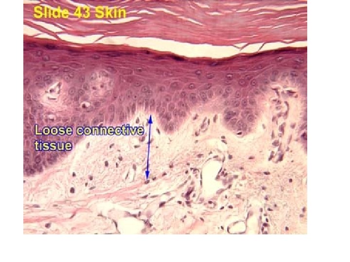
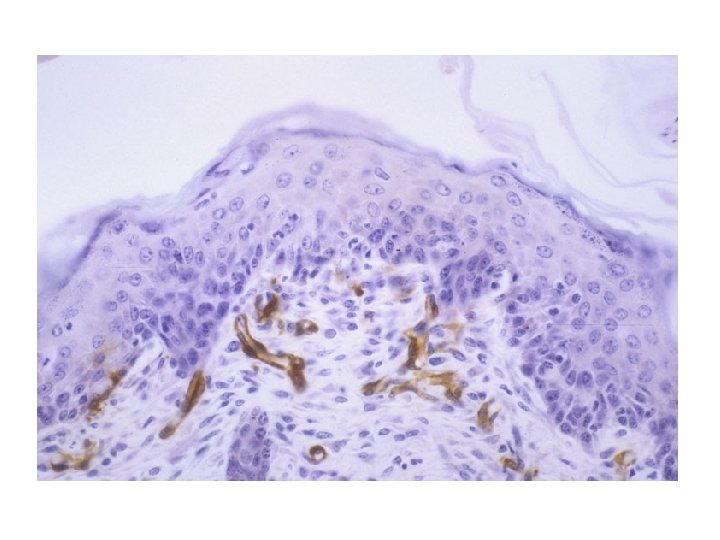
Stratified Squamous Epithelium • Specific types – Keratinized – contain the protective protein keratin • Surface cells are dead and full of keratin – Non-keratinized – forms moist lining of body openings • Function – Protects underlying tissues in areas subject to abrasion • Location – Keratinized – forms epidermis – Non-keratinized – forms lining of esophagus, mouth, and vagina
Çok tabakalı yassı epitelin yüzeyden görünüşü (Kurbağa derisi)
Çok tabakalı keratinleşmiş yassı epitel
Keratin Epitel Bağ dokusu Çok tabakalı keratinleşmiş yassı epitel, Deri.
Keratin tabakası Epitel Bağ dokusu Çok tabakalı keratinleşmiş yassı epitel
Epitel Keratin Bağ dokusu Çok tabakalı keratinleşmiş yassı epitel (Deri)
Çok tabakalı keratinleşmiş yassı epitel (Deri)
Çok tabakalı keratinleşmemiş yassı epitel (İnsan özofagus)
Yassı epitel hücreleri Çok tabakalı yassı epitel Bağ dokusu Bazal hücreler Bazal lamina Çok tabakalı keratinleşmemiş yassı epitel (Resimde seçilmiyor)
Çok tabakalı keratinleşmemiş yassı epitel (Dil)
Çok tabakalı keratinleşmemiş yassı epitel (İnsan özofagus)
Çok tabakalı keratinleşmemiş yassı epitel (H&E) (x 350)
Çok tabakalı keratinleşmemiş yassı epitel (Vajina) (H & E)
Çok tabakalı keratinleşmemiş yassı epitel , Vajina duvarı (H&E) http: //www. city. ac. uk/optometry/Biolabs/Tissue/
Stratified Cuboidal • Features – Rare – Several sequential cubic cells • Location – In the fetus epithelium, the male seminiferous tubules – Some sweat and salivary glands drain – The mammary gland's wide channel – Ovarian follicle
Çok tabakalı kübik epitel (Ovaryum folikülü)
Çok tabakalı kübik epitel, meme süt bezi kanalı kesidi (H&E) http: //www. city. ac. uk/optometry/Biolabs/Tissue/
Çok tabakalı kübik epitel , tükürük bezi enine kesit (H&E) http: //www. city. ac. uk/optometry/Biolabs/Tissue/
Stratified Columnar Features • Not common, • The underlying layered cells on the basal lamina are irregularly shaped and multifaced • The cells on the surface are cylindrical
Stratified Columnar Location • The arch-shaped bottom zone (Fornix conjunctiva) of the conjunctiva covering the eyelids, • Part of the male urethra, • Some areas of the anal mucous membranes • Farinks, epiglottis (part of the larynx) • Large drains of some cloths • Nose part of soft ear, larynx • Temporary fetal esophagus
Kanal lümeni Çok tabakalı silindirik epitel, Parotid Bezi (H&E) Bağ dokusu
Transitional Epithelium • Description – Basal cells usually cuboidal or columnar – Superficial cells domeshaped or squamous • Function – stretches and permits distension of urinary bladder • Location – Lines ureters, urinary bladder and part of urethra
Değişken epitel Üreter (H&E)
Değişken Epitel
Değişken epitel (İdrar kesesi)
Değişken epitel (İnsan, üreter ve renal pelvis)
Değişken Epitel