AP Histology Tissues Histology What is histology Histology


- Slides: 37

A&P Histology Tissues

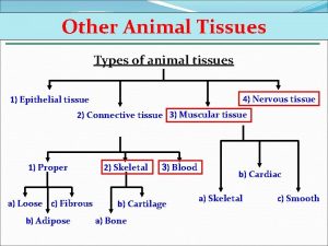
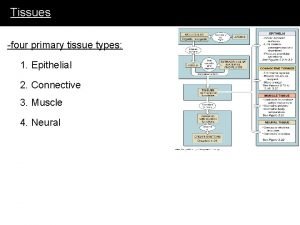
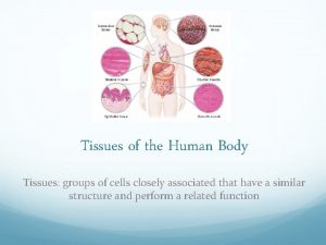
Histology • What is histology? Histology is the study of tissues • What is a tissue? A group of similar cells Ususally have a common embryonic origin Work together to carry out specialized activities

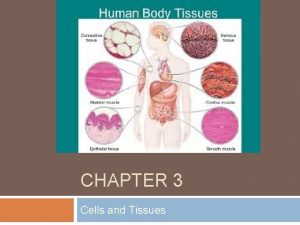
Histology • What types of tissues are there? Epithelial: ü covers body surfaces ü Lines hollow organs and body cavities ü Forms glands

Histology • What types of tissues are there? Connective tisssue ü protects and supports the body and organs ü Binds organs together ü Stores energy (fat) ü Provide immunity

Histology • What types of tissues are there? Muscular tissue ü generates force needed to make the body move

Histology • What types of tissues are there? Nervous tissue ü detects changes inside and outside body ü Responds by generating action potentials ü Helps maintain homeostasis (constant internal environment)

Histology • Cell junctions Contact points between the plasma membranes of tissue cells Joins cells into functional units

Histology • Adherens junctions Contain plaque- a dense layer of proteins inside the plasma membrane Plaque attaches to transmembrane proteins and the cytoskeleton Help epithelial cells resist separation

Classwork/homework Classwork: Copy figure 4. 2 on page 85 in blue book (This is 4. 1 on page 109 in red book). Include the definitions of tight junction, gap junction, desmosome, hemidesmosome Homework: Read pages 108 -110. Do #1, 2 on page 108 and #3, 4 on page 110.

Epithelium • Structural features of epithelium Cells are closely packed Cell junctions secure cells tightly to each other at lateral (side to side) surfaces Avascular: lacks blood vessels

Epithelium • Structural features of epithelium Has nerve supply Microvilli may be present for secretion or absorption Cilia may be present for moving substances along

Epithelium • Basement membrane Basal (bottom) surfaces of epithelial cells attach by a basement membrane to underlying connective tissue Basal lamina: membrane made of collagen and laminin, right under epithelial cells Reticular lamina: below basal lamina

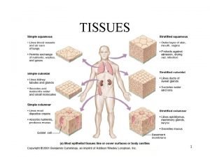
Epithelium Single layer of flat cells • Simple squamous epithelia Centrally located nucleus Function: filtration, diffusion, secretion Found where absorption takes place Also reduces friction (slick, slippery surface)

Epithelium • Simple cuboidal epithelia Single layer of cube shaped cells Centrally located nucleus Function: secretion, absorption Found in pancreas, kidney, ovary

Epithelium • Simple columnar epithelia Single layer of column like cells Nuclei near base of cells Function: secretion and absorption

Epithelium • Ciliated simple columnar epithelium Single layer of column like cells Has cilia Nuclei near base Function: moves mucus and other substances

Epithelium • Pseudostratified columnar epithelium Not really stratified (layered) All cells attached to basement membrane Nuclei are anywhere Function: secretion, move mucus

Epithelium Several layers • Stratified squamous epithelia Basal (bottom) layers are cuboidal Apical layers are squamous Function: protection

Classwork/homework Classwork: paste in figure of epithelial cells and label. Include the name of each tissue, one or more location, one or more function. Color (code for yourself) these structures: nucleus, cytoplasm, basement membrane Homework: read pages 110 -121. Do page 121 #7

Connective Tissue Most abundant tissue in body • Features Consists of cells and extracellular matrix Extracellular matrix : protein fibers and ground substance (material between cells and fibers). Extracellular matrix is secreted by the connective tissue, helps determine properties of the tissue

Connective Tissue binds together • Functions supports strengthens other tissues protects, insulates transport system

Connective Tissue. Types Made of collagen fibers, mast cells, fat cells, fibroblasts, macrophages, elastic fibers • Loose Connective Tissue Found beneath dermis of skin, digestive/respiratory/urinary tracts, between muscles, around blood vessels, around joints Function: protection (physical, immunity) and support

Connective Tissue. Types Made of adipocytes (fat cells) • Adipose Found beneath dermis of skin, behind eyeballs, around kidneys. Function: protection (physical), insulation, energy storage

Connective Tissue. Types Made of collagen fibers • Dense connective tissue Found in tendons, ligaments, covering skeletal muscles and organs Function: attachment, movement, reduce friction, stabilization

Connective Tissue. Types • Cartilage Made of ground substance, collagen fibers, chondrocytes (cartilage cells) Found around bones Function: support, reduces friction, prevents bone-tobone contact

Connective Tissue. Types • Bone Made of osteocytes (bone cells), blood vessels Found in skeletal system, ear Function: support, protection, blood formation, movement

Connective Tissue. Types • Liquid connective tissue Made of blood plasma, red blood cells, white blood cells, platelets Found within blood vessels Function: transport gases, immunity, repair

Classwork/Homework Classwork: Label figures with name of tissue, one or more location, one or more function. Color (you decide color code) these structures: nucleus, cytoplasm, fat globule (H only) Homework: read pages 125 -132. Do page 132 #13

Muscle Tissue • Features Elongated cells called muscle fibers. • Functions Produces body movements, maintains posture, generates heat, provides protection

Muscle Tissue • Skeletal muscle tissue Attached to bones Striationsalternating light and dark bands Voluntary

Muscle Tissue • Cardiac muscle tissue Forms most of the wall of the heart Muscle fibers are branched Striations Involuntary

Muscle Tissue • Smooth muscle tissue Found in the walls of hollow internal structures (blood vessels, airways, the stomach, etc) Lack striations Involuntary Function: control flow of fluids through these areas

Nervous Tissue • Nervous tissue Made of neurons (nerve cells) and neuroglia (support cells). Found throughout the nervous system Function: convert stimuli/responses to action potentials

Classwork/Homework Classwork: paste in figure of muscle and nerve cells and label. Include the name of each tissue, one or more location, one or more function. Homework: RED BOOK read pages 134 -137. Do pages 136 #12, 18 and 127 #19

Additional Vocabulary • Areolar Connective Tissue A type of loose connective tissue. It contains many types of cells (fibroblasts, white blood cells, etc) as well as fibers. Forms the subcutaneous layer with addipose.

Additional Vocabulary • Hyaline cartilage Ground substance, gel. Appears bluish white, shiny.

Additional Vocabulary • Dense Connective Tissue (Regular vs Irregular) Regular: arranged in parallel patterns. Found in tendons and ligaments. Irregular: not parallel. Found beneath skin, around muscles and organs.
 Chapter 3 cells and tissues
 Body tissues chapter 3 cells and tissues
 Body tissues chapter 3 cells and tissues
 Cells form tissues. tissues form __________.
 Body tissue
 Plant tissues
 Types of tissues
 Cell membrane phospholipids
 Genetic effects on gene expression across human tissues
 Periradicular tissues
 Four principal types of tissue
 What is this tissue
 3 tissues of a plant
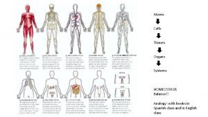
 Analogy of tissues
 Tissue is a group of cells having
 Four primary tissues
 Hyaline cartilage location
 Tissues causes of civil war
 Tissues are groups of similar cells working together to:
 What macromolecule is a prominent part of animal tissues
 Specialized connective tissue
 Tissues definition
 Elastic tissue location and function
 Pearson endocrine system
 Tissues
 Chapter 3 cells and tissues figure 3-1
 Predominant cell type
 Batang dikotil
 Cohesion bond
 Circulatory system tissue
 Tissues are groups of similar cells working together to
 Tissues group together to form
 Epithelium location
 Meristematic tissue
 Periradicular
 Cells-tissues-organ-systems-organism
 Tissue types in the body
 Four major tissues
