Lysosomes Lysosomes derived from the Greek words lysis

 are")


")


 enter lysosomal membrane and release enzymes lead to gouty arthritis")


 heteropolysaccharides having i. amino sugar: (N-acetylglucosamine or Nactylgalactosamine) ii. Uronic")


 are degraded in lysosomes by acid hydrolases, p. H 5. 0 Low")


 is caused by deficiency of any enzyme α -mannosidosis")


- Slides: 65
Lysosomes
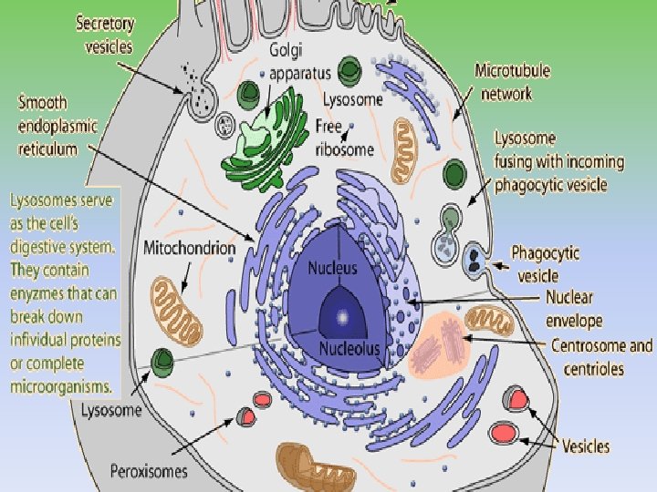
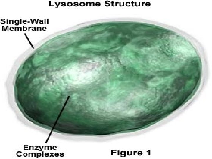
Lysosomes (derived from the Greek words lysis, meaning "to separate", and soma, "body") are the cell's waste disposal system and can digest some compounds. Characteristic: - Heterogeneous subcellular organelles in cytosol, bounded by cell membrane - Minute bodies, spherical or ellipsoidal (0. 8 -0. 9 μm) -found in cells having greater metabolic activity and in phagocytes.
The lysosomal membrane protects the cytosol, and therefore the rest of the cell, from the degradative enzymes within the lysosome At p. H 4. 8, the interior of the lysosomes is acidic compared to the slightly basic cytosol (p. H 7. 2). The lysosome maintains this p. H differential by pumping protons (H+ ions) from the cytosol across the membrane via proton pumps and chloride ion channels
Lysosomal production involves ongoing synthesis of enzymes more than 40 in number, which are acid hydrolases: i. Proteinases ii. Lipases iii. Carbohydrases iv. Estrases v. Nucleases Enzymes are heavily glycosylated, maintained at low p. H
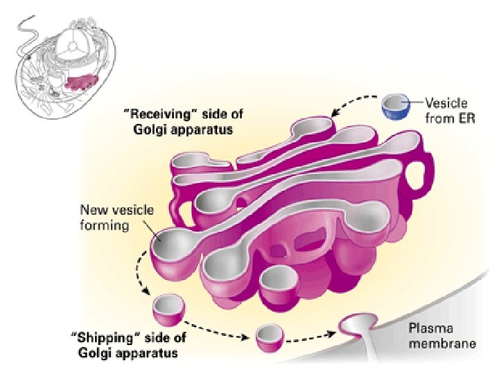
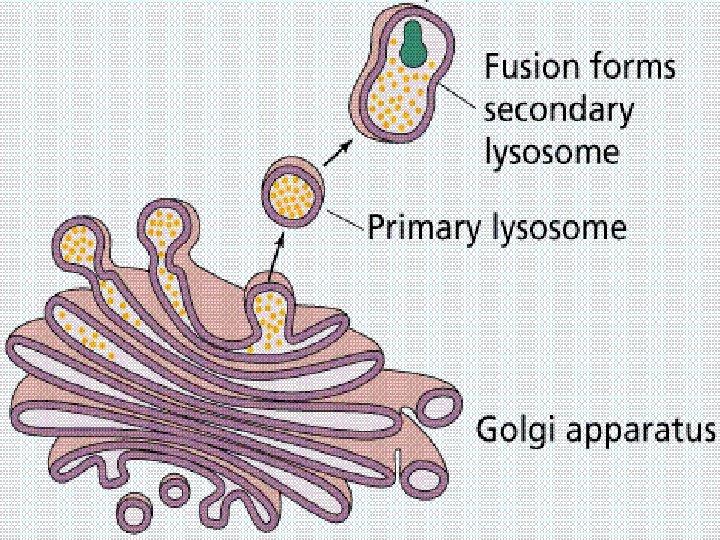
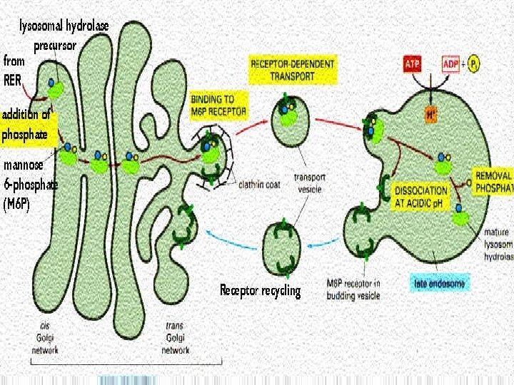
Lysosomal contents are first formed • in rough endoplasmic reticulum, here some CHO is added to enzymes • They then pass on Golgi complex for further changes and packaging in vesicular form. • Final processing is fusion of vesicles with endosome
Enzymes to enter lysosomes possess mannose at their end and can not move into lysosome. A change in mannose moiety occurs to mannose-6 phosphate , which can be recognized by specific receptors on lysosomal membrane Mannose 6 phosphate-enzyme complex readily pass into lysosomes.
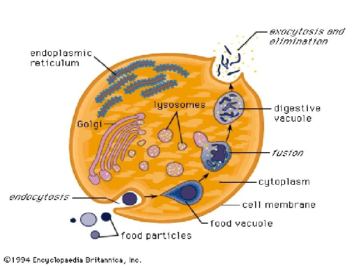
Functions of lysosomes: A. Autophagy: hydrolytic digestion of cells own worn out, damaged or unwanted cytoplasmic material. The material is segregated within a membranevacuole which becomes fused with lysosomes for digestion. B. Hetrophagy: Digestion of exogenous material i. Phagocytosis ii. Pinocytosis iii. Endocytosis
Phagocytosis: cells take up large particles e. g bacteria( vesicle containing phago-cytosed material is called phagosomes) Pinocytosis: taking up of fluid materil (pinosome: Endocytosis: taking up of smaller particulate material (endosomes) After digestion residual, insoluble digested contents are ejected from cell by exocytosis OR may remain in cell as residual bodies.
Residual bodies stay in non-dividing cells throughout life, become prominent in old age in form of lipofuscin (lipid rich pigment) within cell.
C. Role in protein degradation: The protein that undergo turn over (replaced by new) is digested. It is completed by proteinases and require no ATP. D. Role during embryonic life: Lysosomes help in causing APOPTOSIS ( programmed death of cell)
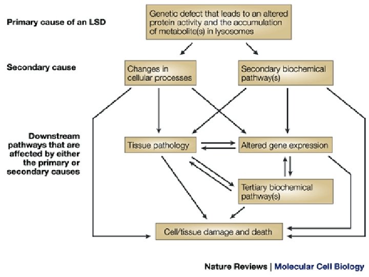
Relationship of lysosomes with diseases
Lysosomal membranes may 1. Release their hydrolases in response to i. Asbestos dust ii. Ion. radiation iii. Hypoxia iv. Silica particles v. Heat vi. Drugs Cell death may take place of mutations in genome occur.
2. Uric acid (hyperuricemia) enter lysosomal membrane and release enzymes lead to gouty arthritis 3. Osteoclasts of bone may release hydrolases cause erosion of bones. More than 50 lysosomal diseases, classified on nature of stored material
Salient features are involvement of Kidneys, muscles, liver, spleen, heart and bones Specific deficiencies of lysosomal enzymes result in non-digestion of their substrate that accumulate in lysosomes in large amount
Classification of LSDs A. Diseases affecting carbohydrate metabolism a. Mucopolysaccharidoses b. Glycoproteinases B. Lipid storage diseases a. Gangliosidoses b. Neutral glycosphingoliiposes c. Mucolipidoses d. Disorders of neutral lipids e. leukodystrophies
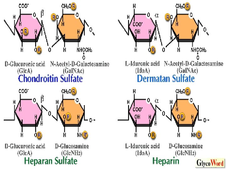
Glycos. Amino. Glycans (GAGs) heteropolysaccharides having i. amino sugar: (N-acetylglucosamine or Nactylgalactosamine) ii. Uronic acid: (D-glucuronic acid or L-iduronic acid) Both of these sugar acids may be present. iii. Amino sugars may have sulfate group. Attached to –OH group.
Examples : 1. Hyaluronic acid -jelly like , lubrication of joints of animals. skin, blood vessels, Connective tissues, corneas, tendon. 2. Chondroitin sulfate: cartilages, ligaments, tendons, aortic wall. 3. Heparin: anti-coagulant found in mast cells lining liver, spleen, lungs, thymus, blood 4. Others : keratan sulfate, dermatan sulfate, heparan sulfate,
Proteoglycans
Glycosaminoglycans (GAG) are degraded in lysosomes by acid hydrolases, p. H 5. 0 Low optimum p. H required to prevent enzymes leaking into cytosol and causing cell death. GAGS have shorter half life except keratan (120 days) They are engulfed by phagocytosis forming vesicle which fuses with lysosome for degradation.
Enzymes cleave polysaccharide chain – endoglycosidases, producing oligosaccharides. Further degradation occurs and last groups SO 4 or sugars are removed.
A. Mucopolysaccharidoses hereditary diseases, Caused by deficiency of hydrolases enzymes involved in hydrolysis of heparan and dermatan sulfates. Progressive disorders –accumulation of GAGs in cells causing i. Skelatal and extra-cellular deformities ii. Mental retardation All are autosomal except Hunter disease which is X-linked
Homozygous children are normal at birth but later deteriorate and in severe cases death result. Incomplete hydrolysis of GAGs cause accumulation of oligo-saccharides in urine and thus specific diseases can be diagnosed. Diagnosis: measuring cellular level of hydrolases.
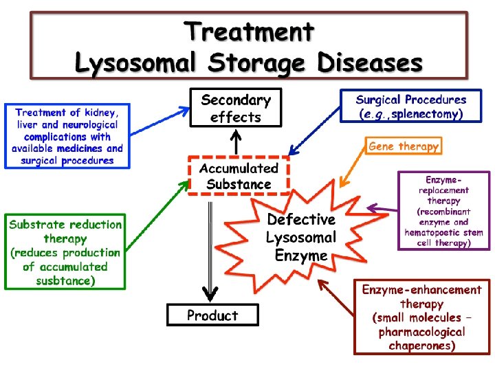
Bone marrow and blood cord transplant is treatment. Transplanted macrophages produce hydrolases Enzymes replacement is also available R/ a. Hurler Syndrome: MPS-1 Α-L Iduronidase deficiency Corneal clouding, dwarfism, mental retardation, coarse facial features, hearing loss Degradation of heparan and dermatan sulfate affected.
b. Hunter syndrome: MPS-II The syndrome has X-linked recessive Iduronate-2 -sulfatase deficiency wide range of severity No corneal clouding but physical and mental 2 replacement treatment Degradation of heparan and dermatan sulfate affected.
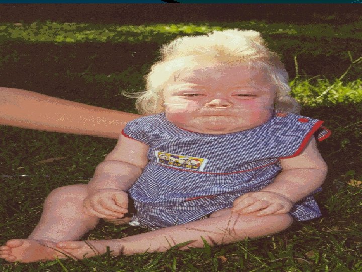
Sanfilippo syndrome: MPS-III severe nervous disorder and mental retardation Defect in removal of N-sulfate or N-acetyl-glucosamine residues. Symptoms appear after the first year of life Behavioral problems Coarse facial features Heavy eyebrows that meet in the middle of the face above the nose Sleep difficulties, full lips Stiff joints that may not extend fully Walking problems
d. SLY syndrome: MPS-VII Β-glucuronidase deficiency Hepato-spleno-megaly Skeletal derormity Short strature Corneal clouding Mental retardation
B. Glycoproteinosis : Acid hydrolysis are specific for removal of one component of glyco-proteins Hydrolases in these respect are exo-enzymes remove respective groups in sequence reverse of incorporation If any degrative enzyme is missing, degradation by other enzymes stop.
C. Glycoprotein storage disease (oligosacchadoses) is caused by deficiency of any enzyme α -mannosidosis type-I is progressive deficiency of α-mannosidase Mannose rich oligosaccharides fragments appear in urine. Diagnosis is by enzyme assays.
Lipidoses are those in which certain lipids accumulate within cells of various tissues. A. Neutral glycosphingolipidoses 1. Niemann Pick Disease Autosomal recessive , affected cells contain sphingomyelin and cholesterol, occur in many organs chiefly in brain. Tissues are deficient in sphingomyelinase(sphingomyelin--ceramide+phosphocholine
Autosomal recessive disease
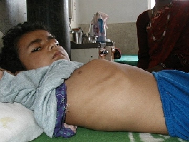
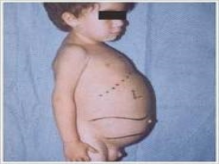
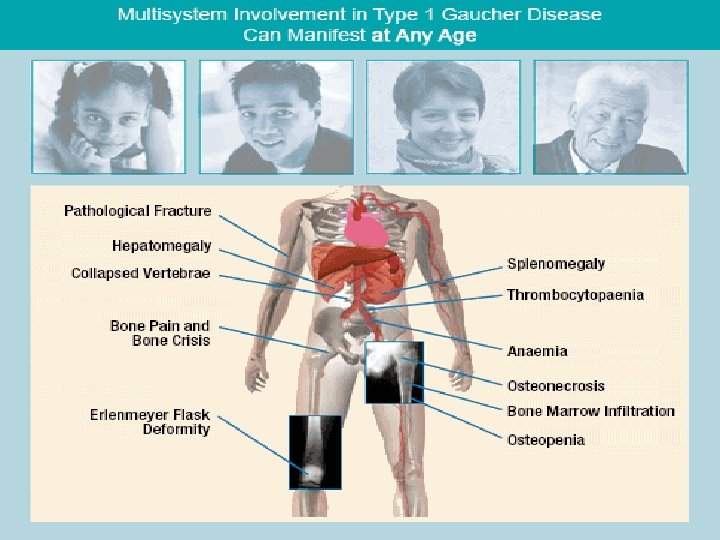
2. Gaucher disease: Genetic disorder. Autosomal recessive Affects fewer than 10, 000 people worldwide. Gaucher affects all racial and ethnic groups; prevalence is higher among Ashkenazi Jews. More common than Tay-Sachs disease. Cells laden with cerebrosides which contain glucose.
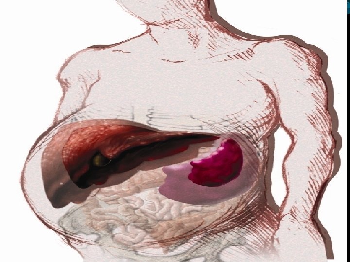
Spleen, liver are much enlarged , lungs, lymph nodes and bones are also affected (phagocytic cells affected)
Deficiency : Enzyme β-glucosidase Ceramide-glucose---- ceramide+glucose Ceramide-glucose is is glucocerebroside Gaucher is a progressive, debilitating and sometimes life-threatening disease. Symptoms can include: easy bleeding and bruising, fatigue, anemia, weak bones, bone and joint pain, and enlargement of the spleen or liver. Symptoms can appear at any age.
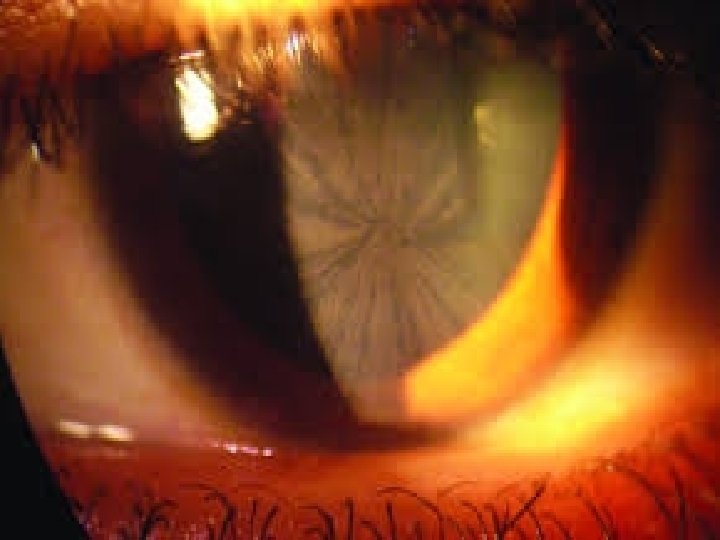
3. Fabry’s disease: Transmitted by X-chromosome Skin, CNS, Heart, muscles, kidneys involved Death due to heart/kidneys defect lies in Trihexosylceramide α-galactosidase. A. Ceramide-glucose-galactose (ceramide trihexoside) ------ceramide-glucosegalactose+galactose (ceramide dihexoside)
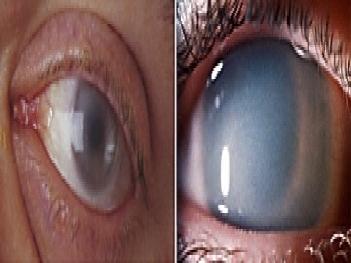
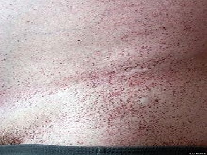
Symptoms: i. Pain whole body ii. Proteinuria iii. Cardiomyopathy iv. Angiokeratoma v. ocular involvement showing cornea verticillata (also known as vortex keratopathy), i. e. clouding of the corneas
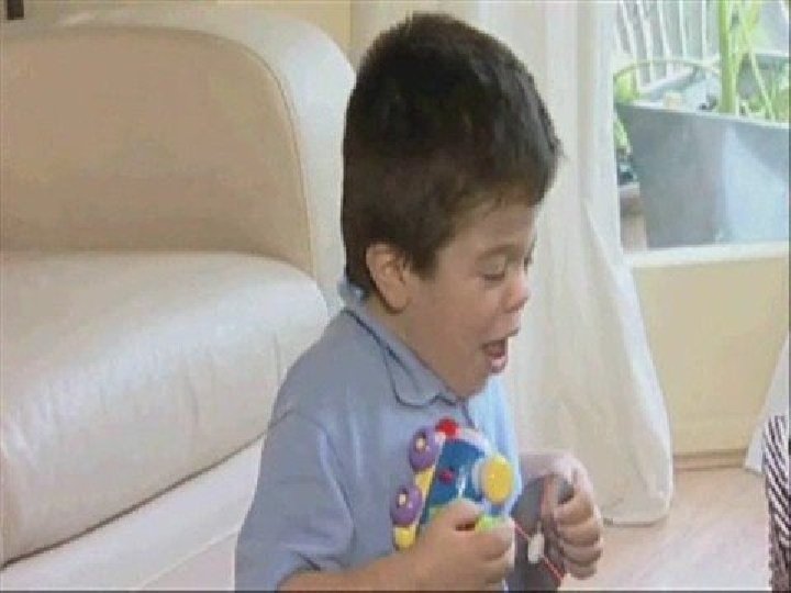
B. Gangliosidoses: 1. Tay-Sach’s Disease: Prevalant in jews, affects young Autosomal recessive Features- optic atrophy and idiocy Due to deficiency β-hexoseminidase A Enzyme responsible for breakdown of gangliosides. Gangliosides in large amount deposits in cells. (CNS, Liver, heart) but symptoms confined to CNS
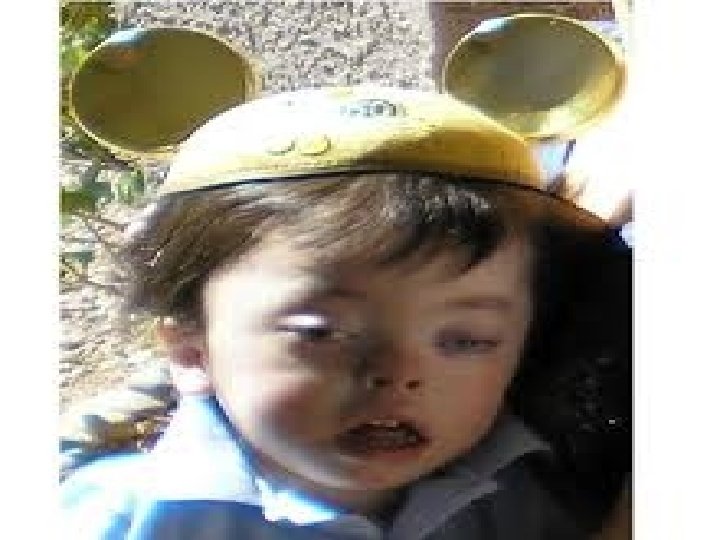
Optic atrophy
2. Sandhoff’s disease: Due to deficiency β-hexoseminidase A but in addition B type is deficient. Disease affects infants
C. Mucolipidoses: i- I. cell disease: Inclusion cell disease Deficiency of UDP-N-Acetylglucosamine-1 phosphotransferase Glycolipids accumulate in cells alongwith glycoproteins such as abnormal skeletal development, coarse facial features, restricted joint movement, may be present at birth. enlargement of certain organs, such as the liver (hepatomegaly) or spleen (splenomegaly), and sometimes even the heart valves
D. Disorders of neutral fats: 1. Wolman disease: deficiency of acid lysosome lipase accumulation of cholestero esters, and Tgs in the cells Feeding difficulties with frequent vomiting Diarrhea (loose frequent stools) Swelling of the abdomen (abdominal distention) Enlargement of the liver (hepatomegaly) and spleen (splenomegaly) Failure to gain weight or sometimes weight loss
2. Cholesterol ester storage disease deficiency of acid lysosome lipase accumulation of cholestero esters E. Leukodystrophies: 1. Krabbe’s Disease: Deficiency of galactosylceramidase Accumulation of galactosylceramide and galactosphingosine