
OPTIC NEURITIS DR ADNAN OPTIC NEURITIS v Optic







- Slides: 35
OPTIC NEURITIS DR ADNAN
OPTIC NEURITIS v Optic neuritis is defined as inflammation of the optic nerve.
ETIOLOGY v Isolated/idiopathic v Infectious and Para infectious v Post vaccination v Inflammatory v Toxic optic neuritis
LOCAL CAUSES v Endophthalmitis v Orbital cellulitis v Sinusitis v Contaguous spread from meninges, brain base of skull
ENDOGENOUS INFECTIONS v Viral – influenza, measles, mumps, chickenpox, herpes zoster v Bacterial – TB, syphilis v Fungal – Cryptococcosis, histoplasmosis v Protozoal – Toxoplasmosis v Parasitic - cysticercosis
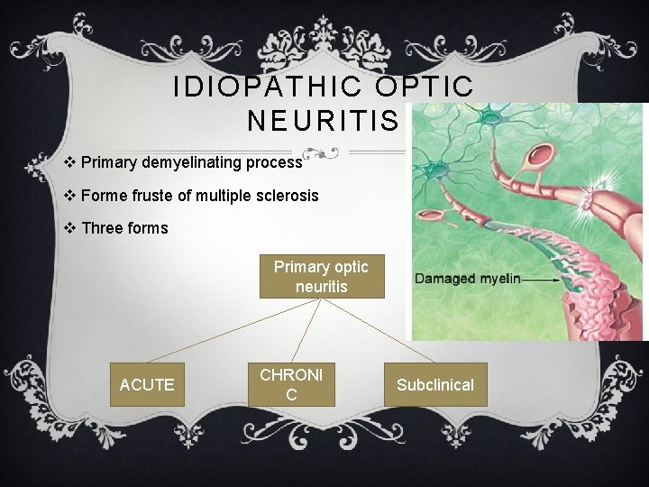
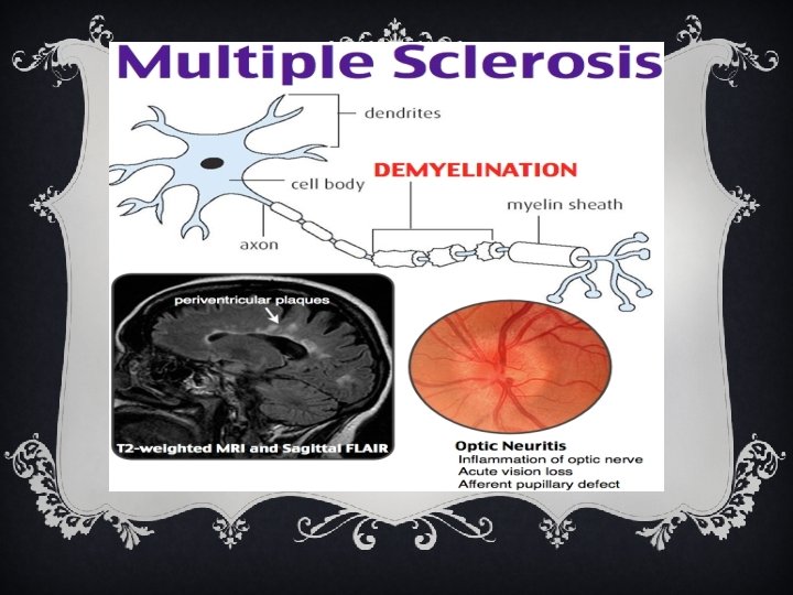
IDIOPATHIC OPTIC NEURITIS v Primary demyelinating process v Forme fruste of multiple sclerosis v Three forms Primary optic neuritis ACUTE CHRONI C Subclinical
EPIDEMIOLOGY v Acute form most common v Annual incidence-1 -5 per 100, 000 per year v Age -20 -50 yrs v Females more affected
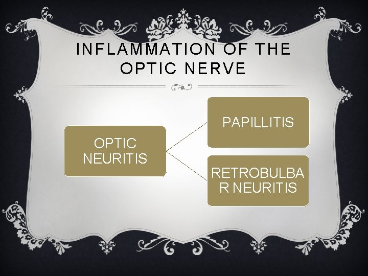
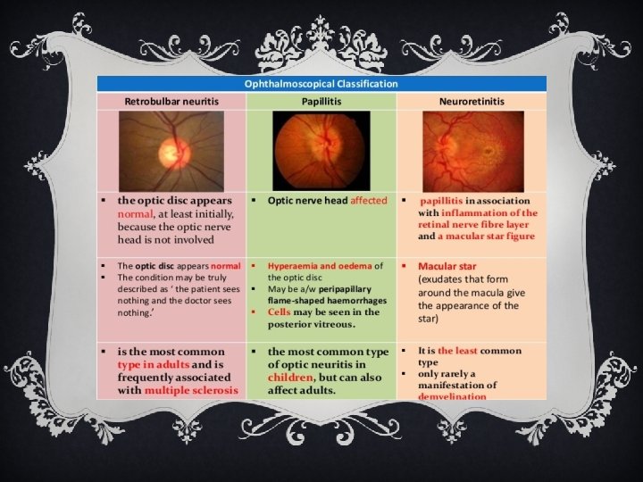
INFLAMMATION OF THE OPTIC NERVE PAPILLITIS OPTIC NEURITIS RETROBULBA R NEURITIS
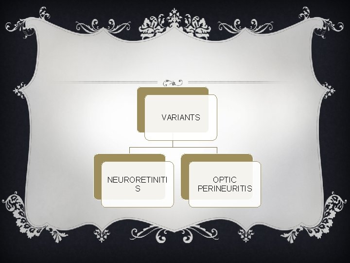
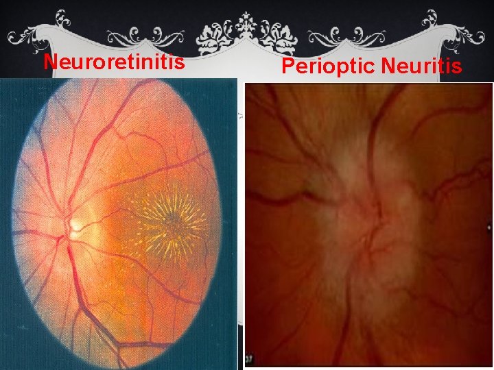
VARIANTS NEURORETINITI S OPTIC PERINEURITIS
Neuroretinitis Perioptic Neuritis
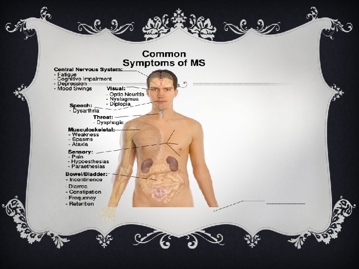
SYMPTOMS v VISUAL LOSS v PAIN v IMPAIRMENT OF COLOUR VISION v MOVEMENT OF PHOSPHENES v EPISODIC TRANSIENT OBSCURATION OF VISION (UHTHOFFS symptom) v Impaired depth perception (Pulfrichs phenomenon)
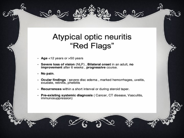
SIGNS v REDUCED VISUAL ACUITY 1. Loss of vision typically over hours to days 2. Unilateral/bilateral 3. 18 to 45 yrs of age 4. Deep orbital, retroocular or brow pain aggravated by eye movement nd increased by pressure on globe 5. Neuralgia and headache
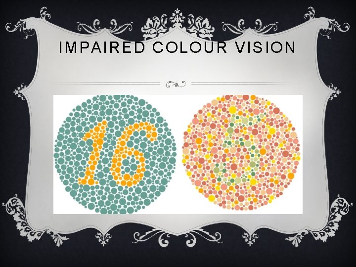
IMPAIRED COLOUR VISION
PUPIL – ILL SUSTAINED CONSTRICTION TO LIGHT MARCUS GUNN PUPIL – RAPD
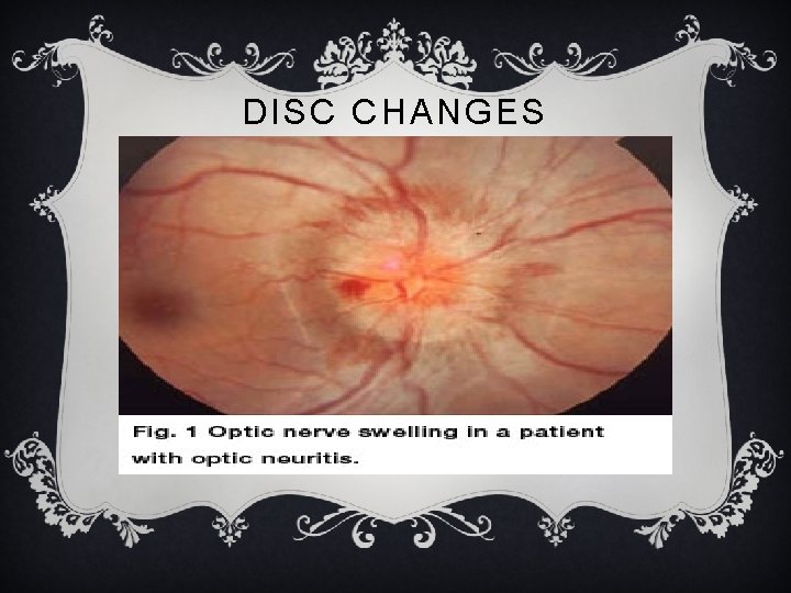
DISC CHANGES
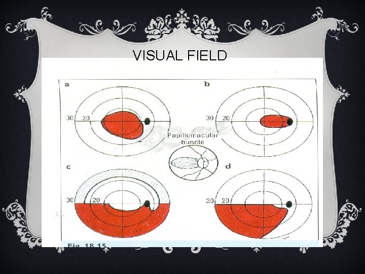
VISUAL FIELD
DIFFERENTIAL DIAGNOSES v Papilledema v Ischemic optic neuropathy v Papillophlebitis , CRVO v Diabetic papillopathy v Hypermetropia v ONH drusen
RECOVERY v Visual recovery is slower than the rate of visual loss & usually starts within 2 weeks & takes between 4 -6 weeks. v 75% to 90% get good visual recovery. v However recurrent attacks of Retrobulbar neuritis are followed by Primary Optic Atrophy
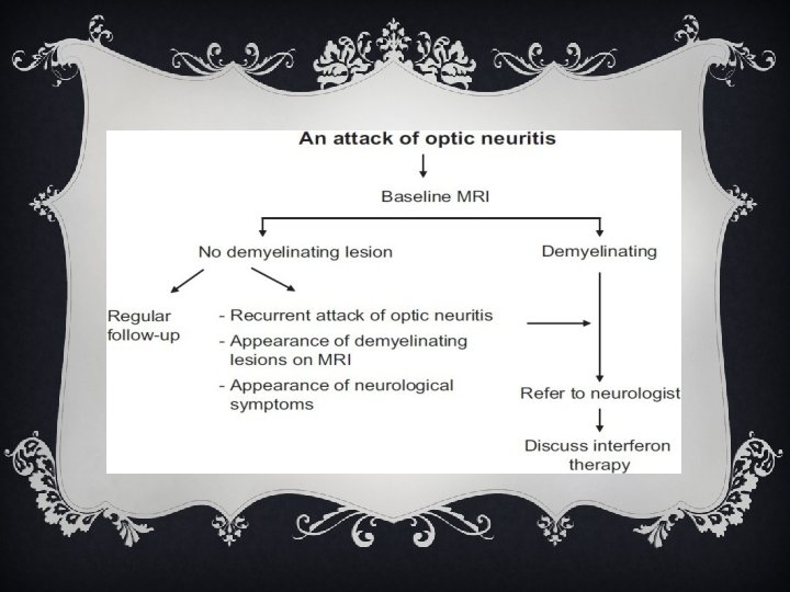
INVESTIGATIONS v. Rule out v Other etiologies e. g. compressive lesions v Other inflammatory causes v Determine visual and neurological prognosis
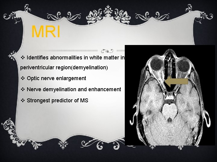
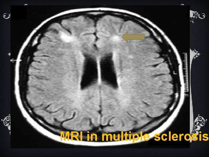
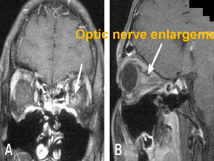
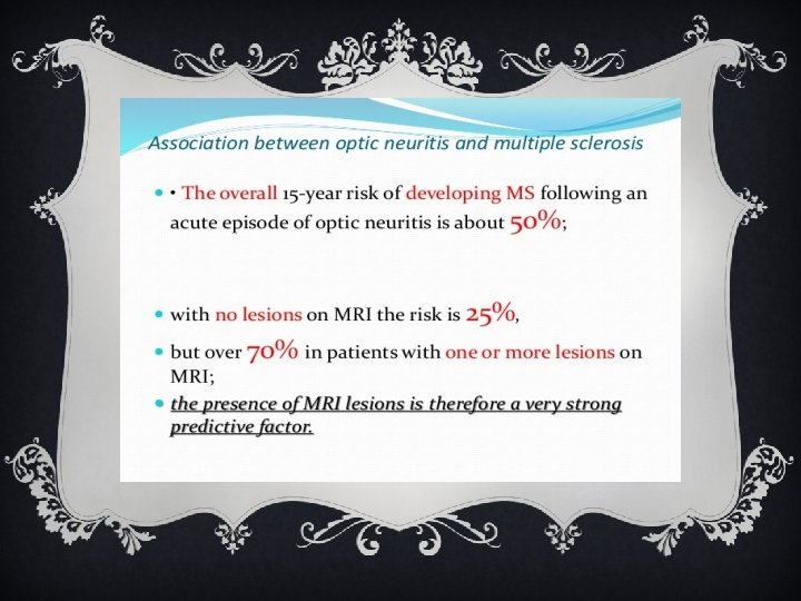
MRI v Identifies abnormalities in white matter in periventricular region(demyelination) v Optic nerve enlargement v Nerve demyelination and enhancement v Strongest predictor of MS
MRI in multiple sclerosis
Optic nerve enlargeme
v Serology v CSF analysis - oligoclonal banding inflammatory / infectious cause
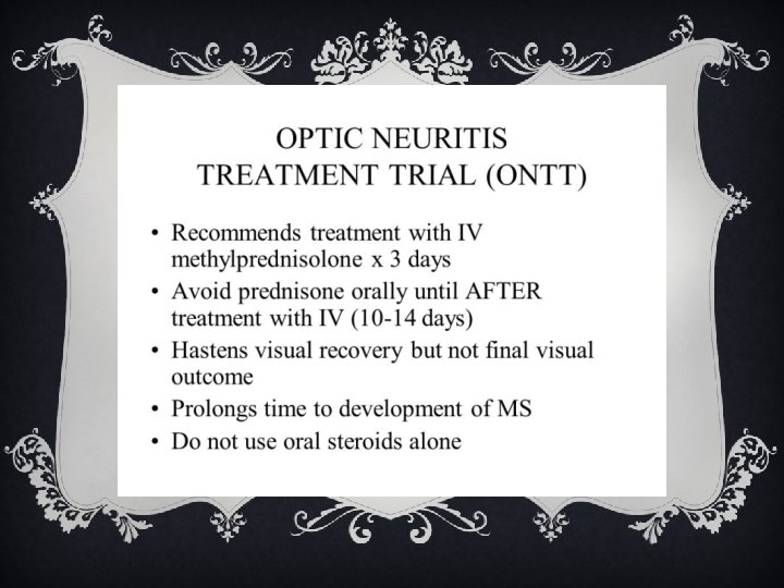
TREATMENT-ONTT Objectives § Assessment of steroid efficacy § Clinical profile of optic neuritis § Natural history § Relationship to MS
§ Intra venous Methyl prednisolone-250 mg 4 times a day for 3 days followed by oral prednisolone 1 mg /kg/day for 11 days § Short oral taper-20 mg on day 15 10 mg on days 16 &18 § Recovery of vision faster greater in 1 st 15 days § No significant difference after 1 yr § Decreases rate of dvpt of MS in 1 st 2 yrs
v Recurrence rate double and increased rate of new attacks in fellow eye if oral steroid alone v Therefore…. . I/v steroids if. . only MRI significant value for MS prevention in long run unproved Interferon beta 1 -a considered in high risk cases
THANK YOU