THE SENSORY ORGANS Anatomy Physiology Page 1 of







- Slides: 167

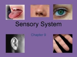
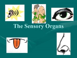
THE SENSORY ORGANS Anatomy & Physiology Page 1 of 67

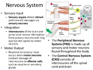
Sensations w Result from stimuli that initiate afferent impulses w Eventually reach a conscious level in the cerebral cortex w All sensations involve receptor organs w Simplest receptor organs are bare nerve endings Page 2 of 67

Sensations w w w Pain Temperature Pressure Touch Special Senses n n n Page 3 of 67 Sight Hearing Taste Smell Orientation in space

Receptors w Exteroceptors n Detect stimuli near outer body surface w Interoceptors n Detect stimuli from inside the body w Proprioceptors n Page 4 of 67 Detect stimuli deep within the body

Exteroceptors w Cold w Warmth w Touch w Pressure w Special senses n n Page 5 of 67 Hearing Vision

Interoceptors w w w Page 6 of 67 Taste Smell p. H Distension Spasm Flow

Proprioceptors w Located in skeletal muscles, tendons, ligaments, and joint capsules. w Provide information to CNS on posture, orientation in space, pressure, etc. w Fibers are heavily myelinated for rapid transmission. Page 7 of 67

Sensory Receptor w Peripheral component of an afferent axon and the centrally located nerve cell body of that axon. w Convert different types of energy into nerve signals (sound, light, thermal, chemical, and mechanical). w Generally receptors are specific and only respond to one form of energy. Page 8 of 67

Graded Responses w Subject to graded responses depending on the intensity of the stimulus. w Receptor can be regarded as a generator in which the amount of voltage produced is determined by stimulus. w Increasing stimulus increases firing rate of receptor. Page 9 of 67

Adaptation w Receptors adapt to continued stimulus. w Receptors vary in their degree of adaptation. w Adaptation involves slowing of firing rate. n Page 10 of 67 Burst of action potentials at a high frequency followed by a decrease in rate that quickly returns to zero.

Phasic Receptor Organ w Quickly accommodates to prolonged stimulation w Example is pacinian corpuscles (sensitive to pressure). w These receptors are best suited for signaling sudden change in environment. Page 11 of 67

Tonic Receptor w Application of a prolonged stimulus elicits a brief volley of action potentials at high frequency followed by an action potential rate that slows to a lower level and is maintained. Page 12 of 67

Pain Reception w Receptors are termed nociceptors n n n Page 13 of 67 Bare nerve endings of pain neurons Pain stimulus causes cell damage resulting in firing of the neuron Essentially a chemoreceptor Either myelinated or unmyelinated Myelinated fibers have a short lag time between stimulus and reaction “bright quality”

Pain Reception w Unmyelinated fibers have a longer lag time, pain is more diffuse, dull, and throbbing w Pain fibers are grouped in a specific tract in the spinal cord w Reaction threshold varies considerably across individuals w Diversion of attention reduces pain reception (e. g. twitch) Page 14 of 67

Visceral Pain w Pain can arise from organs in abdominal cavity (viscera) w Peritonitis and pleuritis are two examples w Intestinal pain is another w Heart can also be a source of pain Page 15 of 67

Referred Pain w Usually felt on surface of the body, but source is deep within viscera w Caused by convergence of cutaneous and visceral pain afferent fibers on the same neuron in the sensory pathway w Example (traumatic pericarditis) hardware w Pressure applied to withers causes pain response Page 16 of 67

Taste w Sense of taste is called gustation w The receptor organ is the taste bud w Taste buds are found on the tongue, palate, pharynx, and larynx. w Taste buds have gustatory cells and supporting cells. w Gustatory cells are receptors for taste. Page 17 of 67

Taste Reception w Taste bud pit communicates with the oral cavity by way of the pore. w Any substance tasted must get into solution and enter the pore of the taste bud. w Hair of the gustatory cell is affected causing stimulation of the gustatory cell. w The impulse is transmitted by cranial nerves VII and IX to the brain. Page 18 of 67

Taste Sensations w Classified as salty, sweet, bitter, or sour. w Each taste sensation is some combination of the above. w Taste perception by animals is based on preference. w Considerable variation within a species Page 19 of 67

Temperature and Taste w In humans, the temperature of a beverage or food markedly affects its taste. Page 20 of 67

Smell w As evolution progressed, nerve cell bodies migrated centrally so that only the nerve fibers remained in a peripheral position. w This provided protection for nerve cells, which do not regenerate. w Central migration did not occur for the nerve cell bodies of Cranial Nerve I (olfactory). w Cell bodies of Cranial Nerve I are found in the mucous membrane of the nasal cavity. Page 21 of 67

Smell w This location is known as the olfactory region. w The size of this region is directly related to the development of the sense of smell. w Dogs can detect substances 1: 1000 of that detectable by humans. w Sensation of smell is known as olfaction. Page 22 of 67

Smell w Animals with greatly developed sense of smell are macrosmatic. w Animals with less developed sense are microsmatic. (e. g. humans and monkeys) w Animals with no sense of smell are anosmatic. (many aquatic animals) Page 23 of 67

Smell w Each olfactory receptor has a cell body and a nerve fiber extending from each end. One is an axon and the other a dendrite. w The dendritic process of the olfactory cell extends to the outside of the olfactory region membrane in crevices between sustentacular cells. Page 24 of 67

Smell w Sustentacular cells provide major support to the dendritic processes and shield the nerve cell body from the olfactory cavity. w Dendritic processes form hair-like structures (olfactory cilia) that extend into the nasal cavity. w Cilia are covered with secretions from the glands of Bowman. Page 25 of 67

Smell w Ducts from the glands of Bowman lead through the epithelium of the nasal cavity to its surface. w Secretions constantly refresh the thin layer of fluid bathing the olfactory cilia. w Sniffing allows for back-and-forth movement of air, providing a greater chance for substances to go into solution. Page 26 of 67

Smell w Once the compound is in solution it binds to olfactory cilia and provides a stimulus for the impulse to be transmitted. w Axons of the olfactory cells join and proceed as fibers and branches of the olfactory nerves. w Basal cells divide and become sustentacular cells or olfactory cells replacing those lost. Page 27 of 67

Smell w It is unlikely that a specific olfactory cell exists for each smell. w It is probable that the basic smells combine to provide the sensation of a particular odor. w Only one odor can be perceived at any one time. w Olfactory cells adapt to odors. Page 28 of 67

Phermones w Animals use odors to communicate with each other. w A chemical secreted by one animal which influences the behavior of another is called a pheromone. w Pheromones are used to identify species, mark territories, emit alarms, mark food location, and identify animals in estrus. Page 29 of 67

ANATOMY OF THE EYE Page 30 of 67

Page 31 of 67

Page 32 of 67

Page 33 of 67

Muscles of the Eye Page 34 of 67

Page 35 of 67

Integumentary System Page 36 of 67

w Objectives: 1 - Describe the functions of the integumentary system. 2 - Identify the major structures found in the three layers of the skin. 3. Describe the anatomy and physiology of hair and nails 4. Define some common dermatopathological disorders. Page 37 of 67

w This system is divided into: 1 - skin 2 - hair 3 - glands 4 - nails 5 - nerve endings I) Skin is an organ because it consists of different tissues that are joined to perform a specific function. Largest organ of the body in surface area and weight. Dermatology is the medical specialty concerning the diagnosing and treatment of skin disorders. Page 38 of 67

Page 39 of 67

Anatomy (structure) Epidermis (thinner outer layer of skin) Dermis (thicker connective tissue layer) Hypodermis (subcutaneous layer or Sub-Q) Muscle and bone Physiology (function) 1 - Protection - physical barrier that protects underlying tissues from injury, UV light and bacterial invasion. - mechanical barrier is part non specific immunity (skin, tears and saliva). Page 40 of 67

2 - Regulation of body temperature - high temperature or strenuous exercise; sweat is evaporated from the skin surface to cool it down. - vasodilation (increases blood flow) and vasoconstriction (decrease in blood flow) regulates body temp. 3 -Sensation - nerve endings and receptor cells that detect stimuli to temp. , pain, pressure and touch. Page 41 of 67

4 - Excretion - sweat removes water and small amounts of salt, uric acid and ammonia from the body surface 5 - Blood reservoir - dermis houses an extensive network of blood vessels carrying 8 -10% of total blood flow in a resting adult. 6 - Synthesis of Vitamin D (cholecalciferol) -UV rays in sunlight stimulate the production of Vit. D. Enzymes in the kidney and liver modify and convert to final form; calcitriol (most active form of Vit. D. ) Calcitriol aids in absorption of calcium from foods and is considered a hormone. Page 42 of 67

w Epidermis: keratinized stratified squamous epithelium with four distinct cell types and five distinct layers. Page 43 of 67

Cells in the epidermis: - keratinoytes - melanocytes - Merkel cells - Langerhans’ cells 1 - Keratinocytes: most abundant - produce keratin (fibrous protein) - protective; waterproofing the skin - continuous mitosis - form in the deepest layer called the stratum basale - cells push their way up to the surface where they are dead cells filled with keratin; will slough off. Regenerates every 25 -45 days. Page 44 of 67

2 - Melanocytes: - cells produce brownish/black pigment called melanin. (8% of epidermal cells) - stratum basale - branching processes (dendrites) - melanin accumulates in melanosomes and transported along dendrites of the melanocytes to keratinocytes. - melanin accumulates on the superficial aspect of the keratinocyte shielding its nucleus from harmful UV light. - lack of melanin: albino Page 45 of 67

Page 46 of 67

3 - Merkel cells: - stratum basale - epidermis of hairless skin - attach to keratinocytes by desmosomes - make contact with a sensory neuron ending called a Merkel disc (touch). 4 - Langerhans’ cells: - star-shaped cells arising from bone marrow that migrate to epidermis. - epidermal dendritic cells (macrophages) - interact with a WBC called a T- helper cell - easily damaged by UV light. Page 47 of 67

Page 48 of 67

Stratum corneum Stratum lucidum Stratum granulosum Stratum spinosum Stratum basale Page 49 of 67

5 layers of the epidermis: 1 - Stratum corneum (horny layer) - layer has many rows of dead cells filled with keratin - continuously shed and replaced (desquamation) - effective barrier against light, heat and bacteria - 20 -30 cell layers thick - dandruff and flakes - 40 lbs. of skin flakes in a lifetime (dust mites!) Page 50 of 67

2 - Stratum lucidum - seen in thick skin of the palms and soles of feet. - 3 -5 rows of clear flat dead cells - keratohyalin (precursor) to keratin 3 - Stratum granulosum - 3 -5 rows of flattened cells - nuclei of cells flatten out - organelles disintegrate cells eventually die - keratohyalin granules (darkly stained) accumulate - lamellated granules secrete glycolipids into extracellular spaces to slow water loss in the epidermis Page 51 of 67

4 - Stratum spinosum: “spiny layer” - 8 -10 rows of polyhedral (many sided) cells - appearance of prickly spines - shrink when prepared for slide - melanin granules and Langerhans’ cell predominate Page 52 of 67

Page 53 of 67

5 - Stratum basale: deepest epidermal layer - attached to dermis - single row of cells - mostly columnar keratinocytes - with rapid mitotic division - stratum germinativum - contain merkel cells and melanocytes - 10 -25% Page 54 of 67

Page 55 of 67

Page 56 of 67

w Dermis: - flexible and strong connective tissue - elastic, reticular and collagen fibers - cells: fibroblasts, macrophages (WBC), mast cells (histamine). - nerves, blood and lymphatic vessels - oil and sweat glands originate - two layers: papillary and reticular Page 57 of 67

1 - Papillary layer: - loose connective tissue with nipple like surface projection called dermal papilla. - capillaries - contain pain receptors - contain touch receptors (Meissner’s corpuscles - dermal ridges- epidermal ridgespattern called fingerprints Page 58 of 67

2 - Reticular layer: - dense irregular c. t. - collagen fibers offer strength - holds water - dermal tearing causes stretch marks. - striae Skin color: attributed to melanin, hemoglobin and carotene. Race is determined by amount of melanin not # of melanocytes. Page 59 of 67

Local accumulation of melanin will result in freckles and pigmented moles. Melanin is made through interaction with tyrosinase present in melanocytes UV light stimulates melanin production. Excessive UV light can damage DNA and cause solar elastosis (elastin fibers clump) Carotene is formed from Vit. A and deposits in stratum corneum and imparts an orange tone to skin Page 60 of 67

Freckles Page 61 of 67

Hemoglobin (blood) will impart pinkish tones to skin. Blushing 1 - Redness (erythema) - reddened skin, embarrassment, fever, hypertension, inflammation, or allergy 2 - Pallor/blanching - pale skin, emotional distress or anemia, low blood pressure 3 - Jaundice - liver disease, bile deposited in tissue 4 - Bronzing - bronze coloration (Addison's disease) hypofunction of adrenal cortex 5 - Black & blue - bruises, escaped blood clots in tissue spaces (clotted blood masses = hematomas) Page 62 of 67

Hair color: Dark hair: mostly melanin Blond and red hair: melanin with Fe and S. Gray hair: loss of pigment (decr. tyrosinase) White hair: air bubbles in the medullary hair shaft. Page 63 of 67

w Hair (pili) - main function is protection - hair root nerve plexus for touch - normal hair loss in adult 70 -100 hairs/day Page 64 of 67

Page 65 of 67

Hair anatomy: - composed of dead columns of keratinized cells. - shaft: is the superficial portion of hair - root: below the surface in the dermis Shaft and root are composed of three layers: inner medulla, middle cortex and outer cuticle. Inner medulla has 2 -3 rows of polyhedral cells where pigment is located Cortex is major portion of shaft Cuticle is scaly and heavily keratinized (shingles) Page 66 of 67

Vellus hair: fine hair Terminal hair : coarser hair; axillary and pubic region. Grow in response to sex hormones Hirsutism: excessive hairiness: incr. androgens Page 67 of 67

Hair follicle surrounds the root. Bulb is the enlargement at the end of the follicle. - Also houses the germinal layer Papilla (nipple like) is located in the bulb and is where the blood supply nourishes the hair. Page 68 of 67

Arrector pili (pl. pilorum) is smooth muscle located in the dermis and is attached to the side of the hair shaft. - fright, cold and emotions will contract muscle and pull hair in vertical position. “Goose bumps”. Page 69 of 67

Glands: Two types of glands exist in the integument. - Sebaceous glands (oil glands) - Sudoriferous glands (sweat glands) Sebaceous glands: (holocrine glands) - connected to hair follicle - not found on palms and soles of feet - secretes sebum (fats, cholesterol and proteins - keep hair from drying out, keeps skin moist - whiteheads, blackheads and acne Page 70 of 67

Page 71 of 67

Whitehead: When the trapped sebum and bacteria stay below the skin surface, a whitehead is formed. Page 72 of 67

Blackhead: A blackhead occurs when the trapped sebum and bacteria partially open to the surface and turn black due to melanin, the skin's pigment. Blackheads can last for a long time because the contents very slowly drain to the surface. Page 73 of 67

Sudoriferous glands: exocrine glands - millions located throughout the skin - two types: - eccrine: more common (merocrine) - originate in sub. Q layer - duct empties on skin surface - palms and soles of feet - sweat is watery (99% H 20) - sweating regulated by sympathetic nervous system Page 74 of 67

Page 75 of 67

- apocrine: axillary and pubic region - duct empties onto hair follicle - viscous fluid - causes body odor (“b-o “) when bacteria break it down Page 76 of 67

Ceruminous glands: located in ear only - modified apocrine glands - originate in Sub Q layer - ducts open onto EAM. - produces cerumen (ear wax) : brown sticky substance that prevents foreign material from entering. Page 77 of 67

Page 78 of 67

Page 79 of 67

Anatomy of the Ear Page 80 of 67

Major Divisions of the Ear Peripheral Mechanism Central Mechanism VIII Outer Middle Inner Crania l Ear Ear Nerve Page 81 of 67 Brain

External ear, Auris external Auricula Meat us acoustics extern us Middle ear, Auris media Cavities tympani Membranes tympanic Ossicula audits Tuba additives Page 82 of 67

Inner ear, Auris interna Labyrinthus membranaceus - Labyrinthus vestibularis - Labyrinthus cochlearis Labyrinthus osseus - Vestibulum - Canales semicirculares ossei - Cochlea - Meatus acusticus internus Page 83 of 67

Outer Ear Pinna Preauricular Tags Preauricular Pits External Auditory Meatus Page 84 of 67 EAM Cerumen

Function of Outer Ear w w w Collects sound Localization Resonator Protection Sensitive (earlobe) Page 85 of 67

Pinna w The visible portion that is commonly referred to as "the ear" w Helps localize sound sources w Directs sound into the ear w Each individual's pinna creates a distinctive imprint on the acoustic wave traveling into the auditory canal Page 86 of 67

External Auditory Meatus w Extends from the pinna to the tympanic membrane n n About 26 mm in length and 7 mm in diameter in adult ear. Size and shape vary among individuals. w Protects the eardrum w Resonator n Provides about 10 decibels (d. B) of gain to the eardrum at around 3, 300 Hertz (Hz). w The net effect of the head, pinna, and ear canal is that sounds in the 2, 000 to 4, 000 Hz region are amplified by 10 to 15 d. B. n n Page 87 of 67 Sensitivity to sounds greatest in this frequency region Noises in this range are the most hazardous to hearing

Outer ear Tissues: elastic cartilage covered with skin A. Meatus acusticus externus besides the hair follicles and fat glands contains: Glandulae ceruminosae – modified sweat glands on the lateral wall of the canal. Сerumеn (ear wax) combination of wax and fat glands secret and desquamated epithelial cells. Page 88 of 67

Middle Ear Tympanic Cavity Tympanic Membrane Ossicles Middle Ear Muscles Eustachian Tube Mastoid Page 89 of 67

Function of Middle Ear w Conduction n Conduct sound from the outer ear to the inner ear w Protection n n Creates a barrier that protects the middle and inner areas from foreign objects Middle ear muscles may provide protection from loud sounds w Transducer n n Converts acoustic energy to mechanical energy Converts mechanical energy to hydraulic energy w Amplifier n n Page 90 of 67 Transformer action of the middle ear only about 1/1000 of the acoustic energy in air would be transmitted to the inner-ear fluids (about 30 d. B hearing loss)

Tympanic cavity • • • Volume – 1. 5 ml Form – flatten drum Structure – six walls: - Lateral - Medial - Anterior - Posterior - Superior - Inferior Page 91 of 67

Lateral wall Page 92 of 67

Tympanic Membrane w Separates outer ear from middle ear w Barrier from foreign objects w Cone-shaped in appearance n about 17. 5 mm in diameter w Vibrates in response to sound waves. w The membrane movement is incredibly small n as little as one-billionth of a centimeter Page 93 of 67

Tympanic membrane Two parts: Pars flaccida – upper, thin, loose Pars tensa – lower, tense Three layers: 1. Outer, cutaneous – continuation of the canal skin. No hairs and glands. 2. Middle, fibrous – elastic fibers. 3. Inner, mucous – tympanic cavity lining Page 94 of 67

Medial wall, paries labyrinthicus Most complex. On this wall are distinguished: -fenestra vestibuli -fenestra cochleae -promontorium -prominentia canalis semicircularis lateralis - prominentia canalis facialis Superior wall, paries tegmentalis Separates tympanic from cranial cavity. Children less than 2 years – infections of the middle ear can pass to the cranial cavity. Page 95 of 67

Inferior wall, paries jugularis Separates tympanic cavity from fossa jugularis Anterior wall, paries caroticus Separates tympanic cavity from canalis caroticus -canalis musculotubularis Posterior wall, paries mastoideus Composed of: • Steroid complex of Procter • Antrum mastoideum • Fossa incudis Page 96 of 67

Auditory (Eustachian) tube Connects tympanic cavity with pharynx Two openings: • ostium pharyngeum tubae • ostium tympanicum tubae. Two parts: • bony • Cartilagenous Function: • Equalizes pressure on both sides of tympanic membrane for optimal hearing. Page 97 of 67

Ossicles w Malleus (hammer) w Incus (anvil) w Stapes (stirrup) smallest bone of the body Page 98 of 67

Inner Ear Auditory Vestibular semicircular canals utricle and saccade Cochlear Vestibular traveling wave pathologies Page 99 of 67

Inner ear Two compartments: (а) Bony labyrinth and (b) Membranous labyrinth. Bony labyrinth: ·complex cavity in dense bone (pars petrosa) · Parts of the bony labyrinth: a. Vestibulum. b. Semicircular canals. c. Cochlea. Page 100 of 67

Bony labyrinth. Labyrinthus osseus Vestibulum and semicircular canals Page 101 of 67

Vestibulum Two walls: External and internal. External wall has • Fenestra vestibuli. Internal wall has: • Recessus ellipticus • Recessus sphericus • Recessus cochlearis • Maculae cribrosae superior, medius, inferior Page 102 of 67

Openings into vestibulum a. Fenestra vestibuli. b. Fenestra cochleae. c. Openings (5) of the semicircular canals d. Aqueductus vestibuli Page 103 of 67

Semicircular canals 3: anterior, posterior and lateral. Have ampulla and crus. Canalis semicircularis lateralis –horizontal. - eminentia canalis semicircularis lateralis on the medial wall of tympanic cavity. Canalis semicircularis anterior –frontal. - eminentia arcuata on pars petrosa of os temporale. Canalis semicircularis posterior –sagittal Page 104 of 67

Labyrinthus osseus. Cochlea Page 105 of 67 Meatus acusticus internus

cochlea • Cone-shaped: base and apex. • Canalis spiralis cochleae - promontorium, on the medial wall of tympanic cavity. • Modiolus - canales longitudinales modioli. • Lamina spiralis ossea -hamulus Divides canalis spiralis cochleae into: • Scala tympani • Scala vestibuli Page 106 of 67

Labyrinth us membranaceus Page 107 of 67

Function of Inner Ear w Converts mechanical sound waves to neural impulses that can be recognized by the brain for: n n Page 108 of 67 Hearing Balance

Membranous labyrinth. Labyrinth us membranaceus • Closed system of sacs and ducts underling the bony labyrinth. • Filled with end lymph. • Two parts: vestibular & cochlear. Page 109 of 67

Vestibular labyrinth Composed of : • Two bags - sacculus et utriculus • Three duct us semicircular • One duct us endolymphaticus. Page 110 of 67

Cristra ampullaris Page 111 of 67

Balance w Linear motion w Rotary motion Page 112 of 67

Sensory cells (Epitheliocytus pilosus) Page 113 of 67

Macula utriculi (sacculi) Otoliths. Statoconia Page 114 of 67

Static balance Maculae react to gravitational forces and participate in maintaining the static balance. Page 115 of 67

Dynamic balance Cristae ampullares react to rotatory movements and paticipate in dynamic balance. Page 116 of 67

Anatomy of Nose and Paranasal Sinus By: Dr. Mohammed aloulah Page 117 of 67

The Nose w The nose consists of the external nose and the nasal cavity, w Both are divided by a septum into right and left halves. Page 118 of 67

External Nose w The external nose has two elliptical orifices called the naris (nostrils), which are separated from each other by the nasal septum. w The lateral margin, the ala nasi, is rounded and mobile. Page 119 of 67

External Nose Page 120 of 67

External Nose w The framework of the external nose is made up above by the nasal bones, the frontal processes of the maxillae, and the nasal part of the frontal bone. w Below, the framework is formed of plates of hyaline cartilage Page 121 of 67

External Nose Page 122 of 67

Blood Supply of the External Nose w The skin of the external nose is supplied by branches of the ophthalmic and the maxillary arteries. w The skin of the ala and the lower part of the septum are supplied by branches from the facial artery. Page 123 of 67

Blood Supply of the External Nose w The infratrochlear and external nasal branches of the ophthalmic nerve (CN V) and the infraorbital branch of the maxillary nerve (CN V). Page 124 of 67

Nasal Cavity w The nasal cavity has n a floor, n a roof, n a lateral wall, n a medial or septal wall. Page 125 of 67

The Floor of Nasal Cavity w Palatine process maxilla w Horizontal plate palatine bone Page 126 of 67

The Roof of Nasal Cavity w Narrow w It is formed n n Page 127 of 67 anteriorly beneath the bridge of the nose by the nasal and frontal bones, in the middle by the cribriform plate of the ethmoid, located beneath the anterior cranial fossa, posteriorly by the downward sloping body of the sphenoid

The Medial Wall of Nasal Cavity w The Nasal Septum w Divides the nasal cavity into right and left halves w It has osseous and cartilaginous parts w Nasal septum consists of the perpendicular plate of the ethmoid bone (superior), the vomer (inferior) and septial cartilage (anterior) Page 128 of 67 Perpendic ular Plate Septal (ethmoid) Cartila Vom ge er

The Nasal Septum Page 129 of 67

The Lateral Walls of Nasal Cavity Marked by 3 projections: n n n Superior concha Middle concha Inferior concha w The space below each concha is called a meatus. Page 130 of 67

The Lateral Walls of Nasal Cavity Page 131 of 67

The Lateral Walls of Nasal Cavity 1. Inferior meatus: nasolacrimal duct 2. Middle meatus: • Maxillary sinus • Frontal sinus • Anterior ethmoid sinuses 3. Superior meatus: ethmoid sinuses 4. Sphenoethmoidal recess: sphenoid sinus Page 132 of 67 posterior

Openings Into the Nasal Cavity Anterior & middle ethmoid air cells, maxillary and frontal sinuses open into middle meatus Page 133 of 67 Nasolacrimal Canal drains into Inferior Sphenoid sinus opens into sphenoethmoidal recess. Posterior ethmoidal air cells open into superior meatus

Blood Supply to the Nasal Cavity w From branches of the maxillary artery, one of the terminal branches of the external carotid artery. w The most important branch is the sphenopalatine artery. w The sphenopalatine artery anastomoses with the septal branch of the superior labial branch of the facial artery in the region of the vestibule. w The submucous venous plexus is drained by veins that accompany the arteries. Page 134 of 67

Blood Supply to the Nasal Cavity Sphenopalatine a. Maxillary a. Page 135 of 67

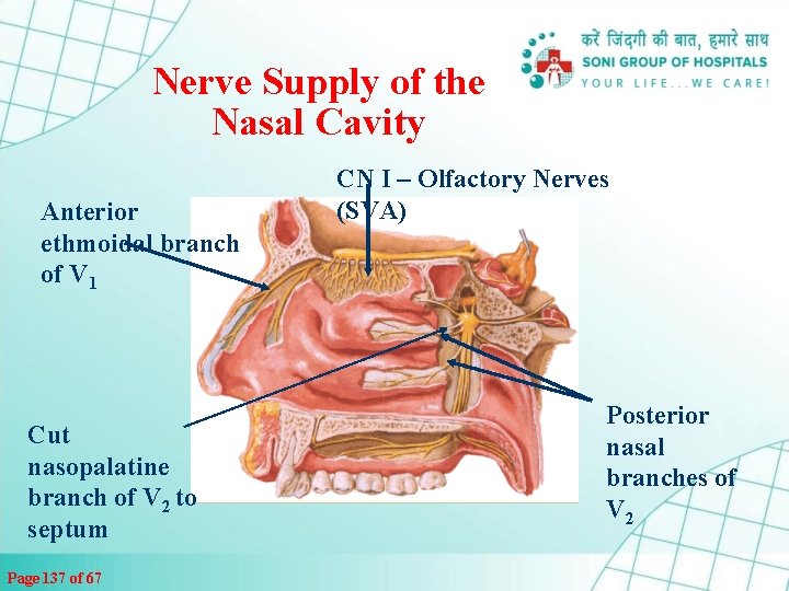
Nerve Supply of the Nasal Cavity w The olfactory nerves from the olfactory mucous membrane ascend through the cribriform plate of the ethmoid bone to the olfactory bulbs. w The nerves of ordinary sensation are branches of the ophthalmic division (V 1) and the maxillary division (V 2) of the trigeminal nerve. Page 136 of 67

Nerve Supply of the Nasal Cavity Anterior ethmoidal branch of V 1 Cut nasopalatine branch of V 2 to septum Page 137 of 67 CN I – Olfactory Nerves (SVA) Posterior nasal branches of V 2

Lymph Drainage of the Nasal Cavity w The lymph vessels draining the vestibule end in the submandibular nodes. w The remainder of the nasal cavity is drained by vessels that pass to the upper deep cervical nodes. Page 138 of 67

Tongue • Pink , Moist , Solid Conical muscular organ , in floor of mouth • Partly oral and partly pharyngeal in position • Covered by mucous membrane Page 139 of 67

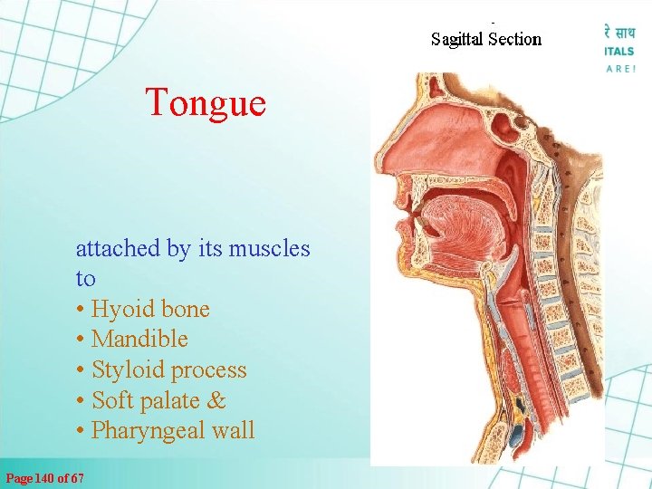
Tongue attached by its muscles to • Hyoid bone • Mandible • Styloid process • Soft palate & • Pharyngeal wall Page 140 of 67

Tongue w Main Functions - Organ of Deglutition w Taste & Speech Page 141 of 67

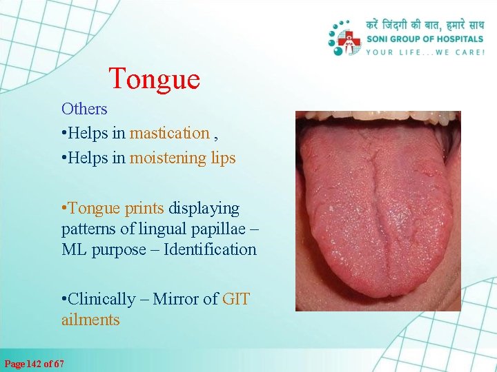
Tongue Others • Helps in mastication , • Helps in moistening lips • Tongue prints displaying patterns of lingual papillae – ML purpose – Identification • Clinically – Mirror of GIT ailments Page 142 of 67

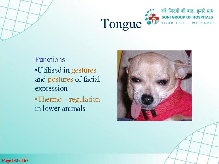
Tongue Functions • Utilised in gestures and postures of facial expression • Thermo – regulation in lower animals Page 143 of 67

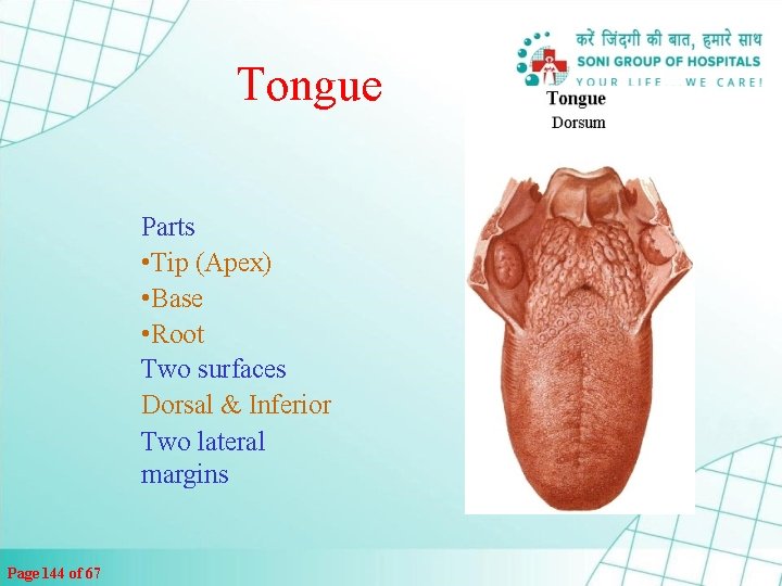
Tongue Parts • Tip (Apex) • Base • Root Two surfaces Dorsal & Inferior Two lateral margins Page 144 of 67

Tongue • Tip – Ant. Free end directed forward in contact with the incisor at rest • Base – directed backward towards oro-pharynx , formed by post 1/3 rd Page 145 of 67

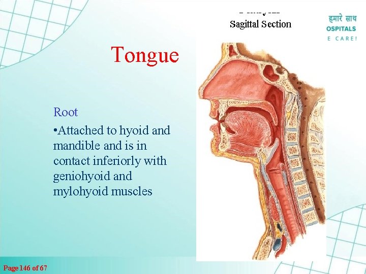
Tongue Root • Attached to hyoid and mandible and is in contact inferiorly with geniohyoid and mylohyoid muscles Page 146 of 67

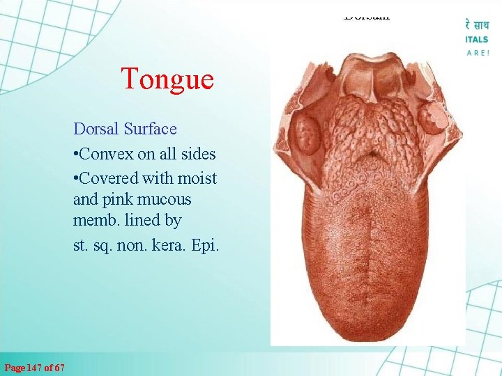
Tongue Dorsal Surface • Convex on all sides • Covered with moist and pink mucous memb. lined by st. sq. non. kera. Epi. Page 147 of 67

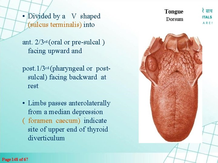
• Divided by a V shaped (sulcus terminalis) into ant. 2/3 rd (oral or pre-sulcal ) facing upward and post. 1/3 rd (pharyngeal or postsulcal) facing backward at rest • Limbs passes anterolaterally from a median depression ( foramen caecum) indicate site of upper end of thyroid diverticulum Page 148 of 67

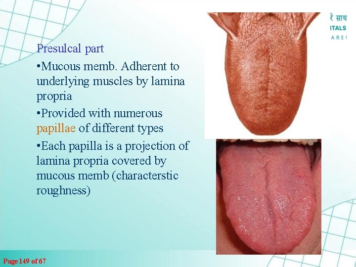
Presulcal part • Mucous memb. Adherent to underlying muscles by lamina propria • Provided with numerous papillae of different types • Each papilla is a projection of lamina propria covered by mucous memb (characterstic roughness) Page 149 of 67

Types of Papilla • Vallate • Fungiform • Foliate • Filliform Page 150 of 67

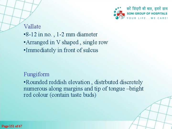
Vallate • 8 -12 in no. , 1 -2 mm diameter • Arranged in V shaped , single row • Immediately in front of sulcus Fungiform • Rounded reddish elevation , distrbuted discretely numerous along margins and tip of tongue –bright red colour (contain taste buds) Page 151 of 67

Filiform Numerous tiny conical projections over the entire dorsal surface of ant. 2/3 rd of tongue (devoid of taste buds) Give velvaty appearance) Page 152 of 67 Foliate 3 -4 vertical mucous folds at margins of tongue in front of sulcus (contain taste buds)

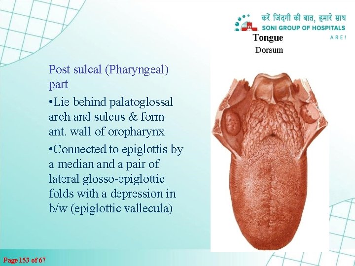
Post sulcal (Pharyngeal) part • Lie behind palatoglossal arch and sulcus & form ant. wall of oropharynx • Connected to epiglottis by a median and a pair of lateral glosso-epiglottic folds with a depression in b/w (epiglottic vallecula) Page 153 of 67

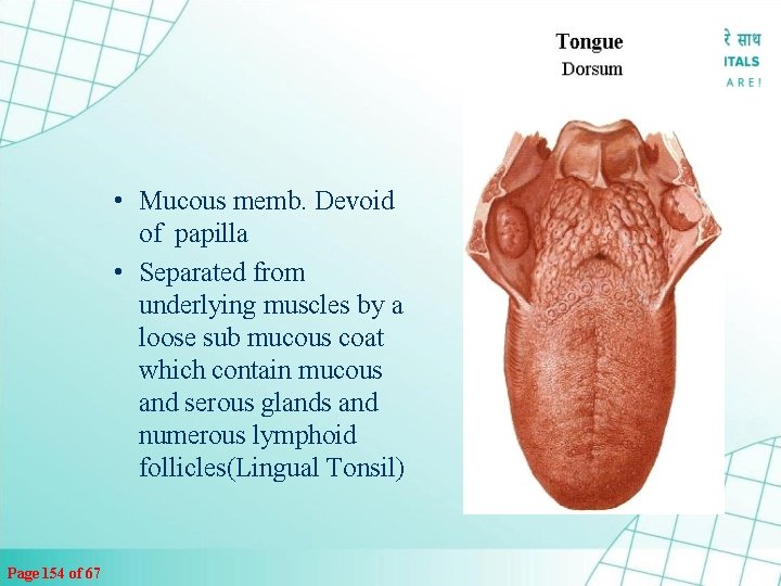
• Mucous memb. Devoid of papilla • Separated from underlying muscles by a loose sub mucous coat which contain mucous and serous glands and numerous lymphoid follicles(Lingual Tonsil) Page 154 of 67

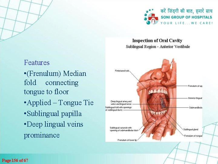
Tongue – Inferior surface • Reflected on floor of mouth • Covered by mucous memb. • devoid of Papillae Page 155 of 67

Features • (Frenulum) Median fold connecting tongue to floor • Applied – Tongue Tie • Sublingual papilla • Deep lingual veins prominance Page 156 of 67

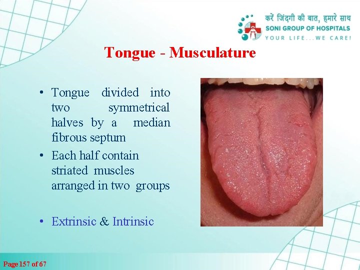
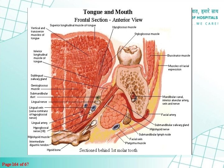
Tongue - Musculature • Tongue divided into two symmetrical halves by a median fibrous septum • Each half contain striated muscles arranged in two groups • Extrinsic & Intrinsic Page 157 of 67

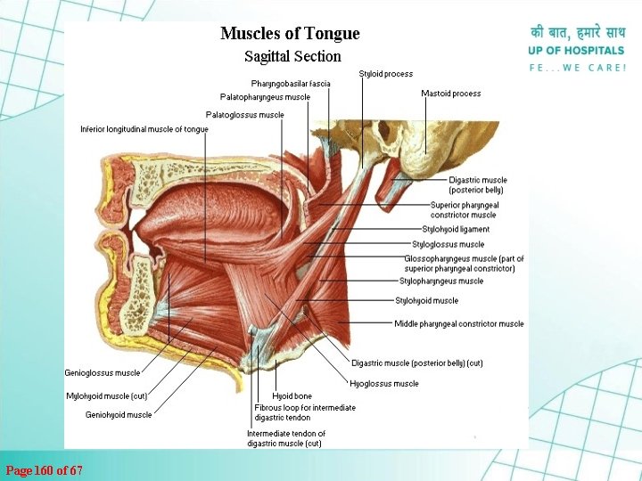
Tongue Musculature Extrinsic – Five Pair Connect to • Genio-glossus (mandible) • Hyo-glossus (Hyoid) • Chondro-glossus • Stylo-glossus (Styloid process) • Palato-glossus (Palate) Alter position of tongue Page 158 of 67

Tongue - Musculature Intrinsic muscles – occupy upper part & are Attached to submucous fibrous layer and to median fibrous septum • Superior Longitudinal • Inferior Longitudinal • Transverse muscle • Vertical muscle Alter shape of Tongue Page 159 of 67

Page 160 of 67

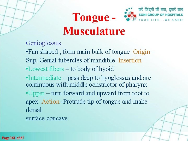
Tongue Musculature Genioglossus • Fan shaped , form main bulk of tongue Origin – Sup. Genial tubercles of mandible Insertion • Lowest fibers – to body of hyoid • Intermediate – pass deep to hyoglossus and are continuous with middle constrictor of pharynx • Upper – turn forward and upward from root to apex Action -Protrude tip of tongue and make dorsal surface concave Page 161 of 67

Hyoglossus • Quadrilateral muscle Origin • Upper surface of greater cornu and partly from body of hyoid • Passes upward & forward under cover of mylohoid Insertion side of tongue b/w styloglossus laterally and inferior longitudinal muscle medially Action Depresses sides of tongue , make dorsal surface Convex Chondroglossus detached part of hyoglossus, seperated by genoiglossus Originate from lesser cornu & attached to side of tongue Page 162 of 67

Page 163 of 67 Styloglossus Arise from tip of styloid process & stylomandibular ligament Passes downward and forward Inserted to side of tongue Oblique fibers interdigitate with hyoglossus Longitudinal fibres continue with inf. Longitudinal muscle Action – retracts tongue backward & upward Antagonist in action to genioglossus

Page 164 of 67

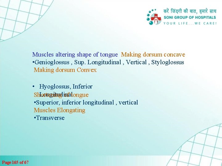
Muscles altering shape of tongue Making dorsum concave • Genioglossus , Sup. Longitudinal , Vertical , Styloglossus Making dorsum Convex • Hyoglossus, Inferior Longitudinal Shortening of tongue • Superior, inferior longitudinal , vertical Muscles Elongating • Transverse Page 165 of 67

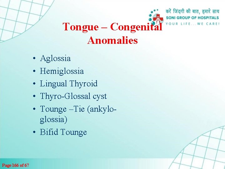
Tongue – Congenital Anomalies • • • Aglossia Hemiglossia Lingual Thyroid Thyro-Glossal cyst Tounge –Tie (ankyloglossia) • Bifid Tounge Page 166 of 67

THANK YOU Page 167 of 67
 Sensory system organs
 Organs of the sensory system
 Chapter 6 general anatomy and physiology
 Integumentary system psoriasis
 Lamina propria
 Anatomy and physiology unit 7 cardiovascular system
 Respiratory system in man
 Epigastric vs hypogastric
 What produces bile
 Irn.org anatomy and physiology
 Chapter 10 blood anatomy and physiology
 Tattoo anatomy and physiology
 Anatomy and physiology
 Agriscience unit 26 self evaluation answers
 Anatomy and physiology
 Chapter 1 introduction to human anatomy and physiology
 Anatomy and physiology exam 1
 Anatomy and physiology
 The speed at which the body consumes energy
 Jeopardy anatomy and physiology game
 Http://anatomy and physiology
 Anatomy and physiology chapter 1
 Anatomy and physiology of the retina
 Pancreas anatomy and physiology
 Gastric ulcer
 Anatomy and physiology revealed
 Anatomy and physiology
 Anatomy and physiology of swine
 Anatomy and physiology
 Cengage anatomy and physiology
 Pse 4
Pse 4 Fish anatomy and physiology
 Gi tract histology
 Physiology trivia
 Google image
 Anatomy and physiology chapter 15
 Anatomy and physiology body parts
 Anatomy science olympiad
 Anatomy and physiology
 Science olympiad forensics cheat sheet
 Biceps muscle names
 Anatomy and physiology of appendicitis
 Welcome to anatomy and physiology
 Horizontal anatomical plane
 Aohs foundations of anatomy and physiology 1
 Anatomy and physiology definition
 Holes anatomy and physiology chapter 1
 Anatomy and physiology of eye
 Chapter 7 anatomy and physiology
 Sheep liver lobes
 Aohs foundations of anatomy and physiology 1
 Figure 11-8 arteries
 Anatomy and physiology chapter 8 special senses
 Anatomy and physiology
 Male vs female skeleton pelvis
 Pancreas histology slide
 Fish anatomy and physiology
 Anatomy and physiology
 Animal tissue
 The central sulcus divides which two lobes? (figure 14-13)
 Cornell notes for anatomy and physiology
 Unit 26 animal anatomy physiology and nutrition
 Chapter 2 basic chemistry anatomy and physiology
 Crown plants examples
 Chapter 2 human reproductive anatomy and physiology
 Aohs foundations of anatomy and physiology 1
 Anatomy and physiology
 Physiology of the foot and ankle
 2012 pearson education inc anatomy and physiology
 Anatomy and physiology
 Holes essential of human anatomy and physiology
 Aohs foundations of anatomy and physiology 1
 Cranial nuclei
 Anatomy and physiology coloring workbook chapter 14
 Difference between anatomy and physiology
 Anatomy and physiology
 Chapter 13 anatomy and physiology of pregnancy
 Thyroid anatomy
 Anatomical structures of the upper respiratory tract
 Anatomy and physiology vocabulary
 Transverse fissure
 Anterior posterior distal proximal
 2012 pearson education inc anatomy and physiology
 Anatomy and physiology of meningitis ppt
 3 layers of muscle
 Anatomy and physiology ninth edition
 Figure 14-1 anatomy and physiology
 Stomach anatomy corpus
 Anatomy and physiology bones
 Paratubular cyst
 Aohs foundations of anatomy and physiology 2
 Body landmarks diagram
 What is a running head in apa style
 Jsp anatomy
 Anatomy of a news article
 Anatomy of jsp page
 Bảng số nguyên tố lớn hơn 1000
 đặc điểm cơ thể của người tối cổ
 Vẽ hình chiếu vuông góc của vật thể sau
 Các châu lục và đại dương trên thế giới
 Thang điểm glasgow
 ưu thế lai là gì
 Sơ đồ cơ thể người
 Tư thế ngồi viết
 Cái miệng nó xinh thế chỉ nói điều hay thôi
 Mật thư anh em như thể tay chân
 Bổ thể
 Tư thế ngồi viết
 Thứ tự các dấu thăng giáng ở hóa biểu
 Thẻ vin
 Thể thơ truyền thống
 Các châu lục và đại dương trên thế giới
 Hát lên người ơi
 Sự nuôi và dạy con của hươu
 Từ ngữ thể hiện lòng nhân hậu
 Diễn thế sinh thái là
 Vẽ hình chiếu vuông góc của vật thể sau
 Phép trừ bù
 Tỉ lệ cơ thể trẻ em
 Lời thề hippocrates
 đại từ thay thế
 Quá trình desamine hóa có thể tạo ra
 Cong thức tính động năng
 Môn thể thao bắt đầu bằng từ chạy
 Hình ảnh bộ gõ cơ thể búng tay
 Sự nuôi và dạy con của hổ
 Thế nào là mạng điện lắp đặt kiểu nổi
 Dot
Dot Vẽ hình chiếu đứng bằng cạnh của vật thể
 Biện pháp chống mỏi cơ
 Phản ứng thế ankan
 Chó sói
 Thiếu nhi thế giới liên hoan
 điện thế nghỉ
 Một số thể thơ truyền thống
 Thế nào là hệ số cao nhất
 Trời xanh đây là của chúng ta thể thơ
 Lp html
 Vestigial organs
 Function of reproductive organs
 Which organs are involved in respiratory system
 Windpipe and food pipe
 Gastric veins
 Germ layers
 Sense organs of amphibians
 There are nine organs given in the word bank
 External anatomy of rat
 Sense organs
 Amiiform
 Plant organs
 Medical imaging
 Transverse mesocolon
 Frog brain diagram
 Batang dikotil
 Organs of speech
 строение горла
 Four organs of a plant
 Generative lymphoid organs
 Organs of extreme perfection
 Images of sense organs with names
 What are the five kingdoms of living things
 Soft paired cone shaped organs
 Gastric gland
 Examples of small plants
 Female organs
 Lymphatic and urinary system
 Malignant neoplasm of the blood-forming organs
 Hemicapsule
