
Chapter 44 Reproduction Female Reproductive Organs ovary oviduct




- Slides: 59
Chapter 44: Reproduction
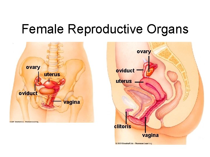
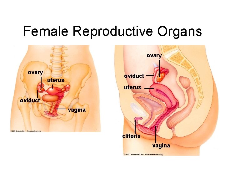
Female Reproductive Organs ovary oviduct uterus oviduct vagina clitoris vagina
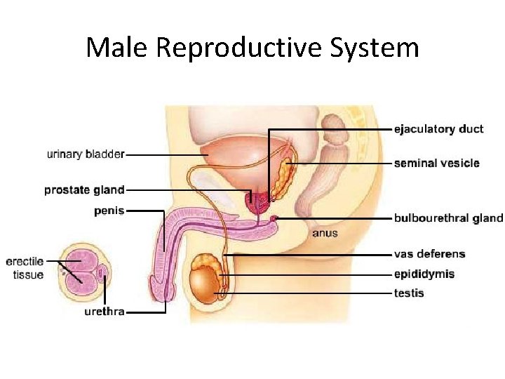
Male Reproductive System Male reproductive system
Human Gonads • Primary sexual organs where genes are packaged into gametes – Male - testes – Female - ovaries • Secrete sex hormones – Regulate secondary sexual traits
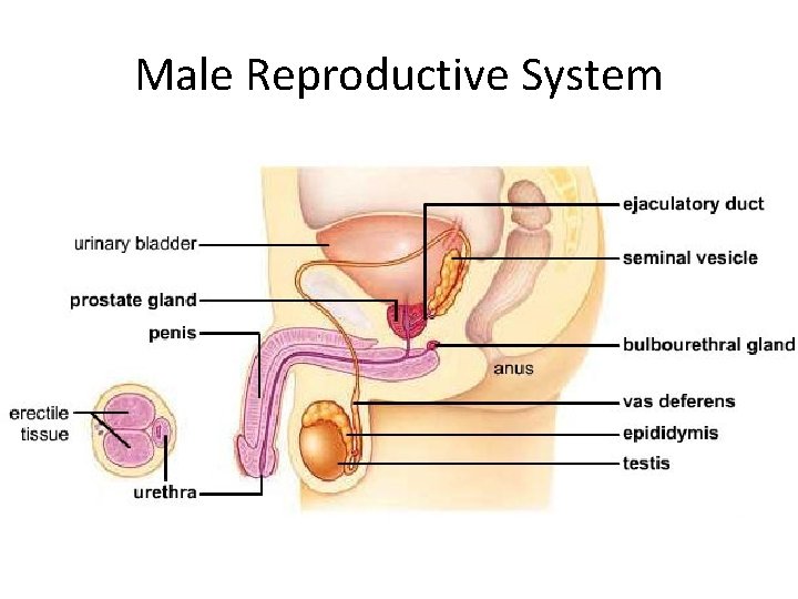
Male Reproductive System Male reproductive system
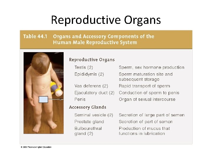
Reproductive Organs
Semen = Sperm + Secretions • Secretions from epididymis aid sperm maturation • Seminal vesicle secretes fructose and prostaglandins • Prostate-gland secretions buffer p. H in the acidic vagina • Bulbourethral gland secretes mucus
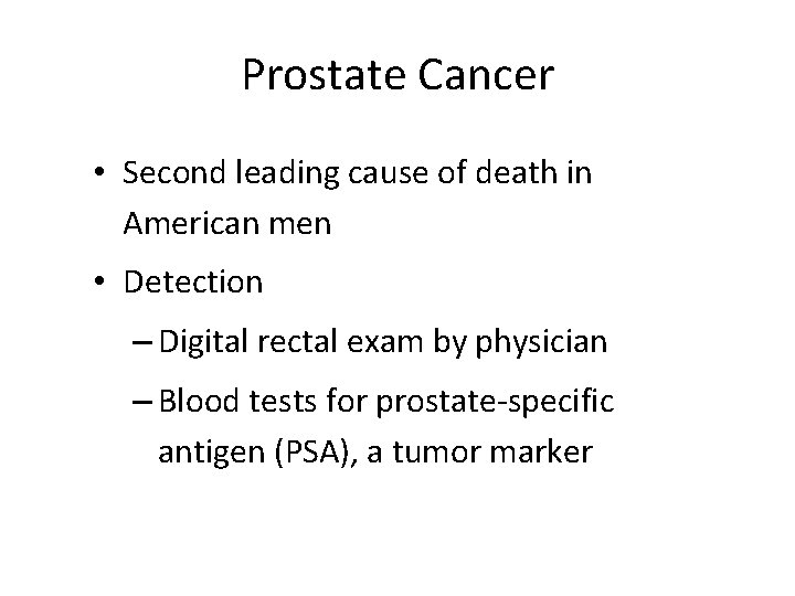
Prostate Cancer • Second leading cause of death in American men • Detection – Digital rectal exam by physician – Blood tests for prostate-specific antigen (PSA), a tumor marker
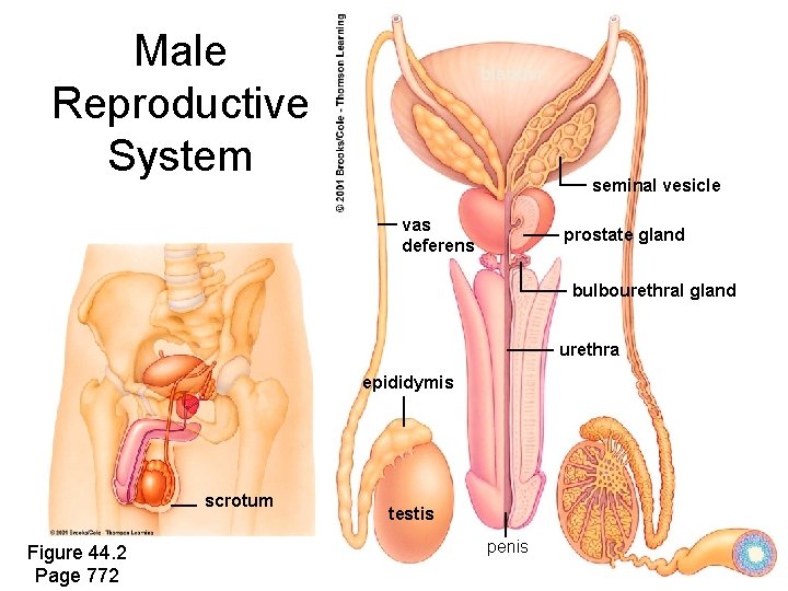
Male Reproductive System bladder seminal vesicle vas deferens prostate gland bulbourethral gland urethra epididymis scrotum Figure 44. 2 Page 772 testis penis
Spermatogenesis • Spermatogonium (2 n) divides by mitosis to form primary spermatocyte (2 n) • Meiosis produces haploid spermatids • Spermatids mature to become sperm Figure 44. 4 Page 775
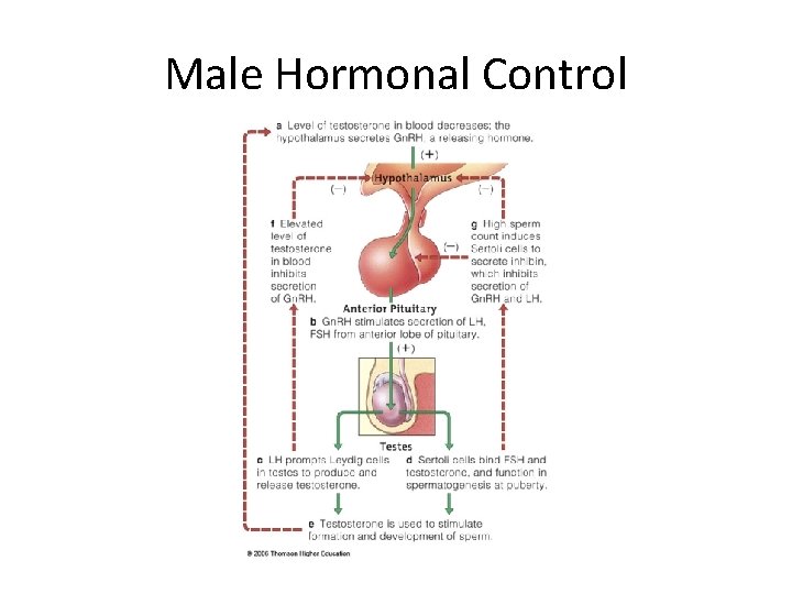
Male Hormonal Control Hypothalamus Gn. RH Inhibin Anterior Pituitary FSH Leydig Cells LH Testes Sertoli Cells Testosterone Formation and Development of Sperm
Male Hormonal Control
Female Reproductive Organs ovary oviduct uterus oviduct vagina clitoris vagina
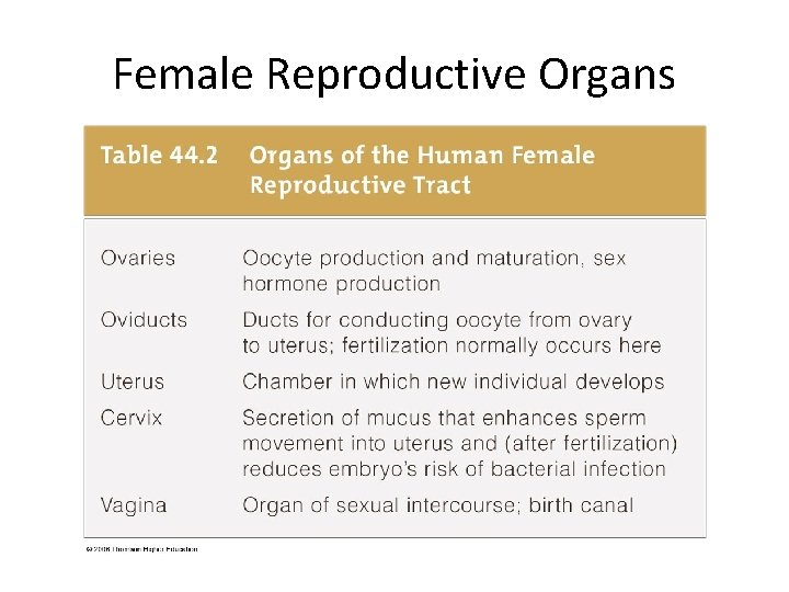
Female Reproductive Organs
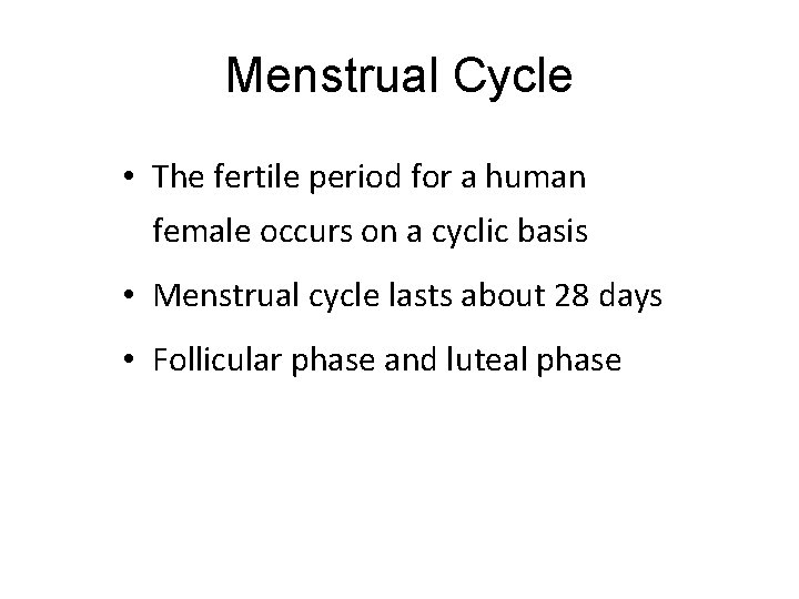
Menstrual Cycle • The fertile period for a human female occurs on a cyclic basis • Menstrual cycle lasts about 28 days • Follicular phase and luteal phase
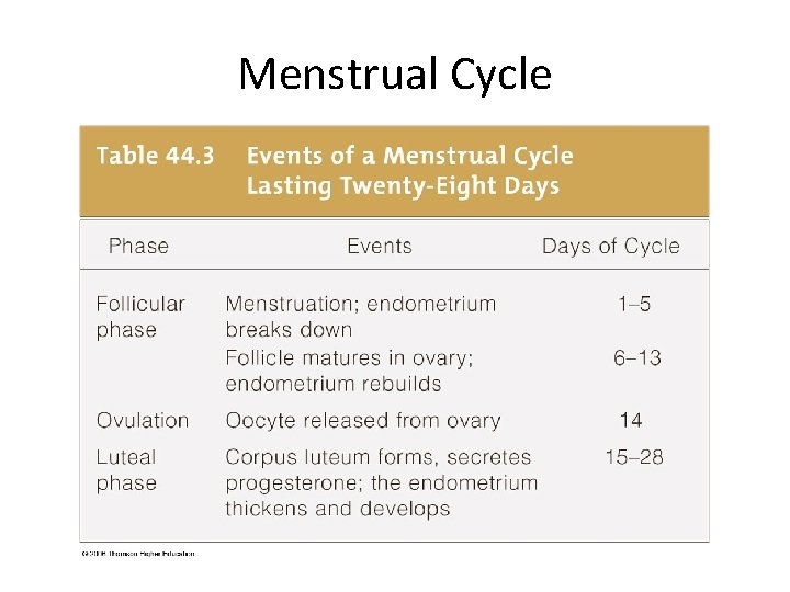
Menstrual Cycle
Oocytes Arrested in Meiosis I • Girl is born with primary oocytes already in ovaries • Each oocyte has entered meiosis I and stopped • Meiosis resumes, one oocyte at a time, with the first menstrual cycle
Menarche to Menopause • First menstruation, or menarche, usually occurs between ages 10 -16 • Menstrual cycles continue until menopause, in a woman’s late 40 s or early 50 s
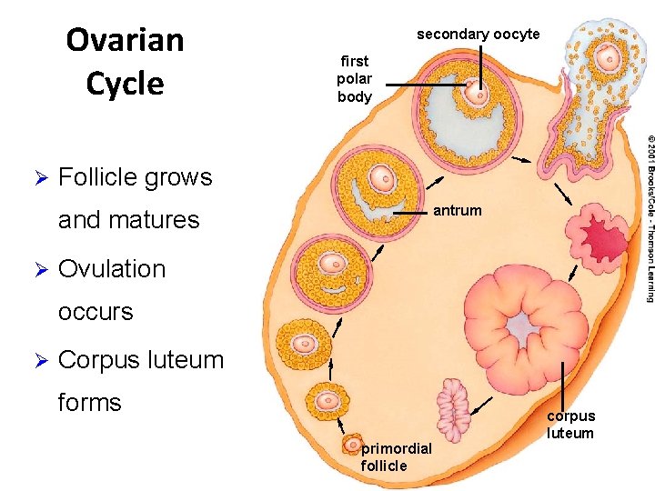
Ovarian Cycle Ø secondary oocyte first polar body Follicle grows antrum and matures Ø Ovulation occurs Ø Corpus luteum forms primordial follicle corpus luteum
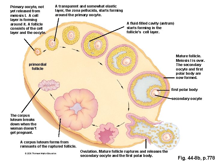
Primary oocyte, not yet released from meiosis I. A cell layer is forming around it. A follicle consists of the cell layer and the oocyte. A transparent and somewhat elastic layer, the zona pellucida, starts forming around the primary oocyte. A fluid-filled cavity (antrum) starts forming in the follicle’s cell layer. Mature follicle. Meiosis I is over. The secondary oocyte and first polar body are now formed. primordial follicle first polar body secondary oocyte The corpus luteum breaks down when the woman doesn’t get pregnant. A corpus luteum forms from remnants of the ruptured follicle. Ovulation. Mature follicle ruptures and releases the secondary oocyte and the first polar body. Fig. 44 -8 b, p. 778
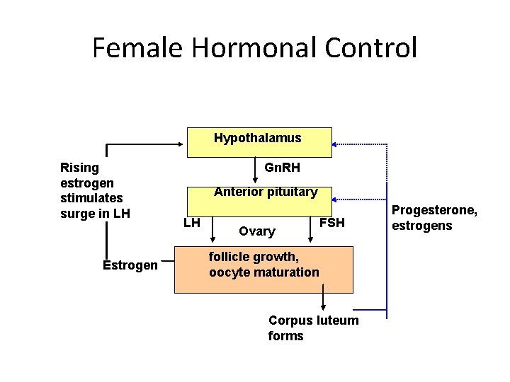
Female Hormonal Control Hypothalamus Rising estrogen stimulates surge in LH Estrogen Gn. RH Anterior pituitary LH Ovary FSH follicle growth, oocyte maturation Corpus luteum forms Progesterone, estrogens
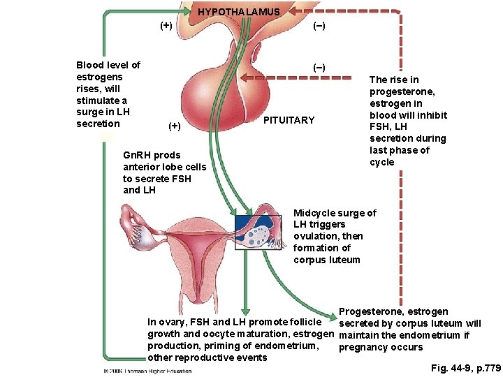
HYPOTHALAMUS (+) Blood level of estrogens rises, will stimulate a surge in LH secretion (–) (+) Gn. RH prods anterior lobe cells to secrete FSH and LH PITUITARY The rise in progesterone, estrogen in blood will inhibit FSH, LH secretion during last phase of cycle Midcycle surge of LH triggers ovulation, then formation of corpus luteum Progesterone, estrogen In ovary, FSH and LH promote follicle secreted by corpus luteum will growth and oocyte maturation, estrogen maintain the endometrium if production, priming of endometrium, pregnancy occurs other reproductive events Fig. 44 -9, p. 779
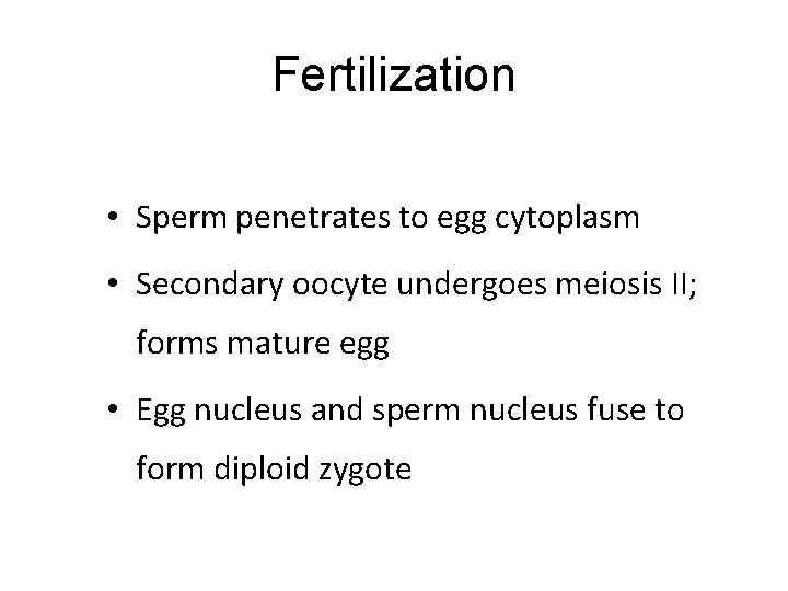
Fertilization • Sperm penetrates to egg cytoplasm • Secondary oocyte undergoes meiosis II; forms mature egg • Egg nucleus and sperm nucleus fuse to form diploid zygote
Birth Control Options Prevent fertilization Prevent ovulation Block implantation
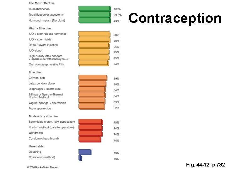
Contraception Fig. 44 -12, p. 782
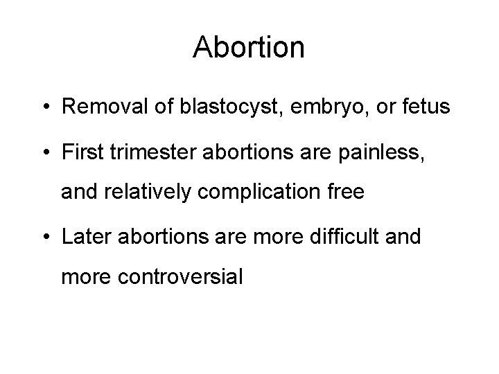
Abortion • Removal of blastocyst, embryo, or fetus • First trimester abortions are painless, and relatively complication free • Later abortions are more difficult and more controversial
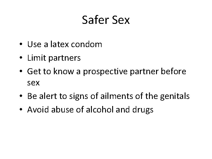
Safer Sex • Use a latex condom • Limit partners • Get to know a prospective partner before sex • Be alert to signs of ailments of the genitals • Avoid abuse of alcohol and drugs
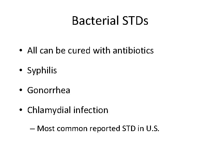
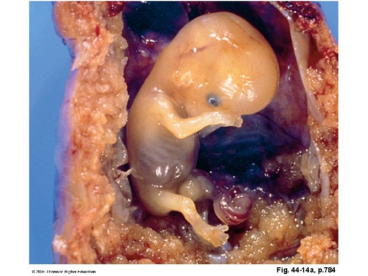
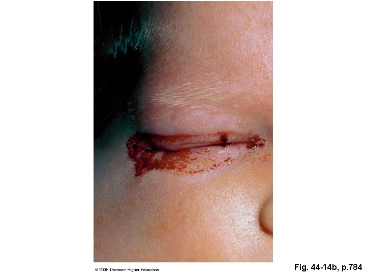
Bacterial STDs • All can be cured with antibiotics • Syphilis • Gonorrhea • Chlamydial infection – Most common reported STD in U. S.
Fig. 44 -14 a, p. 784
Fig. 44 -14 b, p. 784
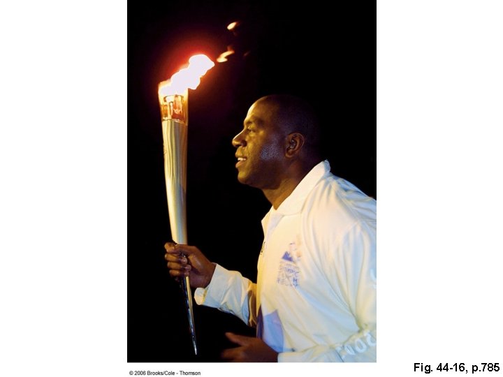
AIDS • Combination of disorders that follows infection with HIV • No vaccine or cure • HIV spreads through anal, vaginal, and oral intercourse and by intravenous drug use
Fig. 44 -16, p. 785
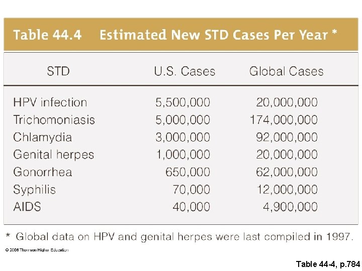
Table 44 -4, p. 784
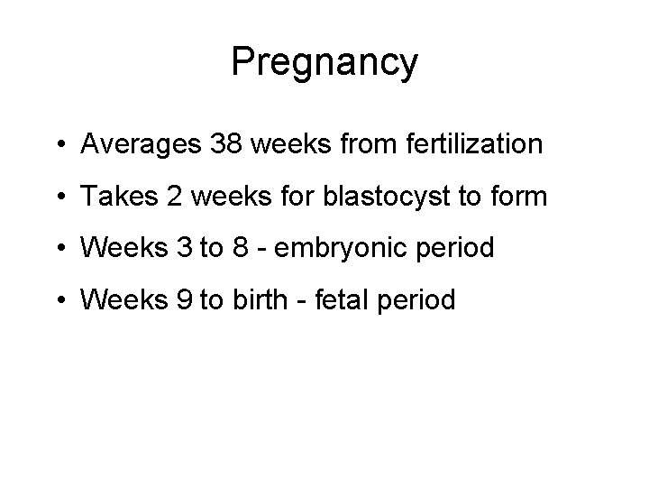
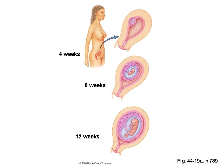
Pregnancy • Averages 38 weeks from fertilization • Takes 2 weeks for blastocyst to form • Weeks 3 to 8 - embryonic period • Weeks 9 to birth - fetal period
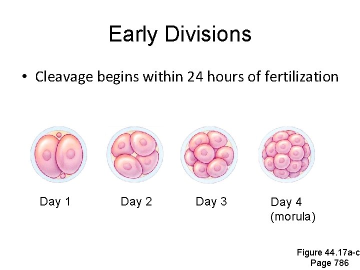
Early Divisions • Cleavage begins within 24 hours of fertilization Day 1 Day 2 Day 3 Day 4 (morula) Figure 44. 17 a-c Page 786
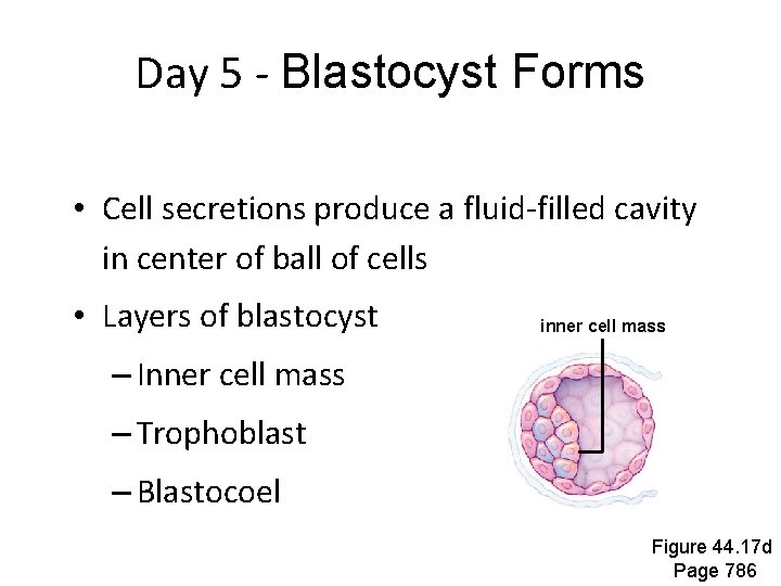
Day 5 - Blastocyst Forms • Cell secretions produce a fluid-filled cavity in center of ball of cells • Layers of blastocyst inner cell mass – Inner cell mass – Trophoblast – Blastocoel Figure 44. 17 d Page 786
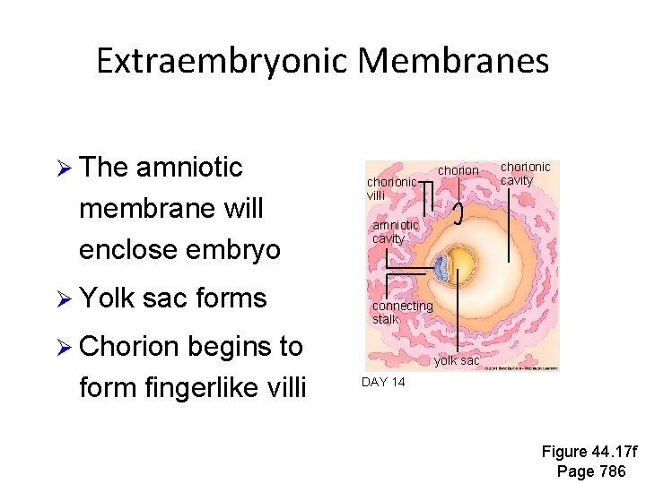
Extraembryonic Membranes Ø The amniotic membrane will enclose embryo Ø Yolk sac forms chorionic villi chorionic cavity amniotic cavity connecting stalk Ø Chorion begins to form fingerlike villi chorion yolk sac DAY 14 Figure 44. 17 f Page 786
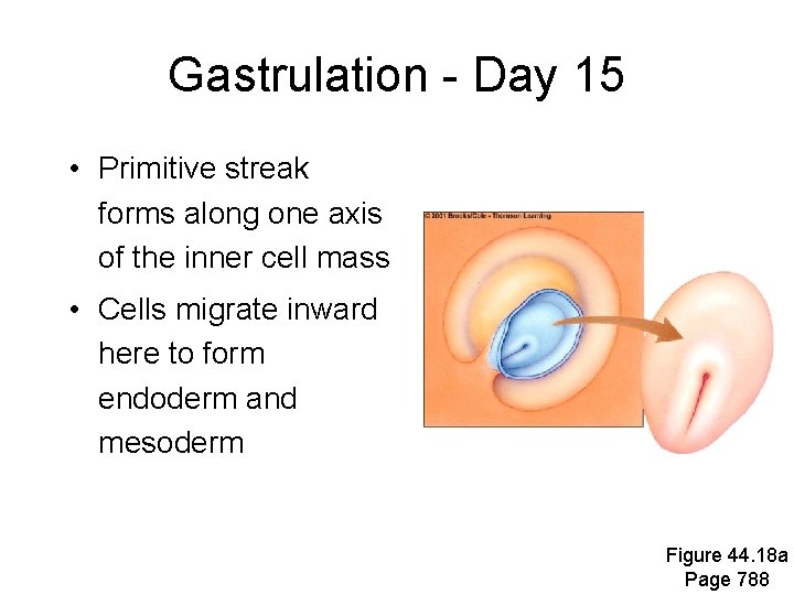
Gastrulation - Day 15 • Primitive streak forms along one axis of the inner cell mass • Cells migrate inward here to form endoderm and mesoderm Figure 44. 18 a Page 788
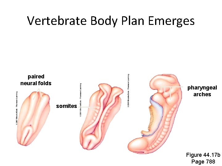
Vertebrate Body Plan Emerges paired neural folds pharyngeal arches somites Figure 44. 17 b Page 788
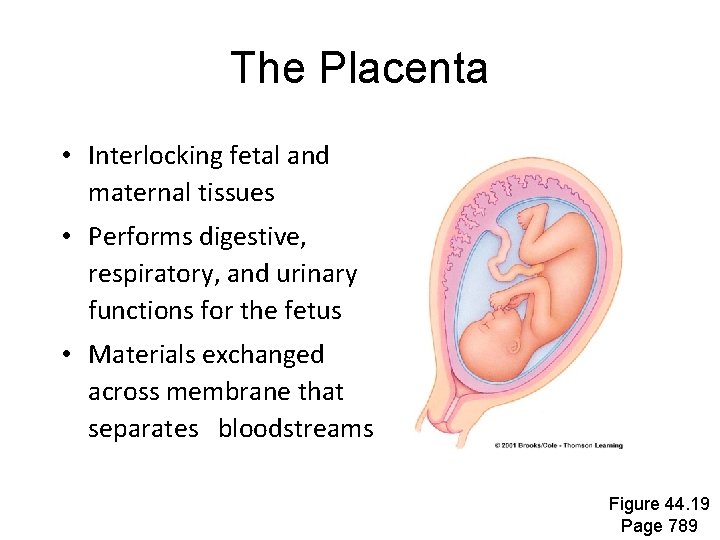
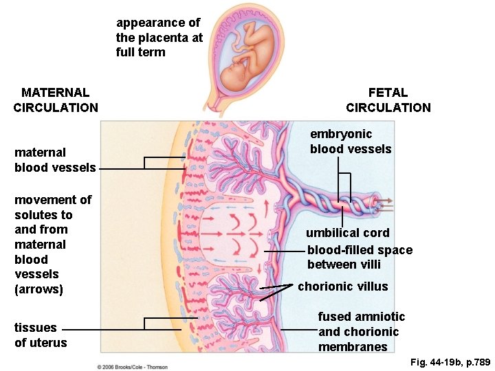
The Placenta • Interlocking fetal and maternal tissues • Performs digestive, respiratory, and urinary functions for the fetus • Materials exchanged across membrane that separates bloodstreams Figure 44. 19 Page 789
4 weeks 8 weeks 12 weeks Fig. 44 -19 a, p. 789
appearance of the placenta at full term MATERNAL CIRCULATION maternal blood vessels movement of solutes to and from maternal blood vessels (arrows) tissues of uterus FETAL CIRCULATION embryonic blood vessels umbilical cord blood-filled space between villi chorionic villus fused amniotic and chorionic membranes Fig. 44 -19 b, p. 789
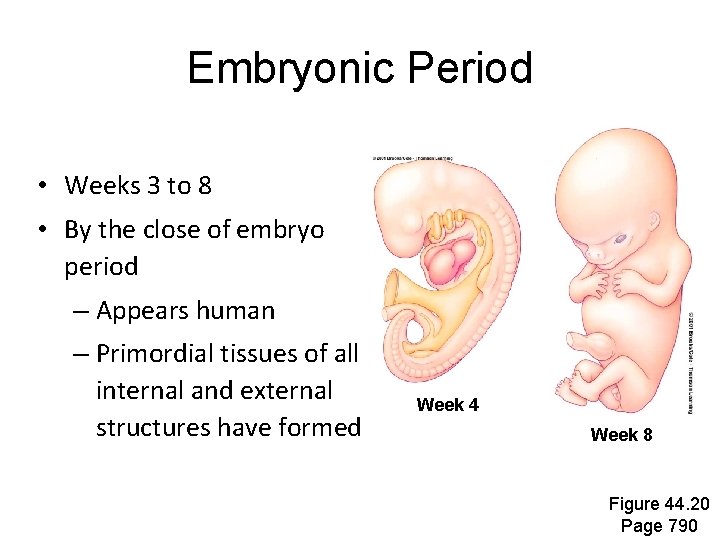
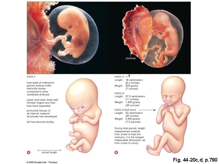
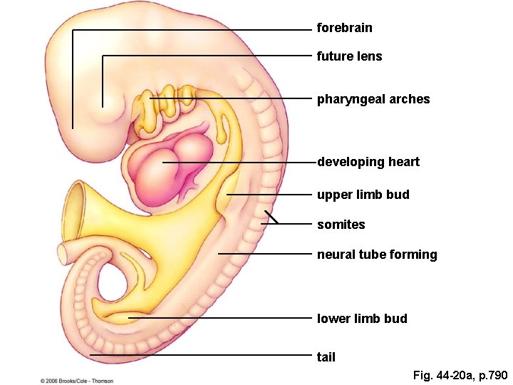
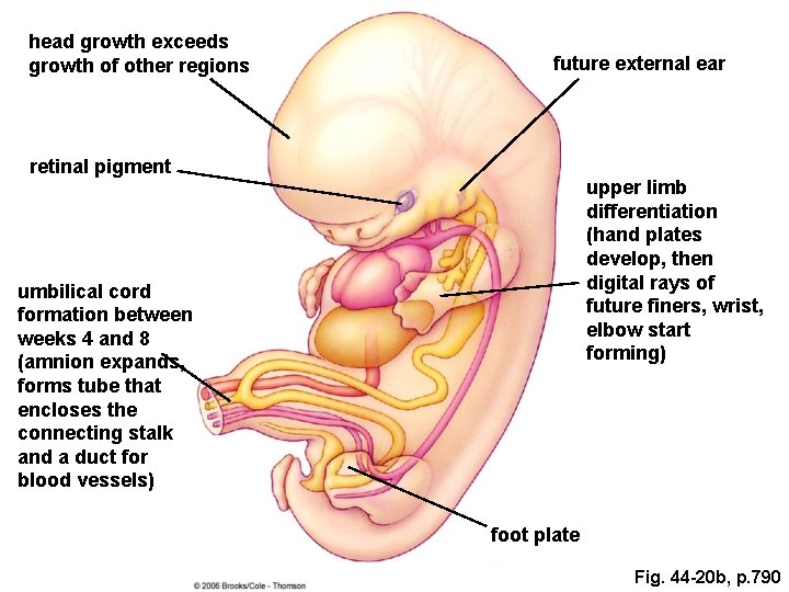
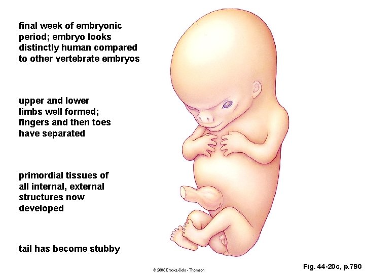
Embryonic Period • Weeks 3 to 8 • By the close of embryo period – Appears human – Primordial tissues of all internal and external structures have formed Week 4 Week 8 Figure 44. 20 Page 790
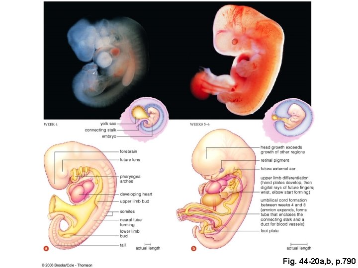
Fig. 44 -20 a, b, p. 790
Fig. 44 -20 c, d, p. 790
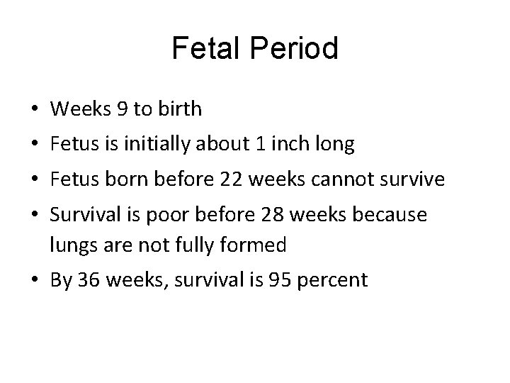
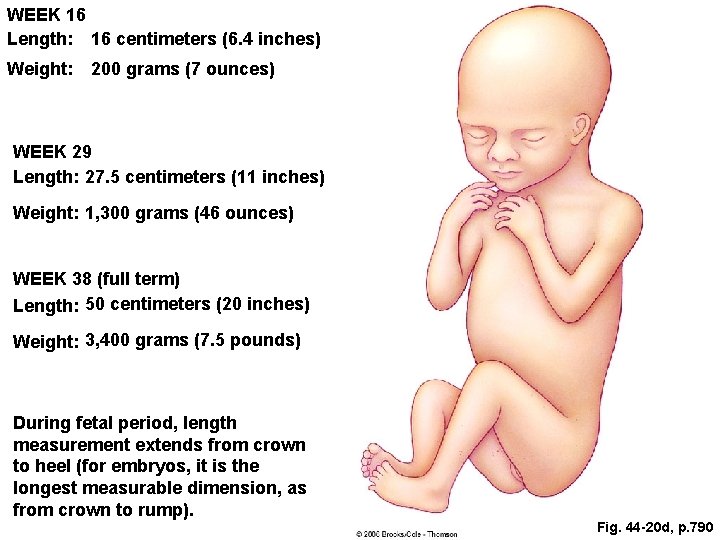
Fetal Period • Weeks 9 to birth • Fetus is initially about 1 inch long • Fetus born before 22 weeks cannot survive • Survival is poor before 28 weeks because lungs are not fully formed • By 36 weeks, survival is 95 percent
forebrain future lens pharyngeal arches developing heart upper limb bud somites neural tube forming lower limb bud tail Fig. 44 -20 a, p. 790
head growth exceeds growth of other regions future external ear retinal pigment upper limb differentiation (hand plates develop, then digital rays of future finers, wrist, elbow start forming) umbilical cord formation between weeks 4 and 8 (amnion expands, forms tube that encloses the connecting stalk and a duct for blood vessels) foot plate Fig. 44 -20 b, p. 790
final week of embryonic period; embryo looks distinctly human compared to other vertebrate embryos upper and lower limbs well formed; fingers and then toes have separated primordial tissues of all internal, external structures now developed tail has become stubby Fig. 44 -20 c, p. 790
WEEK 16 Length: 16 centimeters (6. 4 inches) Weight: 200 grams (7 ounces) WEEK 29 Length: 27. 5 centimeters (11 inches) Weight: 1, 300 grams (46 ounces) WEEK 38 (full term) Length: 50 centimeters (20 inches) Weight: 3, 400 grams (7. 5 pounds) During fetal period, length measurement extends from crown to heel (for embryos, it is the longest measurable dimension, as from crown to rump). Fig. 44 -20 d, p. 790
Fetal Nutrition • All nutrients for fetal growth and development must be delivered via the placenta • Mother’s diet affects fetal health • Smoking may affect ability to absorb nutrients and to pass them to fetus
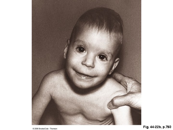
Fig. 44 -22 b, p. 793
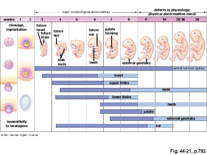
defects in physiology; physical abnormalities minor major morphological abnormalities weeks: 1 cleavage, implantation 2 3 4 future heart future eye brain limb buds 5 6 7 future ear palate forming teeth 8 9 16 20 -36 38 external genitalia central nervous system heart upper limbs eyes lower limbs teeth palate insensitivity to teratogens external genitalia ear Fig. 44 -21, p. 792
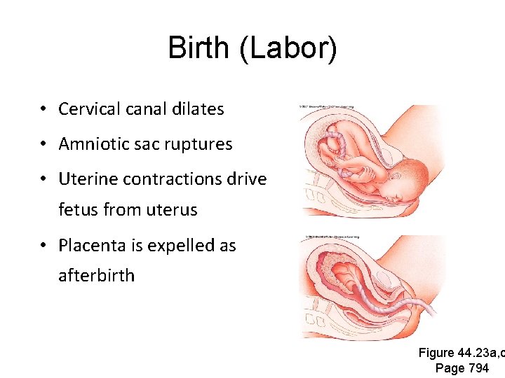
Birth (Labor) • Cervical canal dilates • Amniotic sac ruptures • Uterine contractions drive fetus from uterus • Placenta is expelled as afterbirth Figure 44. 23 a, c Page 794
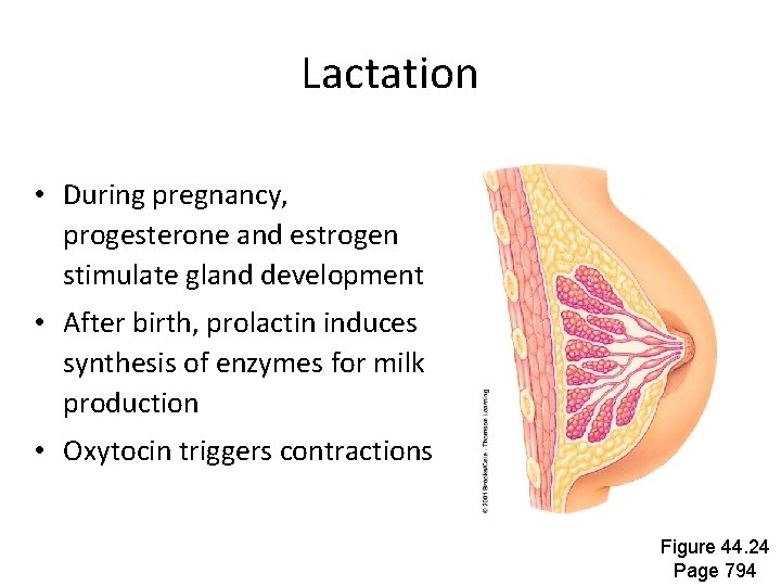
Lactation • During pregnancy, progesterone and estrogen stimulate gland development • After birth, prolactin induces synthesis of enzymes for milk production • Oxytocin triggers contractions Figure 44. 24 Page 794
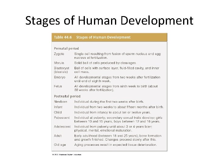
Stages of Human Development Prenatal • Zygote - Single cell • Morula - Solid ball of cells • Blastocyst - Ball with fluid-filled cavity • Embryo - 2 weeks to 8 weeks • Fetus - 9 weeks to birth
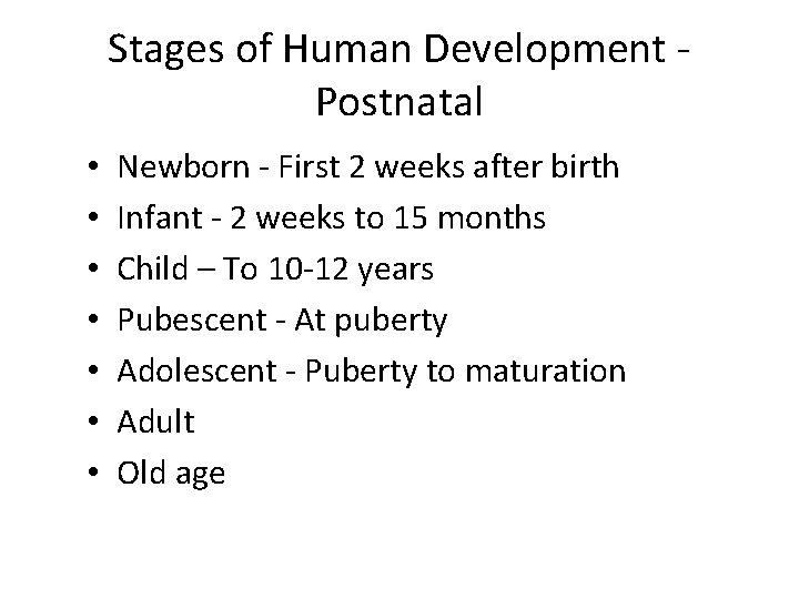
Stages of Human Development Postnatal • • Newborn - First 2 weeks after birth Infant - 2 weeks to 15 months Child – To 10 -12 years Pubescent - At puberty Adolescent - Puberty to maturation Adult Old age
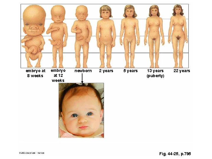
embryo at 8 weeks embryo at 12 weeks newborn 2 years 5 years 13 years (puberty) 22 years Fig. 44 -25, p. 795
Stages of Human Development