Figure 22 2 Gastrointestinal tract activities Ingestion Mechanical
 • Churning")


 Submucosa")


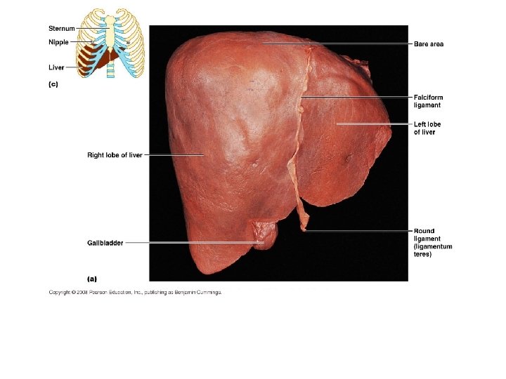
 Celiac plexus Liver")
. Upper lip Gingivae (gums)")

.")
. Soft palate Palatoglossal arch")
. Bolus of food Tongue Uvula Pharynx Bolus Epiglottis Glottis")
 Submucosa")
. (4 of 5) Relaxed muscles 4 Peristalsis moves food")
. (5 of 5) Relaxed muscles 5 The gastroesophageal sphincter")


 Tongue*")


")


 flexure")
 flexure")

 Sensory nerve fibers")


- Slides: 83
Figure 22. 2 Gastrointestinal tract activities. Ingestion Mechanical breakdown • Chewing (mouth) • Churning (stomach) • Segmentation (small intestine) Digestion Food Pharynx Esophagus Propulsion • Swallowing (oropharynx) • Peristalsis (esophagus, stomach, small intestine, large intestine) Stomach Absorption Lymph vessel Small intestine Large intestine Defecation © 2014 Pearson Education, Inc. Blood vessel Mainly H 2 O Feces Anus
Figure 22. 3 a Peristalsis and segmentation. From mouth Peristalsis: Adjacent segments of alimentary tract organs alternately contract and relax, moving food © 2014 Pearson Education, along the tract distally. Inc.
Figure 22. 3 b Peristalsis and segmentation. Segmentation: Nonadjacent segments of alimentary tract organs alternately contract and relax, moving food forward then backward. Food mixing and slow food propulsion occur. © 2014 Pearson Education, Inc.
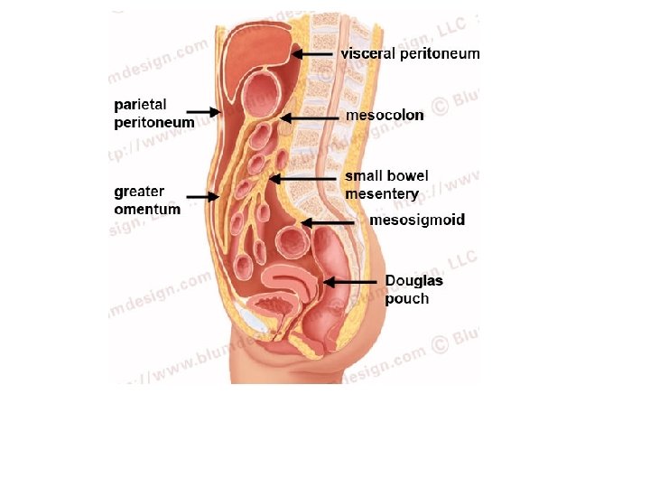
Figure 22. 5 a The peritoneum and the peritoneal cavity. Abdominopelvic cavity Vertebra Dorsal mesentery Parietal peritoneum Ventral mesentery Visceral peritoneum Peritoneal cavity Alimentary canal organ Liver Two schematic cross sections of abdominal cavity illustrate the peritoneums and mesenteries. © 2014 Pearson Education, Inc.
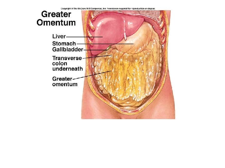
Figure 22. 30 d Mesenteries of the abdominal digestive organs. Liver Lesser omentum Pancreas Stomach Duodenum Transverse mesocolon Transverse colon Mesentery Greater omentum Jejunum Ileum Visceral peritoneum Parietal peritoneum Urinary bladder Rectum © 2014 Pearson Education, Inc.
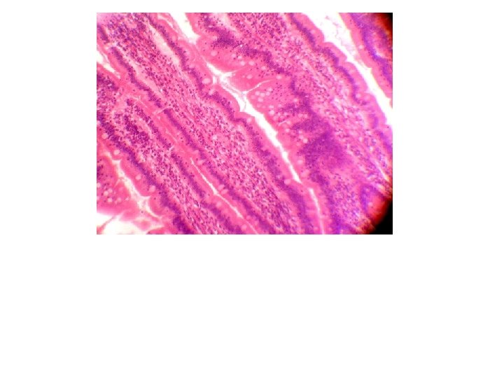
Figure 22. 6 Basic structure of the alimentary canal. Intrinsic nerve plexuses • Myenteric nerve plexus • Submucosal nerve plexus Glands in submucosa Mucosa • Epithelium • Lamina propria • Muscularis mucosae Submucosa Muscularis externa • Longitudinal muscle • Circular muscle Mesentery Nerve Gland in mucosa Artery Vein Duct of gland outside Lymphatic vessel alimentary canal © 2014 Pearson Education, Inc. Serosa • Epithelium (mesothelium) • Connective tissue Lumen Mucosa-associated lymphoid tissue
Figure 19. 2 a The lymphatic system. Regional lymph nodes: Internal jugular vein Entrance of right lymphatic duct into vein Cervical nodes Entrance of thoracic duct into vein Thoracic duct Axillary nodes Cisterna chyli Aorta Collecting lymphatic vessels Drained by the right lymphatic duct Drained by the thoracic duct Inguinal nodes General distribution of collecting lymphatic and regional lymph nodes. © 2014 Pearson Education, vessels Inc.
Figure 22. 12 a Microscopic structure of the esophagus. Mucosa (stratified squamous epithelium) Submucosa (areolar connective tissue) Lumen Muscularis externa • Circular layer • Longitudinal layer © 2014 Pearson Education, Inc. Adventitia (fibrous connective tissue)
Figure 18. 24 a Arteries of the abdomen. Diaphragm Abdominal aorta Inferior phrenic arteries L. gastric artery R. gastric artery Common hepatic artery Hepatic artery proper L Splenic artery Gastroduodenal artery R Celiac trunk R. gastroepiploic artery Middle suprarenal arteries L. gastroepiploic artery Intestinal arteries Middle colic artery Superior mesenteric artery R. colic artery Renal arteries Gonadal arteries Ileocolic artery Sigmoidal arteries Inferior mesenteric artery L. colic artery Lumbar arteries Superior rectal artery Median sacral artery Common iliac arteries © 2014 Pearson Education, Inc. Schematic flowchart.
Figure 18. 29 a Veins of the abdomen. Inferior vena cava Cystic vein Hepatic portal system Inferior phrenic veins Hepatic portal vein Superior mesenteric vein Splenic vein Suprarenal veins Renal veins Inferior mesenteric vein Gonadal veins Lumbar veins R. ascending lumbar vein Common iliac veins L. ascending lumbar vein External iliac vein Internal iliac veins © 2014 Pearson Education, Inc. Schematic flowchart.
Figure 14. 3 The subdivisions of the ANS. Parasympathetic Sympathetic Eye Brain stem Salivary glands Skin* Cranial Sympathetic ganglia Heart Eye Salivary glands Cervical Lungs T 1 Heart Stomach Thoracic Pancreas Liver and gallbladder Pancreas L 1 Liver and gallbladder Adrenal gland Lumbar Bladder © 2014 Pearson Education, Genitals Inc. Bladder Sacral Genitals
Figure 14. 4 Parasympathetic division of the ANS. Eye Ciliary ganglion CN III Lacrimal gland CN VII Pterygopalatine ganglion CN IX CN X Submandibular ganglion Otic ganglion Nasal mucosa Submandibular and sublingual glands Parotid gland Heart Cardiac and pulmonary plexuses Lung Celiac plexus Liver and gallbladder Stomach Pancreas S 2 Large intestine S 4 Small intestine Pelvic splanchnic nerves Inferior hypogastric plexus Rectum Urinary bladder and ureters Genitalia (penis, clitoris, and vagina) © 2014 Pearson Education, Inc. CN S Preganglionic Postganglionic Cranial nerve Sacral nerve
Figure 14. 4 Parasympathetic division of the ANS. (2 of 2) Celiac plexus Liver and gallbladder Stomach Pancreas S 2 S 4 Pelvic splanchnic nerves Inferior hypogastric plexus Large intestine Small intestine Rectum Urinary bladder and ureters Genitalia (penis, clitoris, and vagina) Preganglionic Postganglionic CN Cranial nerve S Sacral nerve © 2014 Pearson Education, Inc.
Figure 22. 7 b Anatomy of the oral cavity (mouth). Upper lip Gingivae (gums) Palatine raphe Hard palate Soft palate Uvula Palatine tonsil Superior labial frenulum Palatoglossal arch Palatopharyngeal arch Posterior wall of oropharynx Tongue Sublingual fold with openings of sublingual ducts Oral vestibule Lower lip © 2014 Pearson Education, Anterior view Inc. Lingual frenulum Opening of Submandibular duct Gingivae (gums) Inferior labial frenulum
Figure 22. 9 The salivary glands. Tongue Teeth Ducts of sublingual gland Frenulum of tongue Sublingual gland Parotid duct Masseter muscle Body of mandible (cut) Posterior belly of digastric muscle Mylohyoid muscle (cut) Submandibular duct Anterior belly of digastric muscle Submandibular gland © 2014 Pearson Education, Inc. Mucous cells Serous cells forming demilunes
Figure 22. 11 Longitudinal section of a canine tooth within its bony socket (alveolus). Enamel Dentin Crown Neck Dentinal tubules Pulp cavity (contains blood vessels and nerves) Gingival sulcus Gingiva (gum) Cement Root canal Periodontal ligament Apical foramen © 2014 Pearson Education, Inc. Bone
Figure 22. 7 a Anatomy of the oral cavity (mouth). Soft palate Palatoglossal arch Uvula Hard palate Oral cavity Palatine tonsil Tongue Oropharynx Lingual tonsil Epiglottis Hyoid bone Laryngopharynx Esophagus Trachea © 2014 Pearson Education, Sagittal section of the oral cavity and pharynx Inc.
Figure 22. 13 Deglutition (swallowing). Bolus of food Tongue Uvula Pharynx Bolus Epiglottis Glottis Trachea Esophagus 1 During the buccal phase, the upper esophageal sphincter is contracted. The tongue presses against the hard palate, forcing the food bolus into the oropharynx. Relaxed muscles 2 The pharyngeal-esophageal phase begins as the uvula and larynx rise to prevent food from entering respiratory passageways. The tongue blocks off the mouth. The upper esophageal sphincter relaxes, allowing food to enter the esophagus. 4 Peristalsis moves food through the esophagus to the stomach. Circular muscles contract Upper esophageal sphincter Bolus 3 The constrictor muscles of the pharynx contract, forcing food into the esophagus inferiorly. The upper esophageal sphincter contracts (closes) after food enters. Relaxed muscles 5 The gastroesophageal sphincter surrounding the cardial oriface opens, and food enters the stomach. Bolus of food Longitudinal muscles contract Circular muscles contract Gastroesophageal sphincter closed Gastroesophageal sphincter opens Stomach © 2014 Pearson Education, Inc.
Figure 22. 12 a Microscopic structure of the esophagus. Mucosa (stratified squamous epithelium) Submucosa (areolar connective tissue) Lumen Muscularis externa • Circular layer • Longitudinal layer © 2014 Pearson Education, Inc. Adventitia (fibrous connective tissue)
Figure 22. 13 Deglutition (swallowing). (4 of 5) Relaxed muscles 4 Peristalsis moves food through the esophagus to the stomach. Circular muscles contract Bolus of food Longitudinal muscles contract Gastroesophageal sphincter closed Stomach © 2014 Pearson Education, Inc.
Figure 22. 13 Deglutition (swallowing). (5 of 5) Relaxed muscles 5 The gastroesophageal sphincter surrounding the cardial oriface opens, and food enters the stomach. Circular muscles contract Gastroesophageal sphincter opens © 2014 Pearson Education, Inc.
Figure 22. 14 a Anatomy of the stomach. Cardia Fundus Esophagus Muscularis externa • Longitudinal layer • Circular layer • Oblique layer Serosa Body Lumen Lesser curvature Rugae of mucosa Greater curvature Duodenum Pyloric sphincter (valve) at pylorus Pyloric canal © 2014 Pearson Education, Inc. Pyloric antrum
Figure 22. 15 b Microscopic anatomy of the stomach. Gastric pits Surface epithelium (mucous cells) Gastric pit Mucous neck cells Parietal cell Gastric gland Chief cell Enteroendocrine cell © 2014 Pearson Education, Inc. Enlarged view of gastric pits and gastric glands
Figure 22. 15 c Microscopic anatomy of the stomach. Pepsinogen HCI Pepsin Mitochondria Parietal cell Chief cell Enteroendocrine cell Location of the HCl-producing parietal cells and pepsin-secreting chief cells in a gastric © 2014 Pearson Education, Inc. gland
Figure 22. 18 Mechanism of HCl secretion by parietal cells. Gastric gland Blood capillary CO 2 Chief cell Stomach lumen CO 2 + H 2 O H+-K+ ATPase H 2 CO 3 Alkaline tide H+ K+ Carbonic anhydrase HCO 3− Parietal cell H+ K+ HCI HCO 3− Cl− Interstitial fluid © 2014 Pearson Education, Inc. HCO 3−- Cl− antiporter Cl−
Figure 22. 15 b Microscopic anatomy of the stomach. Gastric pits Surface epithelium (mucous cells) Gastric pit Mucous neck cells Parietal cell Gastric gland Chief cell Enteroendocrine cell © 2014 Pearson Education, Inc. Enlarged view of gastric pits and gastric glands
Figure 22. 15 c Microscopic anatomy of the stomach. Pepsinogen HCI Pepsin Mitochondria Parietal cell Chief cell Enteroendocrine cell Location of the HCl-producing parietal cells and pepsin-secreting chief cells in a gastric © 2014 Pearson Education, Inc. gland
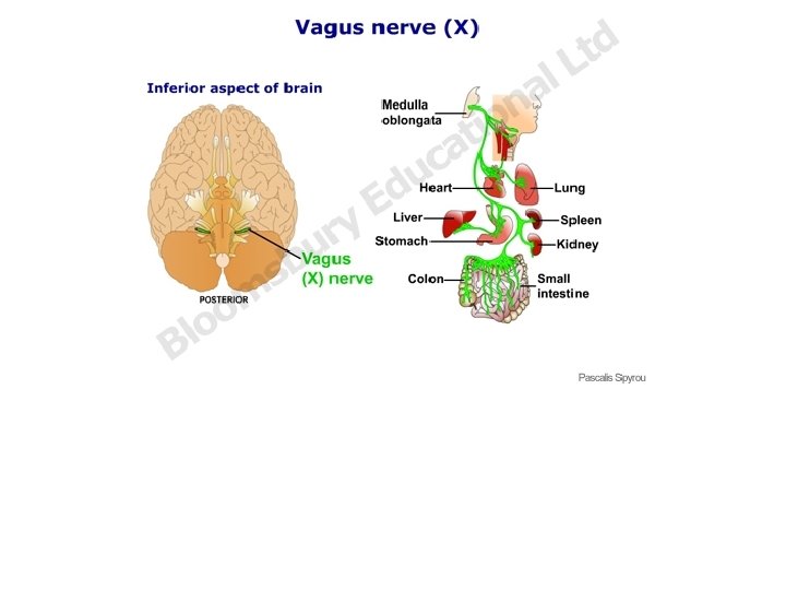
Figure 22. 17 Neural and hormonal mechanisms that regulate release of gastric juice. Inhibitory events Stimulatory events Cephalic phase 1 Sight and thought of food Cerebral cortex Conditioned reflex 2 Stimulation of taste and smell receptors Gastric phase 1 Stomach distension activates stretch receptors Hypothalamus and medulla oblongata Vagovagal reflexes Intestinal phase Stimulate Inhibit © 2014 Pearson Education, Inc. Vagus nerve Local reflexes 2 Food chemicals (especially peptides and caffeine) and rising p. H activate chemoreceptors 1 Presence of partially digested foods in duodenum or distension of the duodenum when stomach begins to empty Medulla G cells Intestinal (enteric) gastrin release to blood Gastrin release to blood Cerebral cortex 1 Loss of appetite, depression Gastrin secretion declines G cells Overrides parasympathetic controls Sympathetic nervous system activation 1 Excessive acidity (p. H < 2) in stomach 2 Emotional stress Lack of stimulatory impulses to parasympathetic center Stomach secretory activity Enterogastric reflex Brief effect Local reflexes Vagal nuclei in medulla Pyloric sphincter Release of enterogastrones (secretin, cholecystokinin, vasoactive intestinal peptide) 1 Distension of duodenum; presence of fatty, acidic, or hypertonic chyme; and/or irritants in the duodenum 2 Distension; presence of fatty, acidic, partially digested food in the duodenum
Figure 22. 15 c Microscopic anatomy of the stomach. Pepsinogen HCI Pepsin Mitochondria Parietal cell Chief cell Enteroendocrine cell Location of the HCl-producing parietal cells and pepsin-secreting chief cells in a gastric © 2014 Pearson Education, Inc. gland
Figure 22. 18 Mechanism of HCl secretion by parietal cells. Gastric gland Blood capillary CO 2 Chief cell Stomach lumen CO 2 + H 2 O H+-K+ ATPase H 2 CO 3 Alkaline tide H+ K+ Carbonic anhydrase HCO 3− Parietal cell H+ K+ HCI HCO 3− Cl− Interstitial fluid © 2014 Pearson Education, Inc. HCO 3−- Cl− antiporter Cl−
Figure 22. 15 c Microscopic anatomy of the stomach. Pepsinogen HCI Pepsin Mitochondria Parietal cell Chief cell Enteroendocrine cell Location of the HCl-producing parietal cells and pepsin-secreting chief cells in a gastric © 2014 Pearson Education, Inc. gland
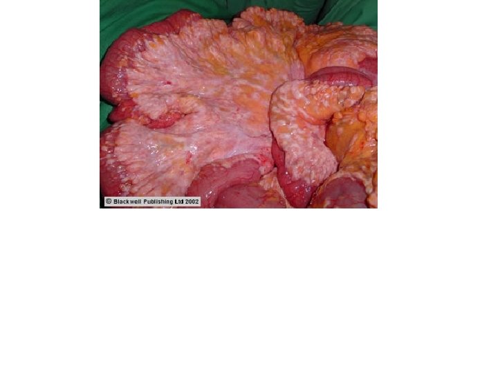
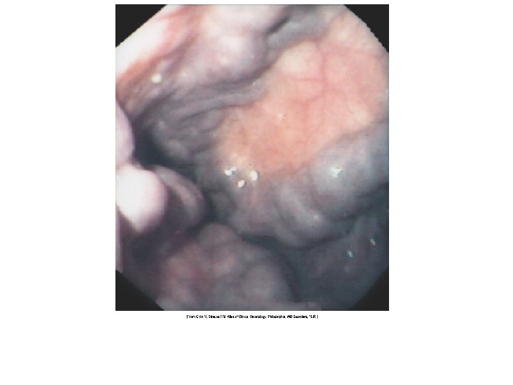
Figure 22. 16 Photographs of a gastric ulcer and the H. pylori bacteria that most commonly cause it. Bacteria Mucosa layer of stomach A gastric ulcer lesion © 2014 Pearson Education, Inc. H. pylori bacteria
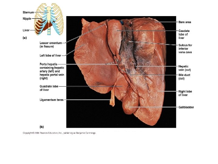
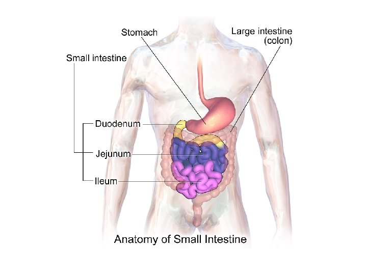
Figure 22. 1 Alimentary canal and related accessory digestive organs. Mouth (oral cavity) Tongue* Parotid gland Sublingual gland Submandibular gland Salivary glands* Pharynx Esophagus Stomach Pancreas* (Spleen) Liver* Gallbladder* Transverse colon Small intestine Anus Duodenum Jejunum Ileum © 2014 Pearson Education, Inc. Descending colon Ascending colon Cecum Sigmoid colon Rectum Appendix Anal canal Large intestine
Figure 22. 21 The duodenum of the small intestine, and related organs. Right and left hepatic ducts of liver Cystic duct Common hepatic duct Bile duct and sphincter Accessory pancreatic duct Mucosa with folds Tail of pancreas Pancreas Jejunum Gallbladder Major duodenal papilla Hepatopancreatic ampulla and sphincter © 2014 Pearson Education, Inc. Main pancreatic duct and sphincter Duodenum Head of pancreas
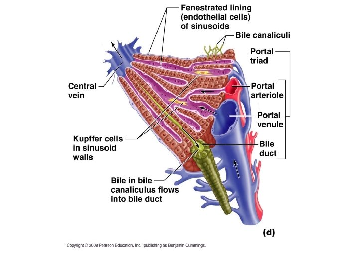
Figure 22. 25 c Microscopic anatomy of the liver. Interlobular veins (to hepatic vein) Central vein Sinusoids Bile canaliculi Plates of hepatocytes Bile duct (receives bile from bile canaliculi) Fenestrated lining (endothelial cells) of sinusoids Stellate macrophages in sinusoid walls Portal vein © 2014 Pearson Education, Inc. Bile duct Portal venule Portal arteriole Portal triad
Figure 22. 21 The duodenum of the small intestine, and related organs. Right and left hepatic ducts of liver Cystic duct Common hepatic duct Bile duct and sphincter Accessory pancreatic duct Mucosa with folds Tail of pancreas Pancreas Jejunum Gallbladder Major duodenal papilla Hepatopancreatic ampulla and sphincter © 2014 Pearson Education, Inc. Main pancreatic duct and sphincter Duodenum Head of pancreas
Figure 22. 21 The duodenum of the small intestine, and related organs. Right and left hepatic ducts of liver Cystic duct Common hepatic duct Bile duct and sphincter Accessory pancreatic duct Mucosa with folds Tail of pancreas Pancreas Jejunum Gallbladder Major duodenal papilla Hepatopancreatic ampulla and sphincter © 2014 Pearson Education, Inc. Main pancreatic duct and sphincter Duodenum Head of pancreas
Figure 22. 26 a Structure of the enzyme-producing tissue of the pancreas. Small duct Acinar cell Basement membrane Zymogen granules Rough endoplasmic reticulum Duct cell One acinus © 2014 Pearson Education, Inc.
Figure 22. 22 a Structural modifications of the small intestine that increase its surface area for digestion and absorption. Vein carrying blood to hepatic portal vessel Muscle layers Circular folds Villi © 2014 Pearson Education, Inc. Lumen
Figure 22. 22 b Structural modifications of the small intestine that increase its surface area for digestion and absorption. Microvilli (brush border) Absorptive cells Lacteal Goblet cell Blood capillaries Mucosaassociated lymphoid tissue Intestinal crypt Muscularis mucosae Duodenal gland © 2014 Pearson Education, Inc. Villus Enteroendocrine cells Venule Lymphatic vessel Submucosa
Figure 22. 22 c Structural modifications of the small intestine that increase its surface area for digestion and absorption. Absorptive cells Goblet cells Villi © 2014 Pearson Education, Inc. Intestinal crypt
Figure 22. 23 Microvilli of the small intestine. Mucus granules Microvilli forming the brush border Absorptive cell © 2014 Pearson Education, Inc.
Figure 22. 28 Mechanisms promoting secretion and release of bile and pancreatic juice. 1 Chyme enter -ing duodenum causes duodenal enteroendocrine cells to release cholecystokinin (CCK) and secretin. 2 CCK (red dots) and secretin (yellow dots) enter the bloodstream. 3 CCK induces secretion of enzyme-rich pancreatic juice. Secretin causes secretion of HCO 3− -rich pancreatic juice. © 2014 Pearson Education, Inc. 4 Bile salts and, to a lesser extent, secretin transported via bloodstream stimulate Liver to produce bile more rapidly. 5 CCK (via blood stream) causes gallbladder to contract and Hepatopancreatic Sphincter to relax. Bile Enters duodenum. 6 During cephalic and gastric phases, vagal Nerve stimulates gallbladder to contract weakly. CCK secretion Secretin secretion
Figure 22. 30 d Mesenteries of the abdominal digestive organs. Liver Lesser omentum Pancreas Stomach Duodenum Transverse mesocolon Transverse colon Mesentery Greater omentum Jejunum Ileum Visceral peritoneum Parietal peritoneum Urinary bladder Rectum © 2014 Pearson Education, Inc.
Figure 22. 30 c Mesenteries of the abdominal digestive organs. Greater omentum Transverse colon Transverse mesocolon Descending colon Jejunum Mesentery Sigmoid mesocolon Sigmoid colon Ileum © 2014 Pearson Education, Inc.
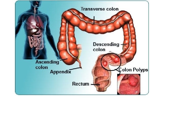
Figure 22. 29 a Gross anatomy of the large intestine. Left colic (splenic) flexure Transverse mesocolon Right colic (hepatic) flexure Epiploic appendages Transverse colon Superior mesenteric artery Haustrum Descending colon Ascending colon IIeum Cut edge of mesentery IIeocecal valve Tenia coli Sigmoid colon Cecum Appendix Rectum Anal canal © 2014 Pearson Education, Inc. External anal sphincter
Figure 22. 29 a Gross anatomy of the large intestine. Left colic (splenic) flexure Transverse mesocolon Right colic (hepatic) flexure Epiploic appendages Transverse colon Superior mesenteric artery Haustrum Descending colon Ascending colon IIeum Cut edge of mesentery IIeocecal valve Tenia coli Sigmoid colon Cecum Appendix Rectum Anal canal © 2014 Pearson Education, Inc. External anal sphincter
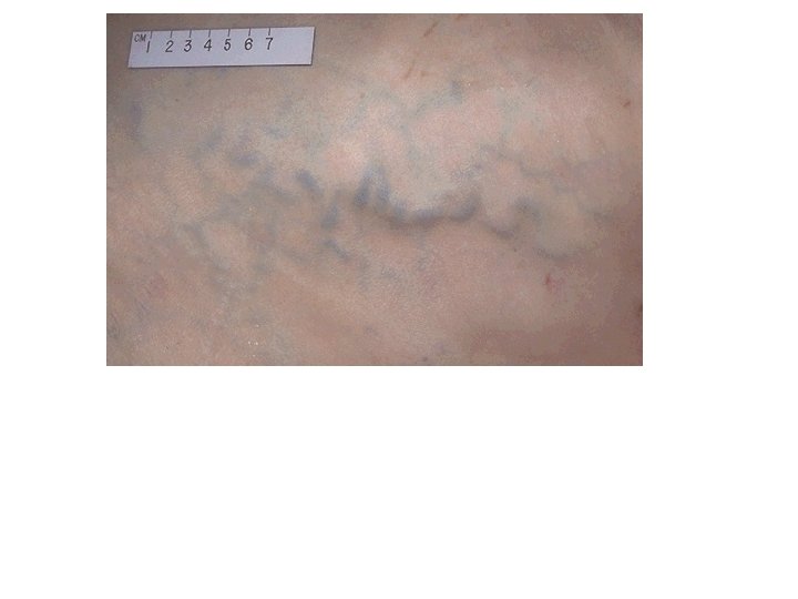
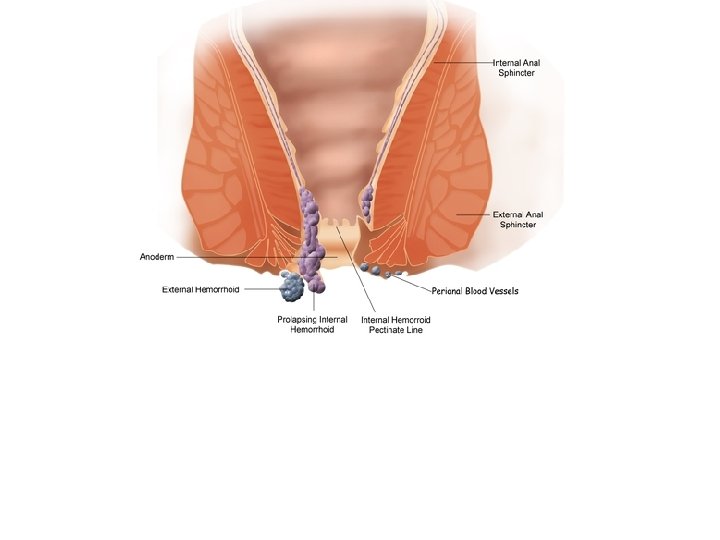
Figure 22. 29 b Gross anatomy of the large intestine. Rectal valve Rectum Hemorrhoidal veins Levator ani muscle Anal canal External anal sphincter Internal anal sphincter Anal columns Pectinate line Anal sinuses Anus © 2014 Pearson Education, Inc.
Figure 22. 31 Defecation reflex. Impulses from cerebral cortex (conscious control) Sensory nerve fibers Voluntary motor nerve to external anal sphincter Sigmoid colon External anal sphincter (skeletal muscle) Rectum Stretch receptors in wall 2 A spinal reflex is initiated in which parasympathetic motor (efferent) fibers stimulate contraction of the rectum and sigmoid colon, and relaxation of the internal anal sphincter. Involuntary motor nerve (parasympathetic division) Internal anal sphincter (smooth muscle) 3 If it is convenient to defecate, voluntary motor neurons are inhibited, allowing the external anal sphincter to relax so feces may pass. © 2014 Pearson Education, Inc. 1 Feces move into and distend the rectum, stimulating stretch receptors there. The receptors transmit signals along afferent fibers to spinal cord neurons.
• ALBUMIN • GLOBULIN • FIBRINOGEN • CLOTTING FACTORS