Histology of Digestive System Dr Haydar AlTukmagi Histology


, jejunum (about")


- Slides: 28
Histology of Digestive System Dr. Haydar Al-Tukmagi
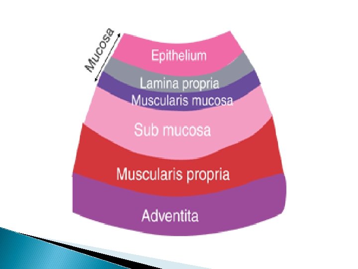
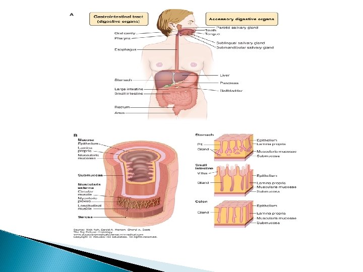
Histology of the Digestive System 1 -Mucosa a. Epithelium b. Lamina Propria c. Muscularis Mucosa 2 -Submucosa a. Submucosal Basic Histological Layersplexus “Plexus of Meissner” Muscularis 3 a. Myenteric plexus “Plexus of Auerbach 4 -Serosa
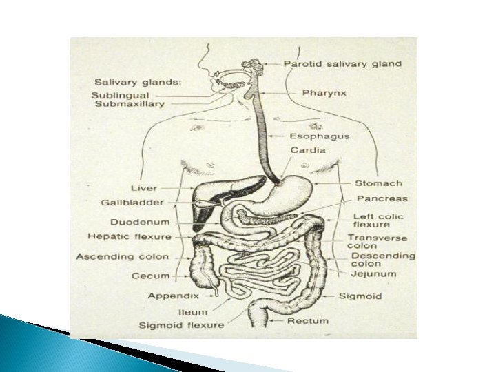
GASTROINTESTINAL TRACT The GI tract is a continuous hollow muscular tube structurally modified into discrete regions and organs that carry out specific functions, as follows: Oral cavity (mouth). Contains the tongue, teeth, and minor salivary glands. The ducts of the major salivary glands open in the oral cavity, where the process of digestion is initiated. Pharynx and esophagus. Transport food from the mouth to the stomach. Stomach. Digests food and secretes hormones. Small intestine (duodenum, jejunum, ileum). Completes the digestive process and absorbs nutrient molecules. Large intestine (cecum, colon, rectum, anus). Absorbs water and electrolytes and compacts and eliminates feces.
ACCESSORY ORGANS The following accessory organs are located outside of the GI tract proper and deliver their secretions, which aid digestion, to the gut via long ducts: Salivary glands (parotid, submandibular, sublingual). Produce saliva. Pancreas. Produces digestive enzymes, which act in the small intestine, and hormones, which are important for glucose and lipid metabolism. Liver. Removes and secretes substances into the blood and produces bile. Gallbladder. Concentrates and stores bile.
Function of the Digestive System Movement of food Secretion of digestive juices Absorption of digested foods, water, and electrolytes
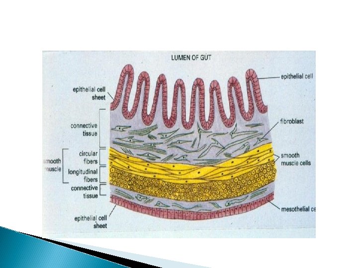
Mucosa A lining epithelium, including glandular tissue, an underlying layer of loose connective tissue called the lamina propria, which provides vascular support for the epithelium, and often contains mucosal glands. Products of digestion pass into these capillaries. Lymphoid follicles, and plasma cells are also often found here. Finally, a thin double layer of smooth muscle is often present - the muscularis mucosa for local movement of the mucosa.
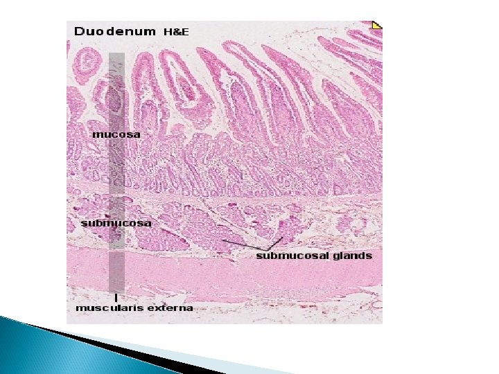
Submucosa A loose connective tissue layer, with larger blood vessels, lymphatics, nerves, and can contain mucous secreting glands. Muscularis propria (externa): smooth muscle layer. There are usually two layers; the inner layer is circular, and the outer layer is longitudinal. These layers of smooth muscle are used for peristalsis (rhythmic waves of contraction), to move food down through the gut. Adventia layer (or serosa) outermost layer of loose connective tissue covered by the visceral peritoneum. Contains blood vessels, lymphatics and nerves.
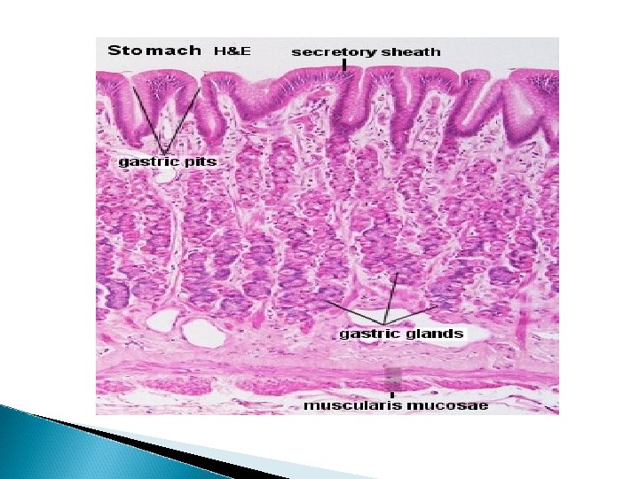
The Stomach The stomach functions both as a reservoir and as a digestive organ. It empties its contents in small portions (suitable for continued digestion) into the small intestine. Anatomically, the stomach is divided into -cardiac part, -fundus, -body or corpus, and -a pyloric part (pyloric antrum and pyloric canal) Histologically, most of the layers of the wall of the stomach appear similar in its different parts. Regional differences are mainly restricted to the appearance of the gastric mucosa.
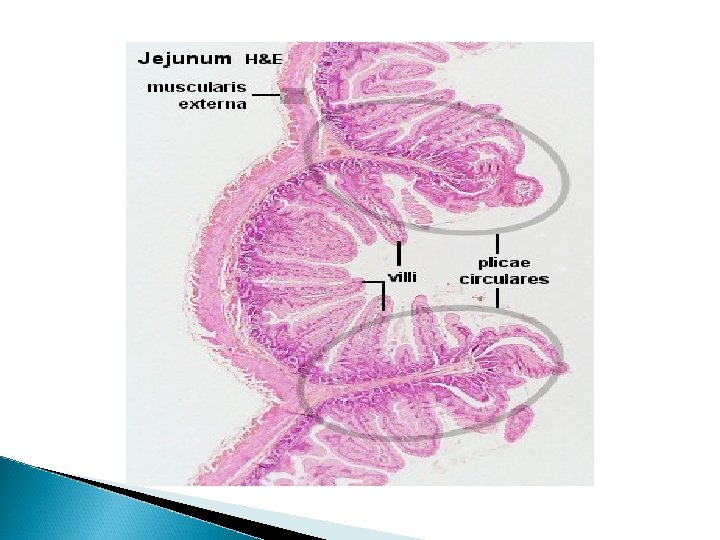
Small Intestine The small intestine is divided into duodenum (25 -30 cm), jejunum (about first two-fifths of the rest) and ileum. The three segments merge imperceptibly and have the same basic histological organization. The entire intestinal mucosa forms intestinal villi (about one mm long), which increase the surface area. The surface of the villi is formed by a simple columnar epithelium. Each absorptive cell or enterocyte of the epithelium forms numerous microvilli
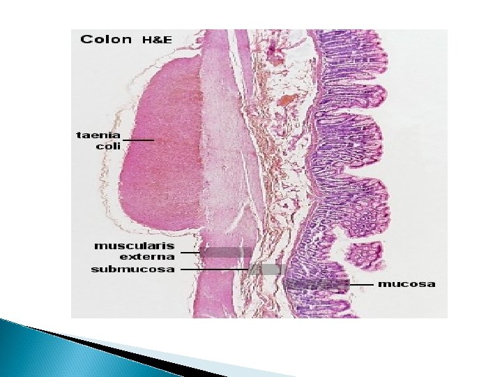
Large Intestine The large intestine constitutes the terminal part of the digestive system. It is divided into three main sections: cecum including the appendix, colon, and rectum with the anal canal. The primary function of the large intestine is the reabsorption of water and inorganic salts. The only secretion of any importance is mucus, which acts as a lubricant during the transport of the intestinal contents.
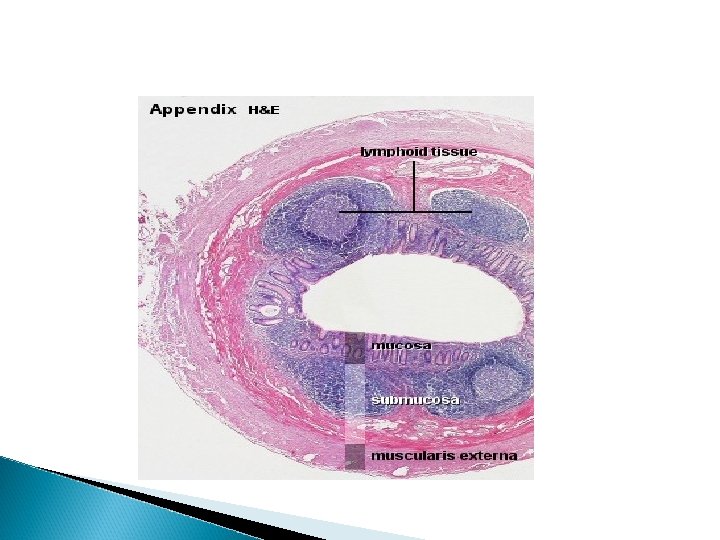
Specialized Sections of the Large Intestine The vermiform appendixis a small blind-ending diverticulum from the cecum. The most important features of the appendix is the thickening of its wall, which is mainly due to large accumulations of lymphoid tissue in the lamina propria and submucosa. Intestinal villi are usually absent, and crypts do not occur as frequently as in the colon. There is often fatty tissue in the submucosa. The muscularis externa is thinner than in the remainder of the large intestine and, the outer, longitudinal smooth muscle layer of the muscularis externa does not aggregate into taenia coli.
The anal canalis the 2. 5 -4 cm long terminal part of the digestive tract. The mucosa has a characteristic surface relief of 5 -10 longitudinal folds, the anal columns. Each column contains a terminal branch of the superior rectal artery and vein.
Histology of the Mucosa Organ Epithelium Mouth Nonkeratinized Stratified Squamous Pharynx Nonkeratinized Stratified Squamous Esophagus Nonkeratinized Stratified Squamous Stomach Simple Columnar Small Intestine Simple Columnar Large Intestine Simple Columnar Anus Nonkeratinized Stratified Squamous
Microscopic View of the Esophagus
Low and High power view of the Stomach Mucosa
High power view of the duodenal Mucosa
High Power View of Villi
Gross view and low-power view of the ileum
Large Intestine