Corticospinal Tract and Other Motor Pathways LundyEkman Dr
- Slides: 89
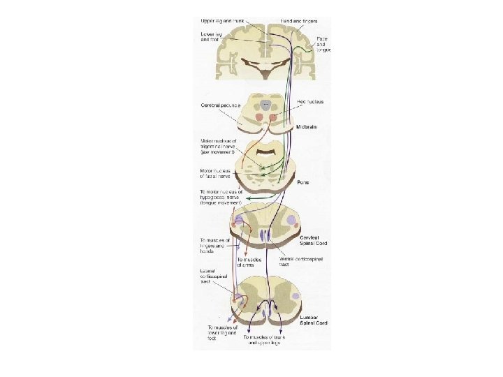
Corticospinal Tract and Other Motor Pathways • Lundy-Ekman • Dr. Donald Allen • Sherrington – Motor systems are the only way we can understand what is happening in the nervous system
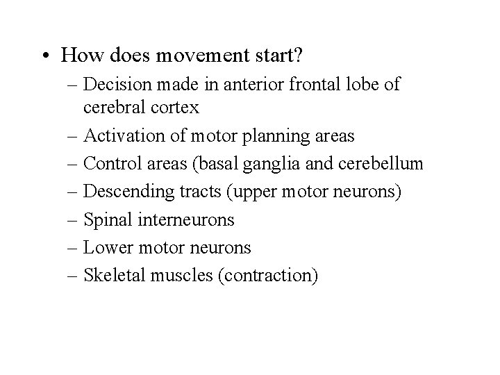
• How does movement start? – Decision made in anterior frontal lobe of cerebral cortex – Activation of motor planning areas – Control areas (basal ganglia and cerebellum – Descending tracts (upper motor neurons) – Spinal interneurons – Lower motor neurons – Skeletal muscles (contraction)
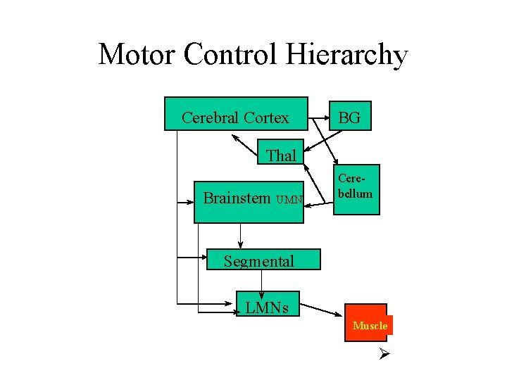
Motor Control Hierarchy Cerebral Cortex BG Thal Brainstem UMN Cerebellum Segmental LMNs Muscle Ø
Muscles • Tone
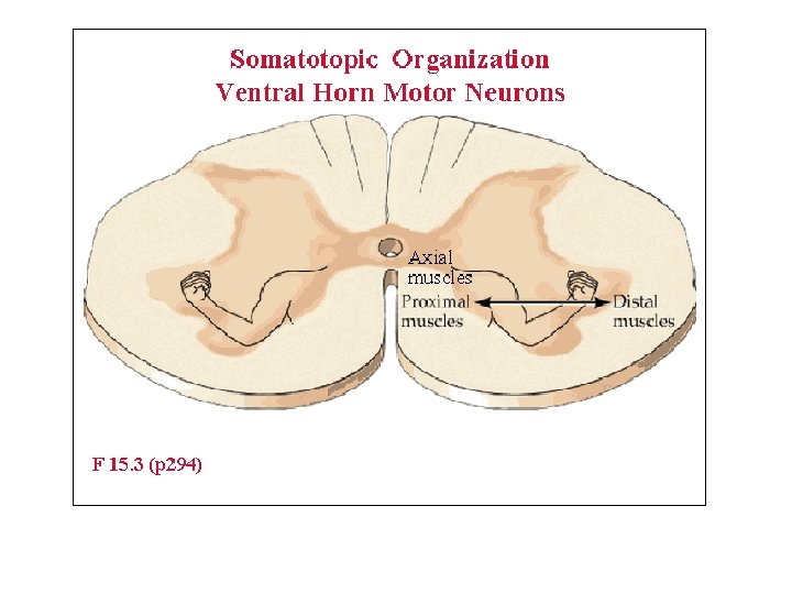
Lower motor neurons • Multipolar neurons • Location – –
Classification of motor neurons • Alpha motor neurons • Gamma motor neurons
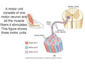
Motor neurons • Synapse • Neurotransmitter • Receptors • Motor Unit
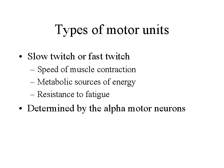
Types of motor units • Slow twitch or fast twitch – Speed of muscle contraction – Metabolic sources of energy – Resistance to fatigue • Determined by the alpha motor neurons
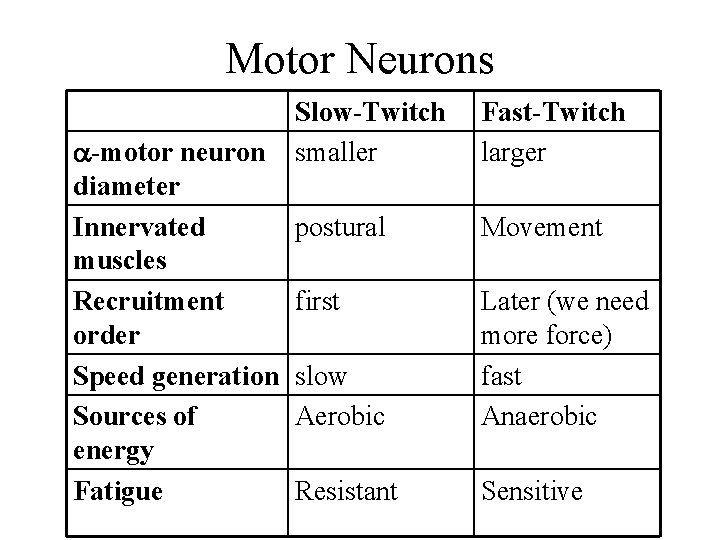
Motor Neurons -motor neuron diameter Innervated muscles Recruitment order Speed generation Sources of energy Fatigue Slow-Twitch smaller Fast-Twitch larger postural Movement first slow Aerobic Later (we need more force) fast Anaerobic Resistant Sensitive
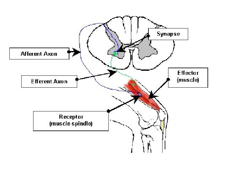
Spinal Reflexes • Components
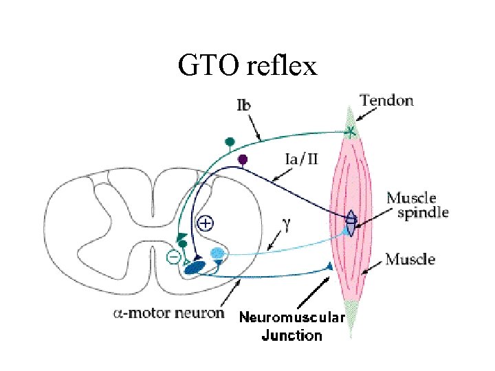
Sensory Receptors • Muscle spindles • Golgi tendon organs • Cutaneous receptors
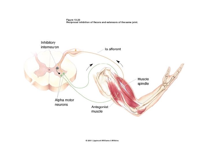
Muscle Spindle Reflexes • Phasic stretch reflex • Tonic stretch reflex
Tonic Stretch Reflex
GTO reflex
Cutaneous Reflexes • Withdrawal reflexes • Respond to nociceptive input
Upper Motor Neurons • Locations in CNS – – • Project to – –
Pathways to spinal cord • Classified based on where they synapse in the ventral horn • Medial activating systems • Lateral activating systems • Non-specific activating systems
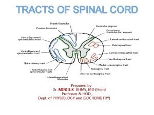
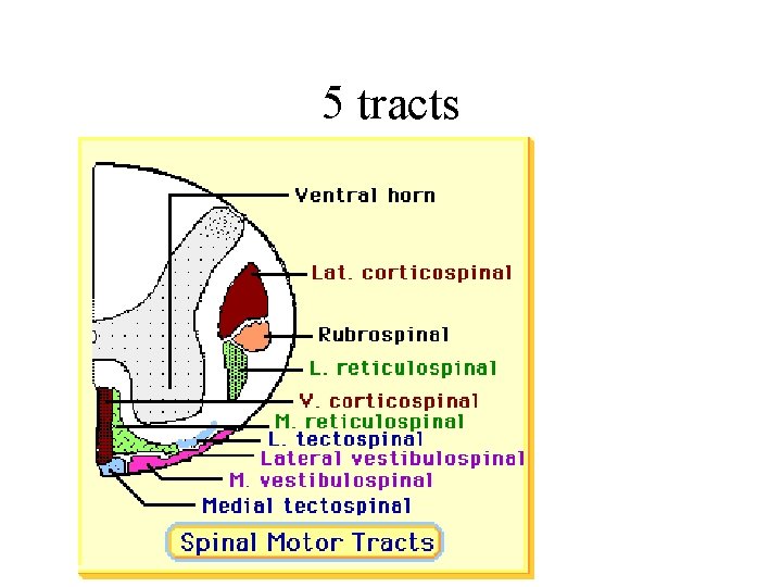
Medial Activating Systems • Anterior Columns of Spinal Cord • Muscles innervated • 5 tracts
5 tracts
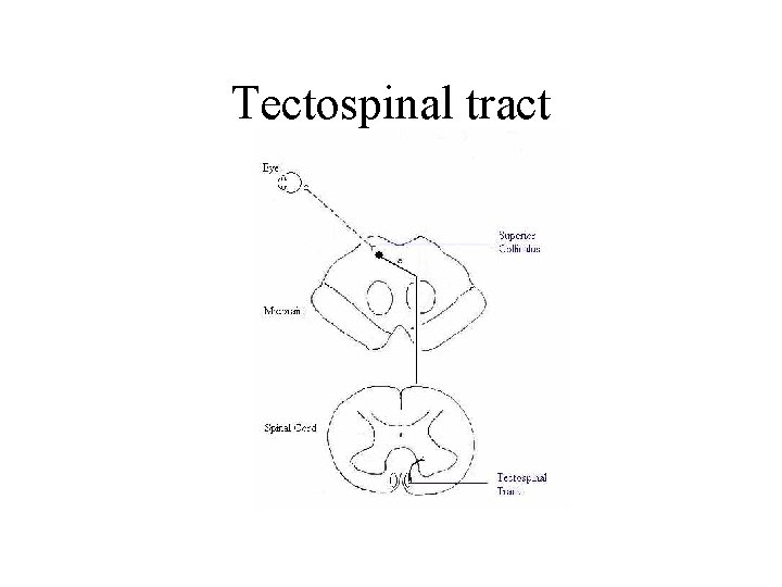
Tectospinal tract
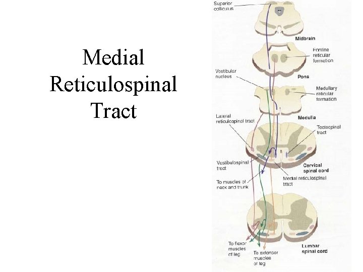
Medial Reticulospinal Tract
Medial vestibulospinal tract
Lateral vestibulospinal tract
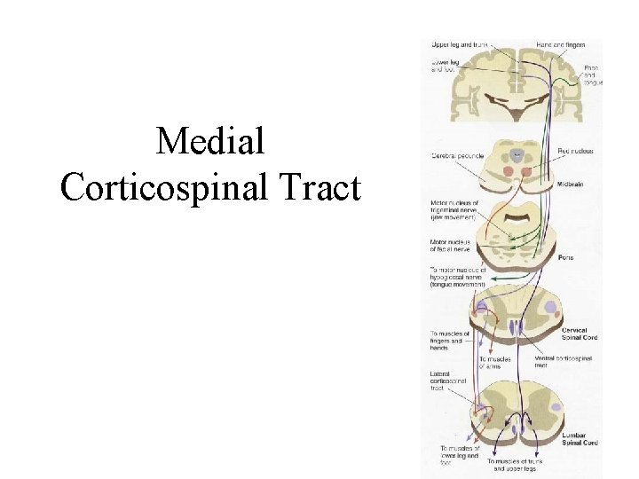
Medial Corticospinal Tract
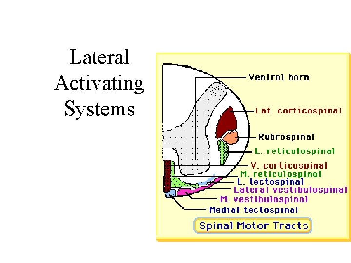
Lateral Activating Systems
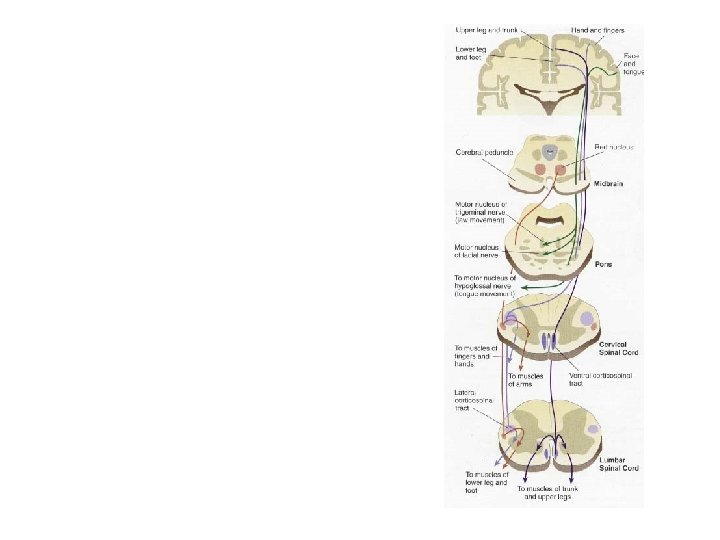
Lateral Corticospinal Tract • AKA: pyramidal tract • Origin (p 215)
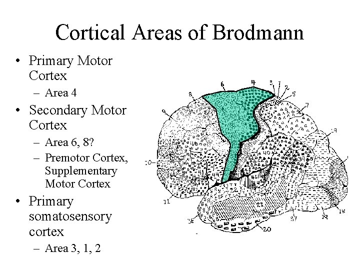
Cortical Areas of Brodmann • Primary Motor Cortex – Area 4 • Secondary Motor Cortex – Area 6, 8? – Premotor Cortex, Supplementary Motor Cortex • Primary somatosensory cortex – Area 3, 1, 2
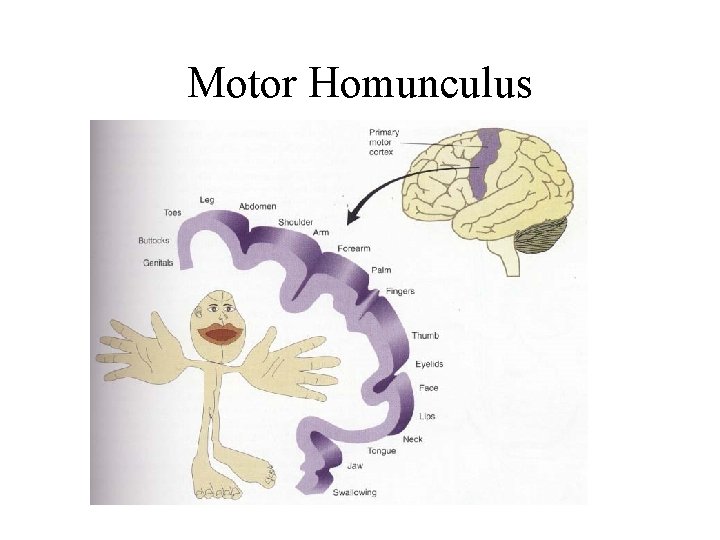
Motor Homunculus
Premotor Cortex
Supplementary Motor Cortex
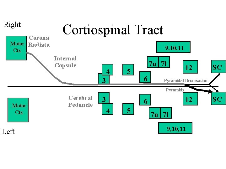
Right Corona Motor Radiata Cortiospinal Tract 9, 10, 11 Ctx Internal Capsule 4 5 3 7 u 7 l 6 12 SC Pyramidal Decussation Pyramids Motor Ctx Left Cerebral Peduncle 3 12 6 4 5 7 u 7 l 9, 10, 11 SC
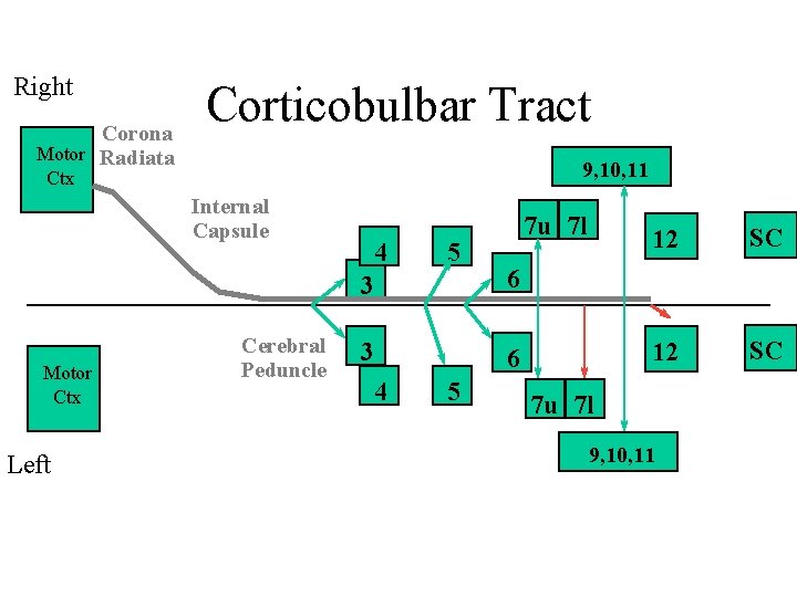
Right Corona Motor Radiata Corticobulbar Tract 9, 10, 11 Ctx Internal Capsule Motor Ctx Left Cerebral Peduncle 4 5 7 u 7 l 3 6 4 5 12 SC 7 u 7 l 9, 10, 11
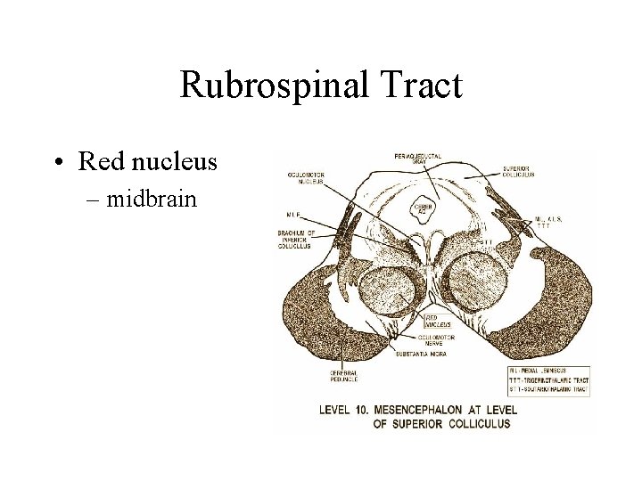
Rubrospinal Tract • Red nucleus – midbrain
Lateral Reticulospinal Tract
Non-specific Motor Pathways • Raphespinal tract • Ceruleospinal tract
Basal Ganglia, Cerebellum and Movement • BG and Cb adjust activity in descending motor tracts • DO NOT have direct connections with lower motor neurons • Connections – –
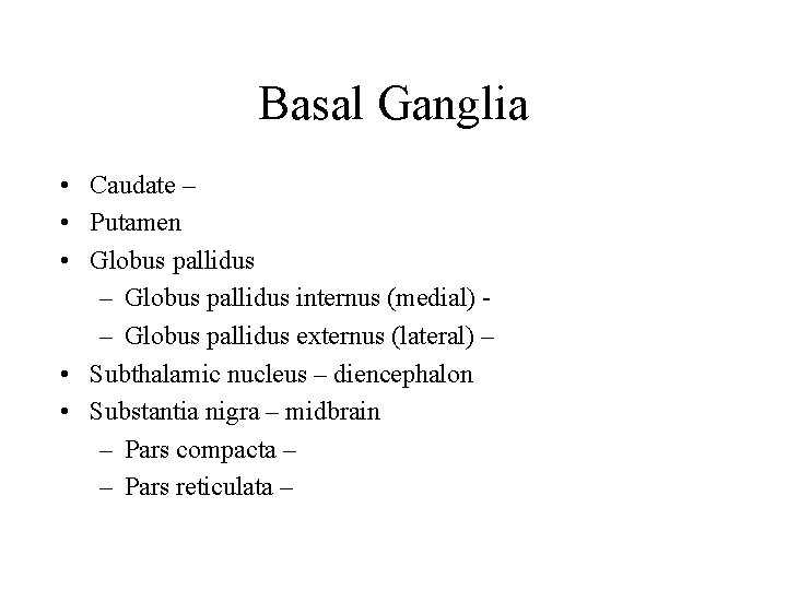
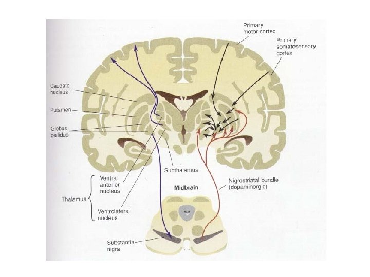
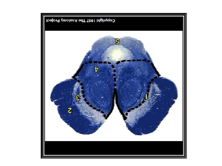
Basal Ganglia • Caudate – • Putamen • Globus pallidus – Globus pallidus internus (medial) – Globus pallidus externus (lateral) – • Subthalamic nucleus – diencephalon • Substantia nigra – midbrain – Pars compacta – – Pars reticulata –
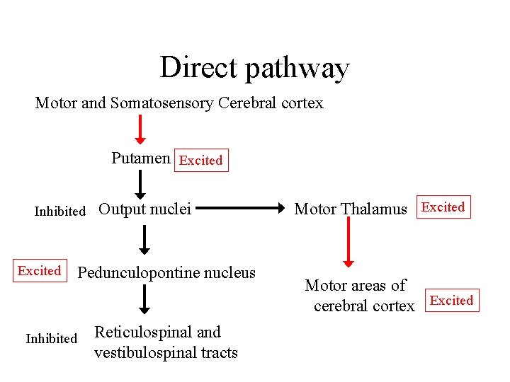
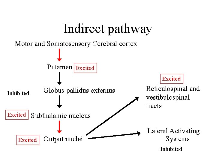
Pathways in BG for motor systems • Input regions • Processing regions – Two pathways through BG • Direct pathway – • Indirect pathway – • Output regions
Direct pathway Motor and Somatosensory Cerebral cortex Putamen Excited Inhibited Excited Output nuclei Pedunculopontine nucleus Inhibited Reticulospinal and vestibulospinal tracts Motor Thalamus Excited Motor areas of cerebral cortex Excited
Indirect pathway Motor and Somatosensory Cerebral cortex Putamen Excited Globus pallidus externus Inhibited Excited Reticulospinal and vestibulospinal tracts Subthalamic nucleus Excited Output nuclei Lateral Activating Systems Inhibited
Substantia Nigra compacta
Output Regions of the BG • Globus pallidus internus • Substantia nigra reticularis
Functions of the Basal Ganglia • Sequencing movements • Regulating muscle tone and force • Selecting synergies (direct pathway) and inhibiting synergies (indirect pathway) • Motor learning
Basal Ganglia Disorders • Hypokinetic – Parkinson’s Disease • Hyperkinetic – Huntington’s disease – Dystonias – Choreoathetotic cerebral palsy – Hemiballismus
Parkinson’s Disease • Most common BG disorder • Neurodegenerative disease – •
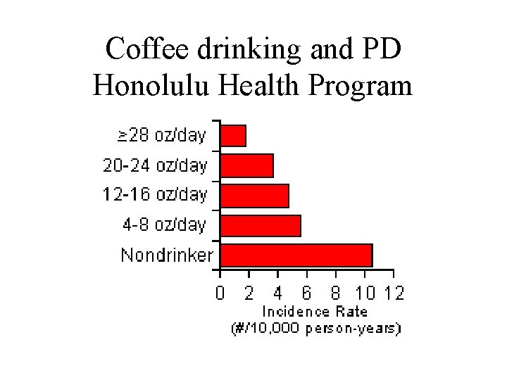
Coffee drinking and PD Honolulu Health Program
Parkinsonism and Parkinsonian Syndrome
Huntington’s Disease
Dystonia • Genetic movement disorder – Dysfunction in basal ganglia – Usually non-progressive • Involuntary sustained muscle contractions – Twisting or repetitive motions or abnormal posture • Focal dystonias are most common type
Focal Dystonia • Affects one part of body • Often limited to a particular activity • Focal dystonia of hand – Writer’s cramp • Deteriation in handwriting due to involuntary muscle contractions in upper limb – Musician’s cramp • Usually 4 th and 5 th fingers flexing involuntarily
Choreoathetotic Cerebral Palsy • Damage to basal ganglia structures • Chorea • Athetotic movements
Hemiballismus • Damage to: • Ballistic movements • Side affected
Cerebellum (Little Brain) • Location: • Functions:
Cb Anatomy • Gray matter • White matter • Deep cerebellar nuclei
Gross anatomy of Cb • Arbor vitae • Folia
Cellular anatomy of the Cb • Three layers of cells – Outer and inner layers • Interneurons – – Granule cells Golgi cells Stellate cells Basket cells – Middle layer • Purkinje cell bodies
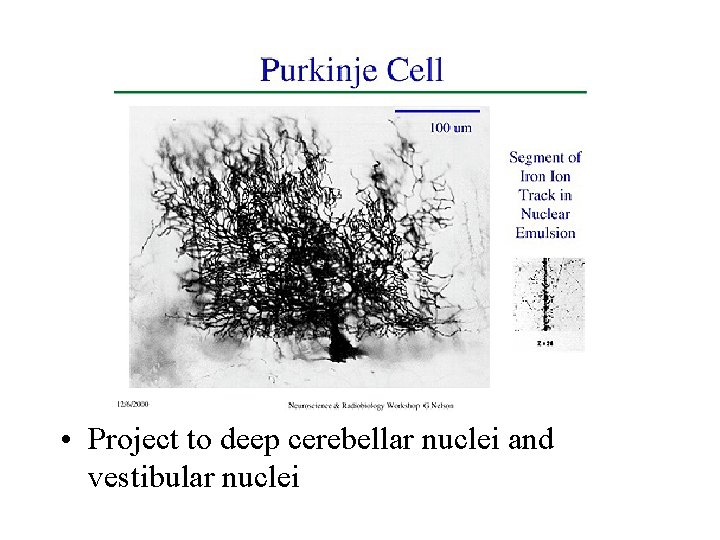
• Project to deep cerebellar nuclei and vestibular nuclei
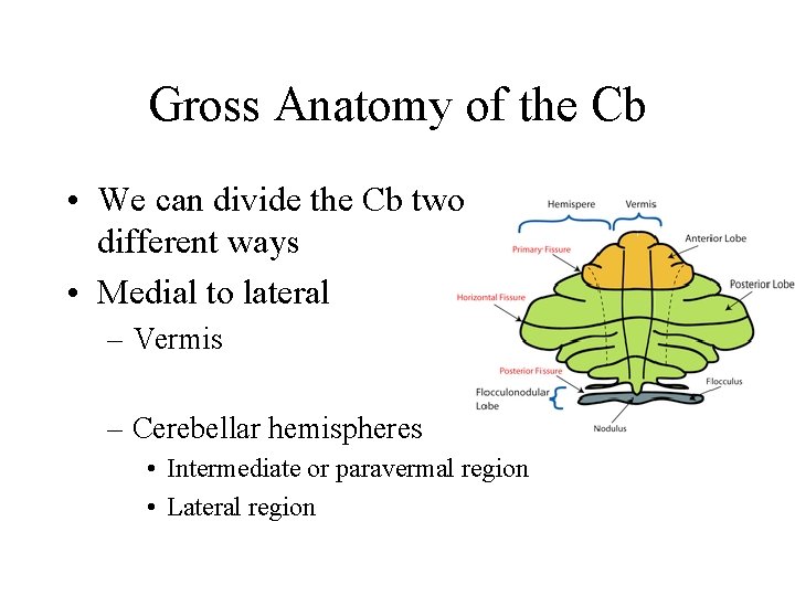
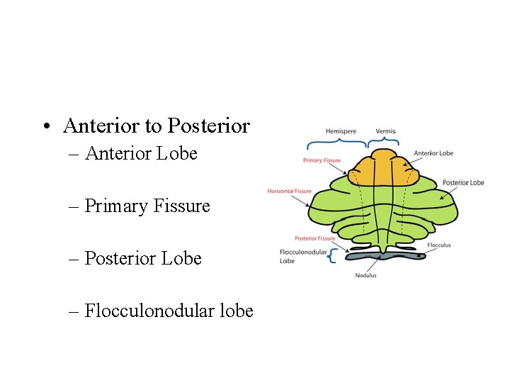
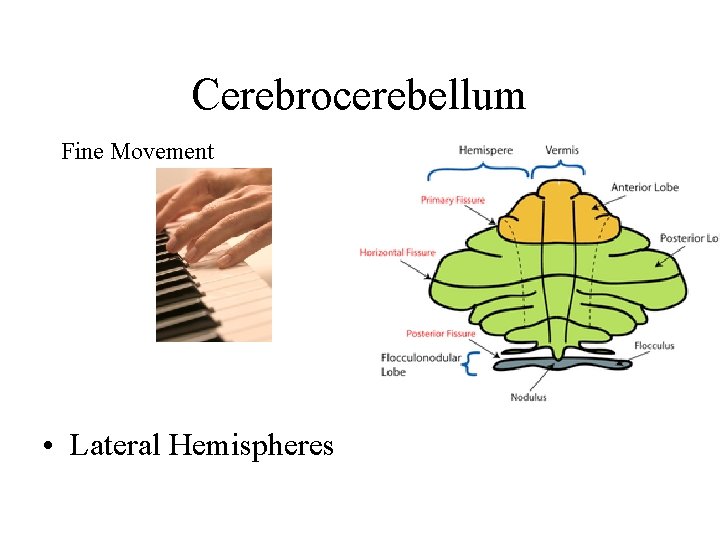
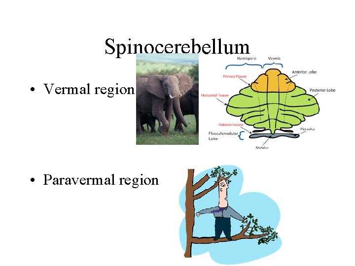
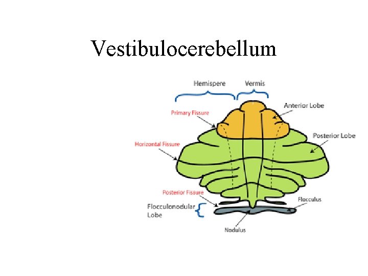
Gross Anatomy of the Cb • We can divide the Cb two different ways • Medial to lateral – Vermis – Cerebellar hemispheres • Intermediate or paravermal region • Lateral region
• Anterior to Posterior – Anterior Lobe – Primary Fissure – Posterior Lobe – Flocculonodular lobe
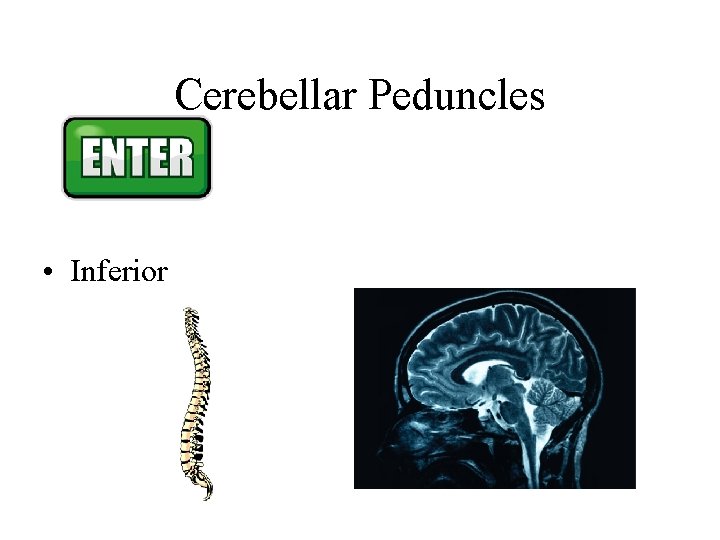
Cerebellar Peduncles • Inferior
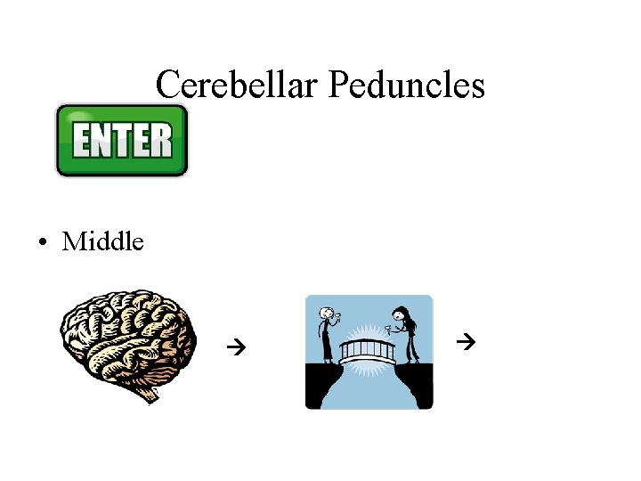
Cerebellar Peduncles • Middle
Cerebellar Peduncles • Superior
Cerebellar Peduncles • Inferior
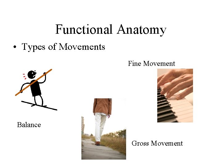
Functional Anatomy • Types of Movements Fine Movement Balance Gross Movement
Cerebrocerebellum Fine Movement • Lateral Hemispheres
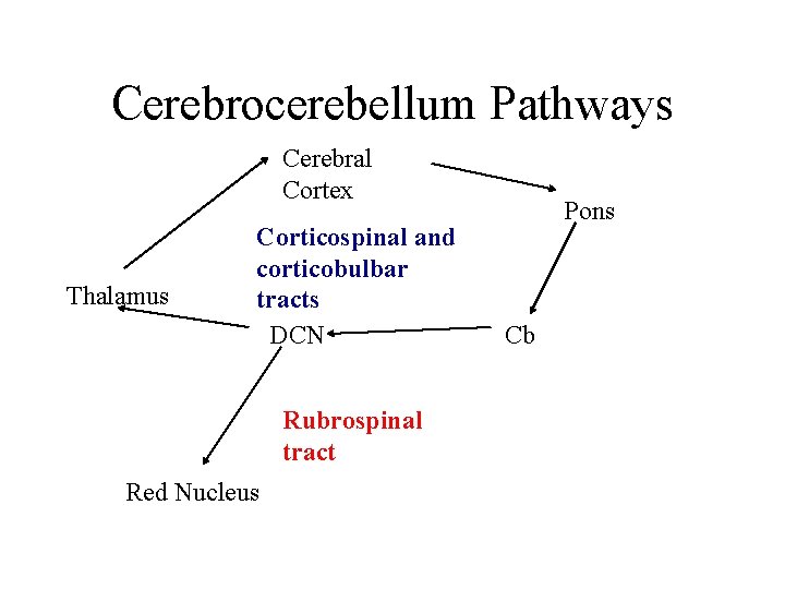
Cerebrocerebellum Pathways Cerebral Cortex Thalamus Corticospinal and corticobulbar tracts DCN Rubrospinal tract Red Nucleus Pons Cb
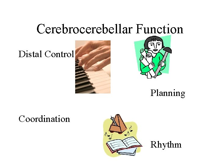
Cerebrocerebellar Function Distal Control Planning Coordination Rhythm
Spinocerebellum • Vermal region • Paravermal region
Vermal • Spinal cord • Vestibular N • Auditory + Vestibular BS
Vermal • Vestibular and Reticular N • Motor Cortex
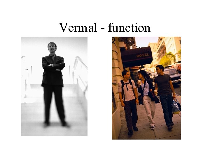
Vermal - function
Paravermal Spinal Cord Motor Cortex Red Nucleus
Vestibulocerebellum
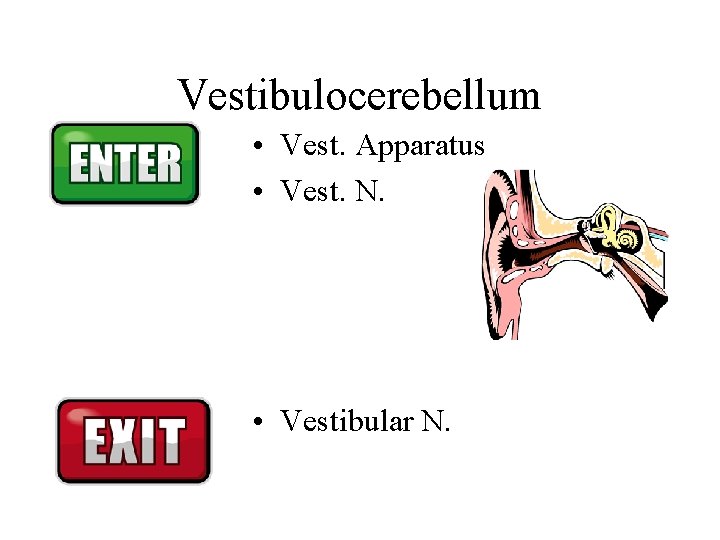
Vestibulocerebellum • Vest. Apparatus • Vest. N. • Vestibular N.
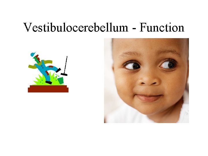
Vestibulocerebellum - Function
Cerebellar Disorders • Side • Tone
Ataxia • Truncal • Limb and Gait • Hand
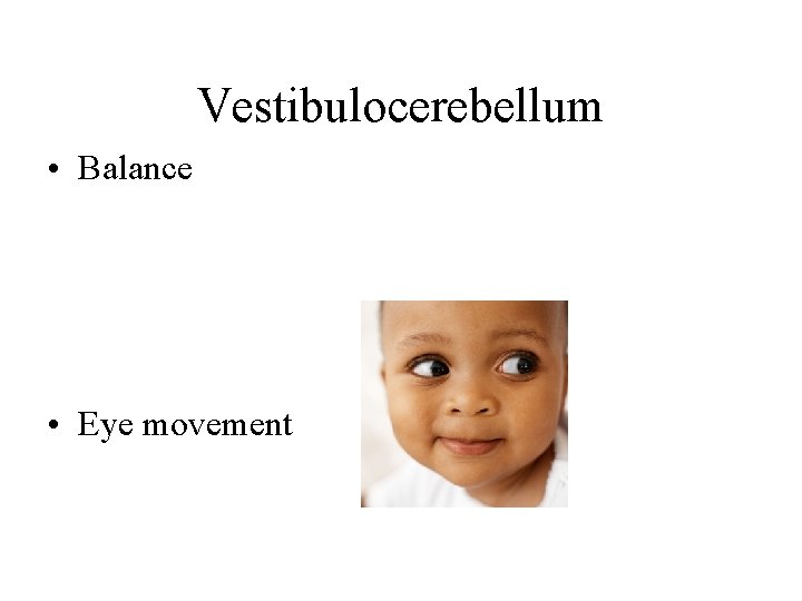
Vestibulocerebellum • Balance • Eye movement
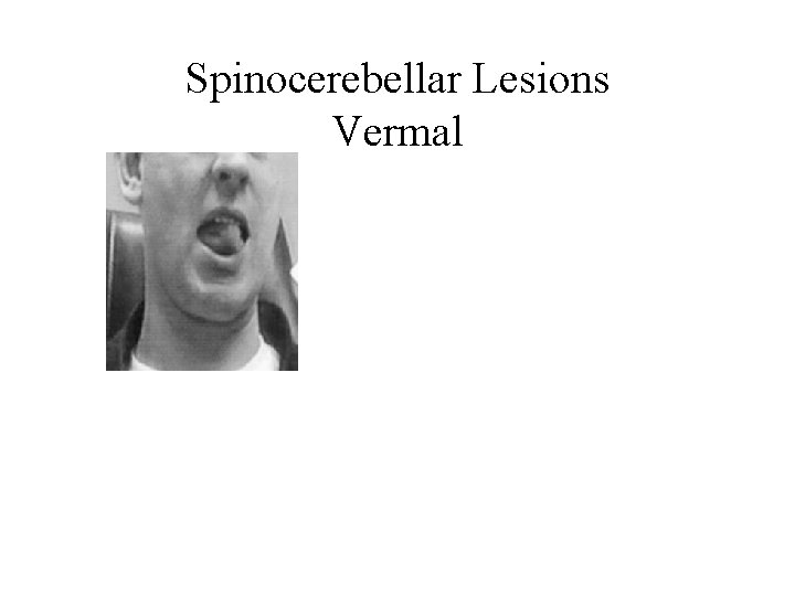
Spinocerebellar Lesions Vermal
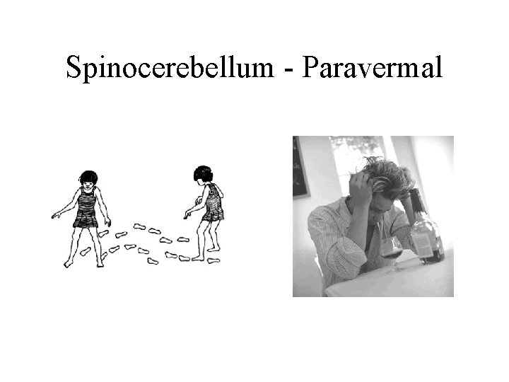
Spinocerebellum - Paravermal
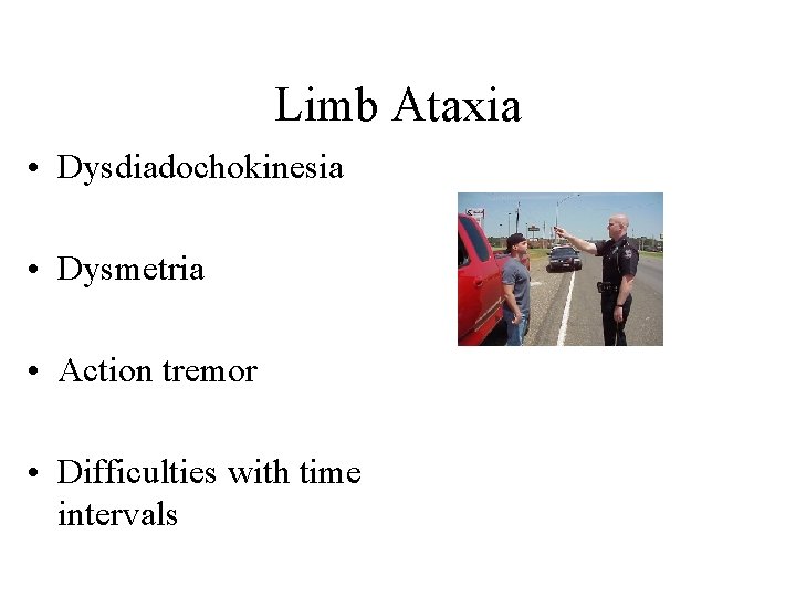
Limb Ataxia • Dysdiadochokinesia • Dysmetria • Action tremor • Difficulties with time intervals
Cerebrocerebellum • Dysarthria • Hand ataxia