A motor unit consists of one motor neuron


- Slides: 16
A motor unit consists of one motor neuron and all the muscle fibers it stimulates. This figure shows three motor units.
Motor units from different muscles will have different numbers of muscle fibers. • Large muscles (e. g. , quadriceps femoris) may have 300 -400 muscle fibers per motor unit. • Small muscles (e. g. external eye muscles) may have 1 muscle fiber per motor unit. • How does the precision of control we have over a muscle relate to the number of muscle fibers in its motor units?
Fig. 6. 17 Striated and smooth muscle.
Mature muscle fibers are very long and result from the fusion of many cells.
• Now go to your notes for some introductory information about the structure of muscle fibers.
Muscle fibers contain myofibrils, which are bundles of myofilaments. Each myofibril is surrounded by the sarcoplasmic reticulum.
Fig. 6. 28 also shows this arrangement.
A myofibril consists of a bundle of thick and thin myofilaments. This bundle is composed of repeating units called sarcomeres. Each myofibril is surrounded by the sarcoplasmic reticulum.
Each sarcomere is bounded by Z lines and consists of overlapping thin and thick myofilaments. The thin myofilaments are attached to the Z line and the thick myofilaments are anchored to the Z line by elastic myofilaments.
Fig. 6. 19 shows the sarcomere in the context of an EM photograph
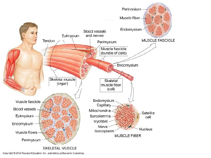
Review of levels of organization of skeletal muscle.
Structure of the thin filament.
Structure of the thick filament.
Fig. 6. 18 shows the structure of both myofilaments.
Muscle fibers shorten as each sarcomere shortens. Look at the distance between the Z lines.