DESCENDING TRACT OF SPINAL CORD 1 BY Dr
 BY Dr. zahid sarfaraz khan A/P ANATOMY KGMC")
DESCENDING TRACT OF SPINAL CORD (1) BY Dr. zahid sarfaraz khan A/P ANATOMY KGMC
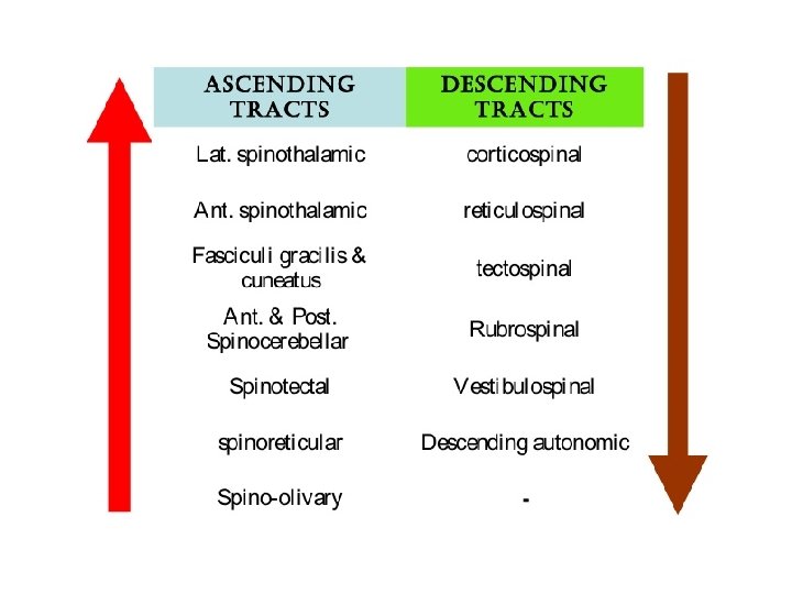

Descending tract of spinal cord

Pyramidal tracts The motor tracts can be functionally divided into two major groups 1 -Pyramidal tracts 2 -Extrapyramidal tracts 1 -Pyramidal tracts – These tracts originate in the cerebral cortex, Ø carrying motor fibres to the spinal cord and brain stem. Ø They are responsible for the voluntary control of the musculature of the body and face.

Extrapyramidal tracts • These tracts originate in the brain stem • Carrying motor fibres to the spinal cord. • They are responsible for the involuntary and automatic control of all musculature, such as muscle tone, balance, posture and locomotion • There are no synapses within the descending pathways. • At the termination of the descending tracts, the neurones synapse with a lower motor neurone. • Thus, all the neurones within the descending motor system are classed as upper motor neurones. Their cell bodies are found in the cerebral cortex or the brain stem, with their axons remaining within the CNS.

Pyramidal Tracts • The pyramidal tracts derive their name from the medullary pyramids of the medulla oblongata, which they pass through. • These pathways are responsible for the voluntary control of the musculature of the body and face. • Functionally, these tracts can be subdivided into two: • Corticospinal tracts – supplies the musculature of the body. • Corticobulbar tracts – supplies the musculature of the head and neck.

Extrapyramidal Tracts • The extrapyramidal tracts originate in the brainstem, carrying motor fibres to the spinal cord. • Responsible for the involuntary and automatic control of all musculature, such as muscle tone, balance, posture and locomotion. • There are four tracts in total. The vestibulospinal and reticulospinal tracts do not decussate, providing ipsilateral innervation. The rubrospinal and tectospinal tracts do decussate, and therefore provide contralateral innervation

The Descending Tracts of the Spinal Cord • The motor neurons situated in the anterior gray columns of the spinal cord • Send axons to innervate skeletal muscle through the anterior roots of the spinal nerves. • These motor neurons are sometimes referred to as the lower motor neurons and constitute the final common pathway to the muscles.

The Descending Tracts of the Spinal Cord • The lower motor neurons are constantly bombarded by nervous impulses • That descend from the medulla, pons, midbrain, and cerebral cortex • As well as those that enter along sensory fibers from the posterior roots. • The nerve fibers that descend in the white matter from different supraspinal nerve centers are segregated into nerve bundles called the descending tracts. • These supraspinal neurons and their tracts are sometimes referred to as the upper motor neurons, and they provide numerous separate pathways that can influence motor activity.

Anatomical Organization • Control of skeletal muscle activity from the cerebral cortex and other higher centers is conducted through the nervous system by a series of neurons. • The descending pathway from the cerebral cortex is often made up of three neurons. The first neuron • the first-order neuron, has its cell body in the cerebral cortex. • Its axon descends to synapse on the second-order neuron, an internuncial neuron, situated in the anterior gray column of the spinal cord.

Anatomical Organization • The axon of the second-order neuron is short • And synapses with the thirdorder neuron, the lower motor neuron, in the anterior gray column. • The axon of the third-order neuron innervates the skeletal muscle through the anterior root and spinal nerve. • In some instances, the axon of the first-order neuron terminates directly on the third order neuron (as in reflex arcs).

Functions of the Descending Tracts • The corticospinal tracts are the pathways concerned with voluntary, discrete, skilled movements, especially those of the distal parts of the limbs. • The reticulospinal tracts may facilitate or inhibit the activity of the alpha and gamma motor neurons in the anterior gray columns • And may, therefore, facilitate or inhibit voluntary movement or reflex activity.

• • Functions of the Descending Tracts The tectospinal tract is concerned with reflex postural movements in response to visual stimuli. Those fibers that are associated with the sympathetic neurons in the lateral gray column are concerned with the pupillodilation reflex in response to darkness. The rubrospinal tract acts on both the alpha and gamma motor neurons in the anterior gray columns And facilitates the activity of flexor muscles and inhibits the activity of extensor or antigravity muscles.

Functions of the Descending Tracts Vestibulospinal tract • by acting on the motor neurons in the anterior gray columns • Facilitates the activity of the extensor muscles • Inhibits the activity of the flexor muscles • And is concerned with the postural activity associated with balance. Olivospinal tract • May play a role in muscular activity, but there is doubt that it exists. Descending autonomic fibers Are concerned with the control of visceral activity.

Corticospinal Tracts • Fibers of the corticospinal tract arise as axons of pyramidal cells situated in the fifth layer of the cerebral cortex. • About one-third of the fibers originate from the primary motor cortex (area 4) • One-third originate from the secondary motor cortex (area 6) • And one-third originate from the parietal lobe (areas 3, 1, and 2); • thus, two-thirds of the fibers arise from the precentral gyrus, and one third of the fibers arise from the postcentral gyrus. • Because electrical stimulation of different parts of the precentral gyrus produces movements of different parts of the opposite side of the body • We can represent the parts of the body in this area of the cortex. Such a homunculus

Corticospinal Tracts • Note that the region controlling the face is situated inferiorly, • And the region controlling the lower limb is situated superiorly and on the medial surface of the hemisphere. • The homunculus is a distorted picture of the body • It is interesting to find that the majority of the corticospinal fibers are myelinated and are relatively slow-conducting, small fibers

Corticospinal Tracts • • The descending fibers converge in the corona radiata And then pass through the posterior limb of the internal capsule. Here, the fibers are organized so that those closest to the genu are concerned with cervical portions of the body While those situated more posteriorly are concerned with the lower extremity. The tract then continues through the middle three-fifths of the basis pedunculi of the midbrain. Here, the fibers concerned with cervical portions of the body are situated medially while those concerned with the leg are placed laterally.

Corticospinal Tracts On entering the pons • The tract is broken into many bundles by the transverse pontocerebellar fibers. In the medulla oblongata • The bundles become grouped together along the anterior border to form a swelling known as the pyramid (hence the alternative name, pyramidal tract). At the junction of the medulla oblongata and the spinal cord Most of the fibers cross the midline at the decussation of the pyramids • And enter the lateral white column of the spinal cord to form the lateral corticospinal tract

Corticospinal Tracts • The remaining fibers do not cross in the decussation • But descend in the anterior white column of the spinal cord as the anterior corticospinal tract. • These fibers eventually cross the midline • And terminate in the anterior gray column of the spinal cord segments in the cervical and upper thoracic regions.

Corticospinal Tracts lateral corticospinal tract q Descends the length of the spinal cord q Its fibers terminate in the anterior gray column of all the spinal cord segments. q Most corticospinal fibers synapse with internuncial neurons, q which, in turn, synapse with alpha motor neurons and some gamma motor neurons.

Corticospinal Tracts Branches 1. Branches are given off early in their descent • And return to the cerebral cortex to inhibit activity in adjacent regions of the cortex. • Branches pass to the caudate and lentiform nuclei, the red nuclei, and the olivary nuclei and the reticular formation. • These branches keep the subcortical regions informed about the cortical motor activity. • Once alerted, the subcortical regions may react • And send their own nervous impulses to the alpha and gamma motor neurons by other descending pathways.

- Slides: 22