TRACTS DR SHUBHANGI SAXENA TRACTS Ascending tracts Which

TRACTS DR. SHUBHANGI SAXENA

TRACTS Ascending tracts Which carry sensory impulse from spinal cord to brain Descending tracts which carry motor impulse from brain to spinal cord

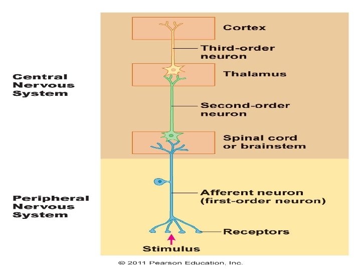
NEURONS • 1 ST Order neuron • 2 nd order neuron • 3 rd order neuron

• 1 st order neuron : receive sensory impulses from the receptors and send them to sensory neurons present in the posterior gray horn of spinal cord through their fibers • The nerve cell bodies of these neurons are located in the posterior nerve root ganglion

• 2 nd order neuron : are the sensory neurons present in the posterior gray horn. • The fibers from these neurons form the ascending tracts of spinal cord. • These fibers carry impulses from spinal cord to different brain areas below cerebral cortex (subcortical areas) such as thalamus. • All ascending tracts are formed by fibers of second order neurons.

• 3 rd order neuron: are in the subcortical areas. • The fibers of these neurons carry the sensory impulse from subcortical areas to cerebral cortex
 white column Anterior white column")
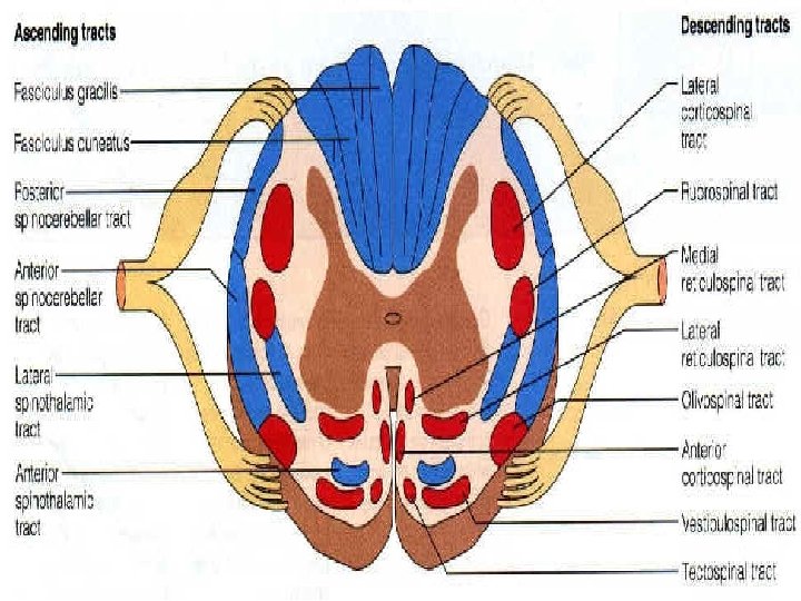
ASCENDING TRACTS Lateral white column Tracts in dorsal (posterior) white column Anterior white column

Lateral white column Lateral spinothalmic tract Ventral spinocerebellar tract Dorsal spinocerebellar tract Spinotectal tract Spinoreticular tract Spinoolivary tract Spinovestibular tract

POSTERIOR WHITE COLUMN • Fasciculus gracilis • Fasciculus cuneatus • Comma tract of schultze

ANTERIOR WHITE COLUMN • Anterior spinothalamic tract

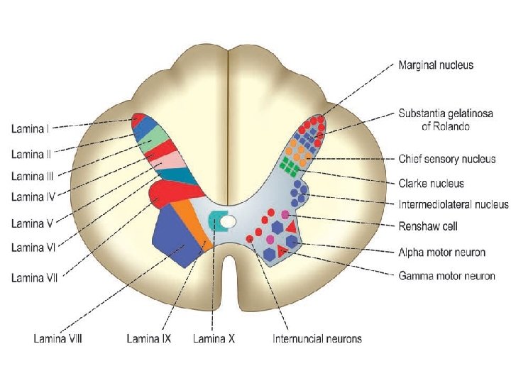
Section of spinal cord

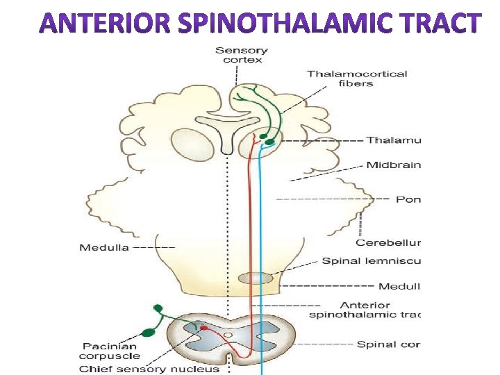
• LOCATION/ SITUATION : anterior white funiculus near the periphery. • ORIGIN : Are formed by the axons of chief sensory cells of posterior grey horn which forms the second order sensory neurons. The 1 st order neurons of this pathway are located in spinal ganglia (post nerve root ganglia)

The neurons receive the impulse from the cutaneous receptors. • COURSE : The tract contain crossed fibers. After taking origin from the chief sensory cells The fibers of anterior spinothalamic tract Cross obliquely in anterior white commissure

And enter the anterior white column of opposite side. Here, the fiber ascends through other segments of spinal cord and brainstem (medulla, pons, midbrain) And reach thalamus.

TERMINATION : fibers of this tract terminate in the ventral posterolateral nucleus of thalamus The cell of this thalamic nucleus form the 3 rd order neuron of the pathway. The fibers from thalamic nucleus carry the impulses to sensory cortex of cerebral cortex.

FUNCTION : carry impulses of crude touch and pressure. EFFECT OF LESIONS : The bilateral lesion of this tract lead to loss of crude touch sensation and loss of sensations like itching and tickling. The unilateral lesion of this tract cause loss of crude touch sensation in opposite side below the level of lesion.
 • LOCATION/ SITUATION : this tract is situated")
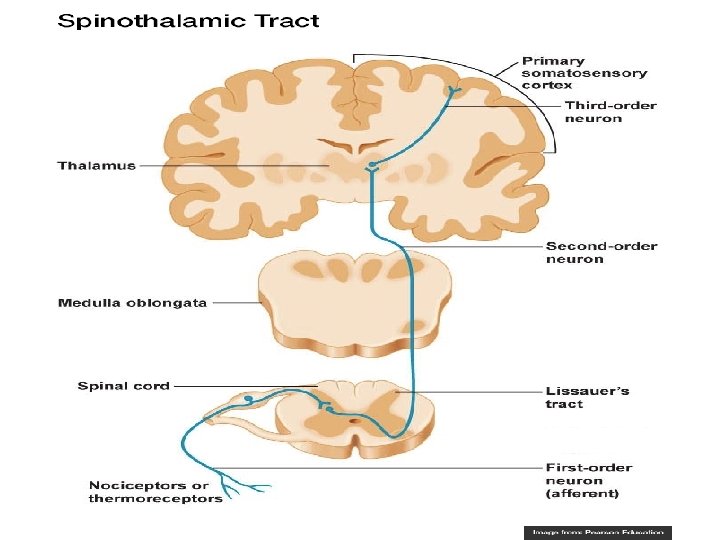
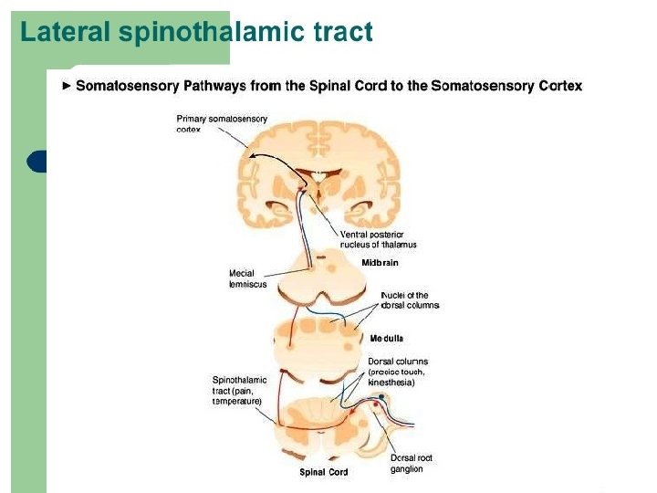
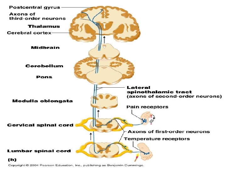
LATERAL SPINOTHALAMIC TRACT (Pain and temperature) • LOCATION/ SITUATION : this tract is situated in lateral column towards medial side. (near the gray matter) • ORIGIN : the fibers of this tract take origin from 2 sources: I. MARGINAL NUCLEUS II. SUBSTANTIA GELATINOSA OF ROLANDO

• COURSE : The tract contain crossed fibers. Axons from marginal nucleus and substantia gelatinosa Cross the opposite side And reach the lateral column of same segment

• Here, the fiber ascends through other segments of spinal cord and brainstem (medulla, pons, midbrain) • And reach thalamus along with the fibers of anterior spinothalamic tract

• TERMINATION: fibers of this tract terminate in the ventral posterolateral nucleus of thalamus The cell of this thalamic nucleus form the 3 rd order neuron of the pathway. The fibers from thalamic nucleus carry the impulses to sensory cortex of cerebral cortex.

• FUNCTION: carry impulse of pain and temperature. • EFFECT OF LESIONS: Bilateral – total loss of pain and temperature sensation on both the sides. Unilateral- loss of pain and temperature sensation on opposite side.
")
Spinothalamic tract (anterior or lateral)
 spinocerebellar tract / Gowers")
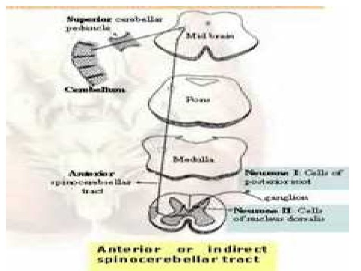
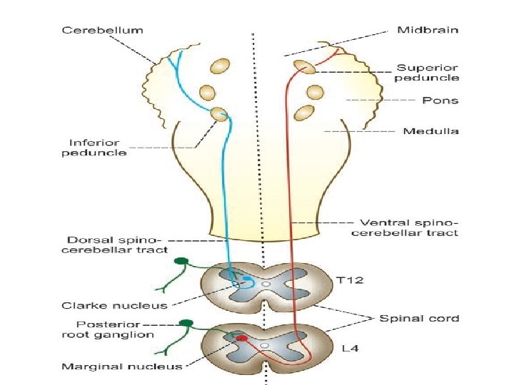
SPINOCEREBELLAR TRACTS • Responsible for subconscious kinesthetic sensation Ventral (anterior) spinocerebellar tract / Gowers tract dorsal (posterior) spinocerebellar tract / Flechsig’s tract

VENTRAL SPINOCEREBELLAR TRACT • LOCATION/ SITUATION: Lateral white funiculus of the spinal cord along the lateral periphery. • ORIGIN: Fibers originate from the marginal nucleus situated in the posterior gray horn of spinal cord.

• First order neuron are in the posterior root ganglia receive the impulses of proprioception from the proprioceptors in muscles, tendons and joints • COURSE: contain both crossed/uncrossed fibres. Majority of fibers from the marginal nucleus cross the midline and ascends in lateral white column of opposite side anterior to the fibers of dorsal spinocerebellar tract ( some fibers ascends in the lateral funiculus of the same side)

• Then these fibers ascends through spinal cord, medulla, pons and mid brain. • Finally, fibers reach the cerebellum through the superior cerebellar peduncle. TERMINATION: Fibers terminate in the cortex of an anterior lobe of cerebellum.

• FUNCTION: carries the impulses of subconscious kinesthetic sensation. • EFFECT OF LESION: loss of kinesthetic sensation in the opposite side

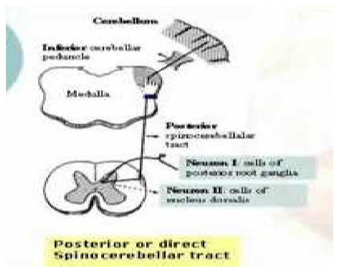
• LOCATION/ SITUATION: Lateral column along the posterolateral periphery of spinal cord. • ORIGIN: Fibers originate from the dorsal nucleus of clarke situated in the posterior gray horn of spinal cord.

• COURSE: contain uncrossed fibres. the axons from the neurons in dorsal nucleus of clarke reach lateral column of same side. Then the fibers ascends through the other spinal segment and reach medulla oblongata.

• Finally, fibers reach the cerebellum through the inferior cerebellar peduncle. TERMINATION: Fibers terminate in the cortex of anterior lobe of cerebellum. FUNCTION: carries the impulses of subconscious kinesthetic sensation.

• EFFECT OF LESION: unilateral loss of kinesthetic sensation occur in the lesion of this tract on the same side, as this tract has uncrossed fibers.
 • Fasciculus cuneatus (tract of Burdach)")
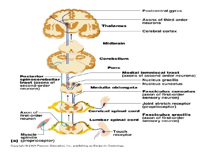
• Fasciculus gracilis ( tract of Goll) • Fasciculus cuneatus (tract of Burdach) • These 2 tracts are together called ascending posterior column tract. LOCATION: posterior white funiculus of spinal cord. Fasciculus gracilis is situated medial to Fasciculus cuneatus

• ORIGIN: These fibers are unique in that they are formed predominantly by the axons of 1 st order neuron. • ARRANGEMENT OF FIBERS: The fibers derived from the lowest ganglia are situated most medially while these from the highest ganglia are most lateral therefore Fasciculus gracilis which lies medially is composed of fibers from the coccygeal, sacreal, lumbar, and lower thoracic ganglia.

Fasciculus cuneatus : which lies laterally is composed of fibers from the upper thoracic and cervical ganglia. • COURSE: uncrossed fibers After entering the spinal cord, the fibers ascends through the posterior white column. these fibers does not synapse in the spinal cord.

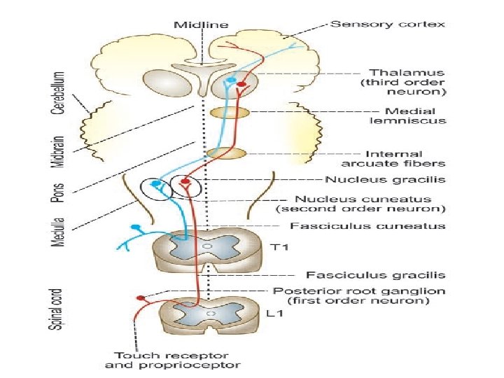
• TERMINATION: These fibers terminate in medulla oblangata. • Fasciculus gracilis- nucleus gracilis in medulla. • Fasciculus cuneatus-nucleus cuneatus. The cells of these medullary nucleus form the 2 nd order neuron. Ascends through pons and midbrain. Terminate in ventral posterolateral nucleus of thalamus

From here fibers of 3 rd order neuron relay to sensory area of cerebral cortex. • FUNCTIONS: v Fine tactile sensation v Tactile localization (ability to locate the area of skin where the tactile stimulus is applied with closed eyes) v Tactile discrimination or two point discrimination (ability to recognize the two stimuli applied over the skin simultaneously with closed eyes). v Sensation of vibration (ability to perceive the vibrations from a vibrating tuning fork placed over bony prominence conducted to deep tissues through skin). It is the synthetic sense produced by combination of touch and pressure sensations.

v. Conscious kinesthetic sensation (sensation or awareness of various muscular activities in different parts of the body) v. Stereognosis (ability to recognize the known objects by touch with closed eyes). It is also a synthetic sense produced by combination of touch and pressure sensations

• EFFECTS OF LESIONS: following symptoms i. Loss of fine tactile sensation; however, crude touch sensation is normal ii. Loss of tactile localization iii. Loss of two point discrimination iv. Loss of sensation of vibration v. Astereognosis (inability to recognize known objects by touch while closing the eyes) vi. Lack of ability to differentiate the weight of different objects vii. Loss of proprioception (inability to appreciate the position and movement of different parts of the body) viii. Sensory ataxia or posterior column ataxia (condition characterized by uncoordinated, slow and clumsy voluntary movements because of the loss of proprioception).

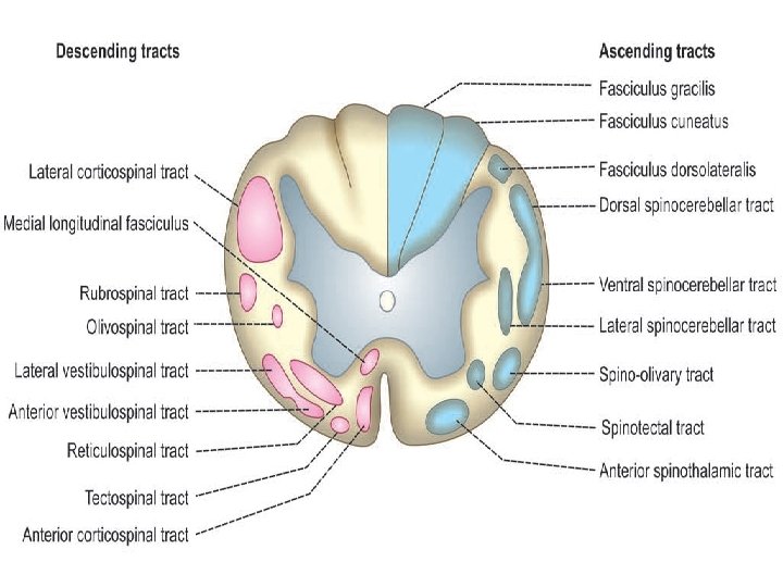
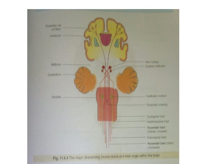
These tracts carry motor impulses from brain to spinal cord. Descending tracts of spinal cord are of two types: A. Pyramidal tracts B. Extrapyramidal tracts

PYRAMIDAL TRACT • Pyramidal tracts of spinal cord are the descending tracts concerned with voluntary motor activities of the body. • These tracts are otherwise known as corticospinal tracts. • While running from cerebral cortex towards spinal cord, the fibers of these two tracts give the appearance of a pyramid on the upper part of anterior surface of medulla oblongata hence the name pyramidal tracts

PYRAMIDAL TRACTS Anterior corticospinal tract Lateral corticospinal tract

EXTRAPYRAMIDAL TRACTS • Descending tracts of spinal cord other than pyramidal tracts are called extrapyramidal tracts.

EXTRA PYRAMIDAL TRACTS Medial longitudinal fasciculus Anterior vestibulospinal tract Lateral vestibulospinal tract Reticulospinal tract Tectospinal tract Rubrospinal tract Olivospinal tract

PYRAMIDAL TRACT EXTRA PYRAMIDAL TRACT ORIGIN: Giant cells or Betz cells ORIGIN: areas in the CNS that in precentral gyrus of the are concerned with muscular motor cortex. movements and postures Premotor area, supplementary motor areas, somatosensory areas. Its axons pass without relay to They have many synapses in the spinal segment levels their descending path where they form synapses with either interneurons in the dorsal horn or directly with motor neuron.

PYRAMIDAL TRACT EXTRA PYRAMIDAL TRACT They have greater influence over motor neurons that control muscle involved in fine movements, particularly those of the fingers and hand They are more involved with coordination of the large muscle group used in the maintainance of opright posture, in locomotion, and in head and body movements when turning towards a specific stimulus Lesions of this tract produces spasticity in the muscle involved. Its lesion produces rigidity of the involved muscles

CORTICOSPINAL TRACT Anterior corticospinal tract lateral corticospinal tract LOCATION: Anterior corticospinal tract : anterior white column Lateral corticospinal tract: lateral white column

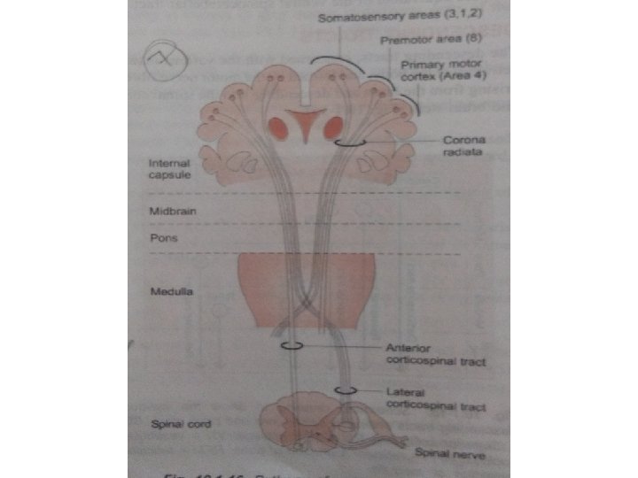
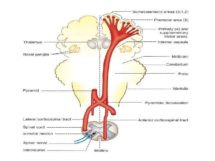
ORIGIN: Fibers of pyramidal tracts arise from following cells or areas of cerebral cortex: • Giant cells or Betz cells in precentral gyrus of the motor cortex. • Premotor area, supplementary motor areas, somatosensory areas. v It is believed that 30% of pyramidal fibers arise from primary motor area (area 4) and supplementary motor areas, v another 30% from premotor area (area 6) and v the remaining 40% of fibers arise from somatosensory areas.

All the above fibers form fibers of upper motor neurons of motor pathway. COURSE: After taking origin, the nerve fibers runs downwards towards brainstem and converge in the form of a fan like structure. (this fan like structure is called Corona radiata) (Corona radiata contain both ascending fibers from thalamus and descending fibers from cerebral cortex)

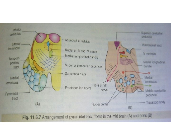
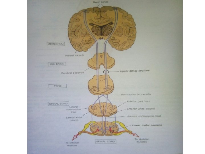
And reach internal capsule (lying between thalamus and caudate nucleus on the medial side and lenticular nucleus and basal ganglia on the lateral side) The fibers converge while descend down through internal capsule. Midbrain pons

While descending through pons, the fibers are divided into different bundles by the nuclei of pons. (means the tract is broken up into a series of scattered bundles). At lower border of pons, the fibers are grouped once again into a compact bundle and then descend down into medulla oblongata.

Medulla (The pyramidal tract were so called because of their shape as they pass along the surface of the medulla) In the lower part of medulla 80 -85% fibers cross to the opposite side, enter the lateral white column and descend down as LATERAL CORTICOSPINAL OR CROSSED/ INDIRECT PYRAMIDAL TRACT 15 -20% fibers do not cross, enter the anterior white column and descends down as ANTERIOR CORTICOSPINAL TRACT OR UNCROSSED/ DIRECT PYRAMIDAL TRACT

While crossing the midline, the fibers of both sides form the pyramidal decussation. TERMINATION: All the fibers of pyramidal tracts, both crossed and uncrossed fibers terminate in the motor neurons of anterior gray horn

Axons of the motor neurons leave the spinal cord as spinal nerves through anterior nerve roots and supply the skeletal muscles. v Neurons giving origin to the fibers of pyramidal tract are called the upper motor neurons. v Anterior motor neurons in the spinal cord are called the lower motor neurons.

• FUNCTIONS: v Lateral corticospinal tract. Concerned with voluntary movements, specially the distal limbs muscles and are concerned with fine, precise movements of the fingers and hands. v Anterior corticospinal tractconcerned with control of muscles of trunk and proximal portions of the limbs to carry out postural adjustment and gross movements.
 and parietal")
Cortico spinal fibers arising from the somatic sensory area (I and II) and parietal lobe association cortex are concerned with sensory motor coordination. eg: aiming the hand towards an object, hand eye coordination etc.

Extra pyramidal system is made up of those areas in the CNS that are concerned with muscular movements and postures. Its fibers having many synapse in their descending path.

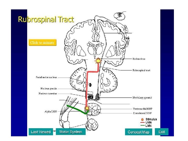
RUBROSPINAL TRACT • LOCATION: lateral white column of spinal cord. • ORIGIN: the fibers of this tract arises from the large cells (nucleus magnocellularis) of red nucleus in midbrain.

• COURSE: After arising from the red nucleus Fibers cross to opposite side in the lower part of tegmentum of mid brain (ventral tegmental decussation) Descend down to pons and medulla

Then follow a course similar to that of lateral corticospinal trat in the lateral luniculus of the spinal cord TERMINATION: Fibers end in the anterior motor neurons of spinal cord. FUNCTION: Exhibits facilitatory influence upon the flexor muscles tone and inhibitory influence on the extensor muscles of the body.

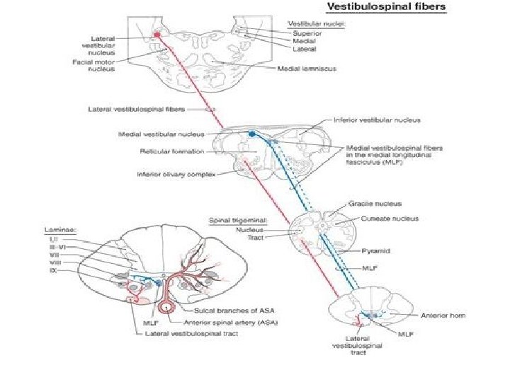
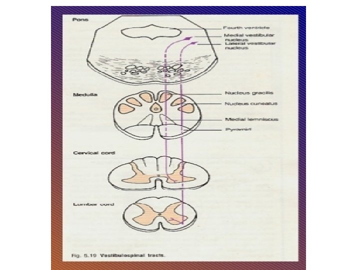
Anterior/medial vestibulospinal tract latersl vestibulospinal tract

ANTERIOR/ MEDIAL VESTIBULOSPINAL TRACT LATERAL VESTIBULOSPINAL TRACT LOCATION Anterior white column along the periphery of spinal cord Lateral white column of spinal cord ORIGIN Fibers arise from medial vestibular nucleus in medulla oblongata Fibers arise from lateral vestibular nucleus in medulla oblongata, this nucleus is also called as DEITER’S NUCLEUS COURSE Fibers run down from medulla into the anterior column of spinal cord Fibers from DEITER’S NUCLEUS run down into the lateral column of spinal cord All the fibers are uncrossed Mainly uncrossed fibers (very few fibers are crossed)

ANTERIOR/ MEDIAL VESTIBULOSPINAL TRACT LATERAL VESTIBULOSPINAL TRACT TERMINATION Anterior motor neuron FUNCTION This tract provides a reflex pathway for the movement of head, neck, eyes in response to visual and auditory stimulus Concerned with adjustment of postural muscles and muscle tone Lateral facilitates activity of extensor muscles and inhibits the activity of flexor muscles in association with the maintenance of balance
 RETICULOSPINAL TRACT LATERAL (MEDULLARY) RETICULO SPINAL TRACT")
RETICULO SPINAL TRACT MEDIAL (PONTINE) RETICULOSPINAL TRACT LATERAL (MEDULLARY) RETICULO SPINAL TRACT
 RETICULO SPINAL TRACT LATERAL (MEDULLARY) RETICULOSPINAL TRACT LOCATION Anterior white column of")
MEDIAL (PONTINE) RETICULO SPINAL TRACT LATERAL (MEDULLARY) RETICULOSPINAL TRACT LOCATION Anterior white column of spinal cord ORIGIN and COURSE Fibers arise from medial pontine reticular formation Fibers arise from gigantocellular component of medullary reticular formation predominantly uncrossed and only few fibers are crossed. These fibers descends in lateral part of anterior column Mostly uncrossed descends in medial part of anterior column TERMINAT Laminae vii and viii of spinal gray ION matter and throug internucial neurons influence alpha and gamma neurons of laminae ix internucial neurons of laminae vii, viii and ix of spinal cord

FUNCTIONS: v The pontine and medullary reticular nuclei mostly function antagonistic to each other. v pontine nuclei are excitatory to antigravity muscles and medullary nuclei are inhibitory. v pontine nuclei facilitate while medullary nuclei inhibits the control of voluntary and reflex movements and control of muscle tone through gamma neurons.

v Pontine reticular formation favours expiration while medullary reticular formation favours inspiration. v pontine nuclei cause vasoconstriction and medullary cause vasodilatation.
- Slides: 87