Figure 22 2 Gastrointestinal tract activities Ingestion Mechanical
 • Churning")


 Submucosa")
. Upper lip Gingivae (gums)")

.")
. Soft palate Palatoglossal arch")
. Bolus of food Tongue Uvula Pharynx Bolus Epiglottis Glottis")
. (4 of 5) Relaxed muscles 4 Peristalsis moves food")
. (5 of 5) Relaxed muscles 5 The gastroesophageal sphincter")


 flexure")

- Slides: 29
Figure 22. 2 Gastrointestinal tract activities. Ingestion Mechanical breakdown • Chewing (mouth) • Churning (stomach) • Segmentation (small intestine) Digestion Food Pharynx Esophagus Propulsion • Swallowing (oropharynx) • Peristalsis (esophagus, stomach, small intestine, large intestine) Stomach Absorption Lymph vessel Small intestine Large intestine Defecation © 2014 Pearson Education, Inc. Blood vessel Mainly H 2 O Feces Anus
Figure 22. 3 a Peristalsis and segmentation. From mouth Peristalsis: Adjacent segments of alimentary tract organs alternately contract and relax, moving food along the tract distally. © 2014 Pearson Education, Inc.
Figure 22. 3 b Peristalsis and segmentation. Segmentation: Nonadjacent segments of alimentary tract organs alternately contract and relax, moving food forward then backward. Food mixing and slow food propulsion occur. © 2014 Pearson Education, Inc.
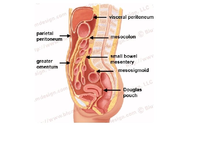
Figure 22. 5 a The peritoneum and the peritoneal cavity. Abdominopelvic cavity Vertebra Dorsal mesentery Parietal peritoneum Ventral mesentery Visceral peritoneum Peritoneal cavity Alimentary canal organ Liver Two schematic cross sections of abdominal cavity illustrate the peritoneums and mesenteries. © 2014 Pearson Education, Inc.
Figure 18. 24 a Arteries of the abdomen. Diaphragm Abdominal aorta Inferior phrenic arteries L. gastric artery R. gastric artery Common hepatic artery Hepatic artery proper L Splenic artery Gastroduodenal artery R Celiac trunk R. gastroepiploic artery Middle suprarenal arteries L. gastroepiploic artery Intestinal arteries Middle colic artery Superior mesenteric artery R. colic artery Renal arteries Gonadal arteries Ileocolic artery Sigmoidal arteries Inferior mesenteric artery L. colic artery Lumbar arteries Superior rectal artery Median sacral artery Common iliac arteries © 2014 Pearson Education, Inc. Schematic flowchart.
Figure 18. 29 a Veins of the abdomen. Inferior vena cava Cystic vein Hepatic portal system Inferior phrenic veins Hepatic portal vein Superior mesenteric vein Splenic vein Suprarenal veins Renal veins Inferior mesenteric vein Gonadal veins Lumbar veins R. ascending lumbar vein Common iliac veins L. ascending lumbar vein External iliac vein Internal iliac veins © 2014 Pearson Education, Inc. Schematic flowchart.
Figure 22. 6 Basic structure of the alimentary canal. Intrinsic nerve plexuses • Myenteric nerve plexus • Submucosal nerve plexus Glands in submucosa Mucosa • Epithelium • Lamina propria • Muscularis mucosae Submucosa Muscularis externa • Longitudinal muscle • Circular muscle Mesentery Nerve Gland in mucosa Artery Vein Duct of gland outside Lymphatic vessel alimentary canal © 2014 Pearson Education, Inc. Serosa • Epithelium (mesothelium) • Connective tissue Lumen Mucosa-associated lymphoid tissue
Figure 19. 2 a The lymphatic system. Regional lymph nodes: Internal jugular vein Entrance of right lymphatic duct into vein Cervical nodes Entrance of thoracic duct into vein Thoracic duct Axillary nodes Cisterna chyli Aorta Collecting lymphatic vessels Drained by the right lymphatic duct Drained by the thoracic duct Inguinal nodes General distribution of collecting lymphatic vessels and regional lymph nodes. © 2014 Pearson Education, Inc.
Figure 22. 12 a Microscopic structure of the esophagus. Mucosa (stratified squamous epithelium) Submucosa (areolar connective tissue) Lumen Muscularis externa • Circular layer • Longitudinal layer © 2014 Pearson Education, Inc. Adventitia (fibrous connective tissue)
Figure 22. 7 b Anatomy of the oral cavity (mouth). Upper lip Gingivae (gums) Palatine raphe Hard palate Soft palate Uvula Palatine tonsil Superior labial frenulum Palatoglossal arch Palatopharyngeal arch Posterior wall of oropharynx Tongue Sublingual fold with openings of sublingual ducts Oral vestibule Lower lip Anterior view © 2014 Pearson Education, Inc. Lingual frenulum Opening of Submandibular duct Gingivae (gums) Inferior labial frenulum
Figure 22. 9 The salivary glands. Tongue Teeth Ducts of sublingual gland Frenulum of tongue Sublingual gland Parotid duct Masseter muscle Body of mandible (cut) Posterior belly of digastric muscle Mylohyoid muscle (cut) Submandibular duct Anterior belly of digastric muscle Submandibular gland © 2014 Pearson Education, Inc. Mucous cells Serous cells forming demilunes
Figure 22. 11 Longitudinal section of a canine tooth within its bony socket (alveolus). Enamel Dentin Crown Neck Dentinal tubules Pulp cavity (contains blood vessels and nerves) Gingival sulcus Gingiva (gum) Cement Root canal Periodontal ligament Apical foramen © 2014 Pearson Education, Inc. Bone
Figure 22. 7 a Anatomy of the oral cavity (mouth). Soft palate Palatoglossal arch Uvula Hard palate Oral cavity Palatine tonsil Tongue Oropharynx Lingual tonsil Epiglottis Hyoid bone Laryngopharynx Esophagus Trachea © 2014 Pearson Education, Inc. Sagittal section of the oral cavity and pharynx
Figure 22. 13 Deglutition (swallowing). Bolus of food Tongue Uvula Pharynx Bolus Epiglottis Glottis Trachea Esophagus 1 During the buccal phase, the upper esophageal sphincter is contracted. The tongue presses against the hard palate, forcing the food bolus into the oropharynx. Relaxed muscles 2 The pharyngeal-esophageal phase begins as the uvula and larynx rise to prevent food from entering respiratory passageways. The tongue blocks off the mouth. The upper esophageal sphincter relaxes, allowing food to enter the esophagus. 4 Peristalsis moves food through the esophagus to the stomach. Circular muscles contract Upper esophageal sphincter Bolus 3 The constrictor muscles of the pharynx contract, forcing food into the esophagus inferiorly. The upper esophageal sphincter contracts (closes) after food enters. Relaxed muscles 5 The gastroesophageal sphincter surrounding the cardial oriface opens, and food enters the stomach. Bolus of food Longitudinal muscles contract Circular muscles contract Gastroesophageal sphincter closed Gastroesophageal sphincter opens Stomach © 2014 Pearson Education, Inc.
Figure 22. 13 Deglutition (swallowing). (4 of 5) Relaxed muscles 4 Peristalsis moves food through the esophagus to the stomach. Circular muscles contract Bolus of food Longitudinal muscles contract Gastroesophageal sphincter closed Stomach © 2014 Pearson Education, Inc.
Figure 22. 13 Deglutition (swallowing). (5 of 5) Relaxed muscles 5 The gastroesophageal sphincter surrounding the cardial oriface opens, and food enters the stomach. Circular muscles contract Gastroesophageal sphincter opens © 2014 Pearson Education, Inc.
Figure 22. 14 a Anatomy of the stomach. Cardia Fundus Esophagus Muscularis externa • Longitudinal layer • Circular layer • Oblique layer Serosa Body Lumen Lesser curvature Rugae of mucosa Greater curvature Duodenum © 2014 Pearson Education, Inc. Pyloric sphincter (valve) at pylorus Pyloric canal Pyloric antrum
Figure 22. 15 b Microscopic anatomy of the stomach. Gastric pits Surface epithelium (mucous cells) Gastric pit Mucous neck cells Parietal cell Gastric gland Chief cell Enteroendocrine cell © 2014 Pearson Education, Inc. Enlarged view of gastric pits and gastric glands
Figure 22. 22 a Structural modifications of the small intestine that increase its surface area for digestion and absorption. Vein carrying blood to hepatic portal vessel Muscle layers Circular folds Villi © 2014 Pearson Education, Inc. Lumen
Figure 22. 22 b Structural modifications of the small intestine that increase its surface area for digestion and absorption. Microvilli (brush border) Absorptive cells Lacteal Goblet cell Blood capillaries Mucosaassociated lymphoid tissue Intestinal crypt Muscularis mucosae Duodenal gland © 2014 Pearson Education, Inc. Villus Enteroendocrine cells Venule Lymphatic vessel Submucosa
Figure 22. 29 a Gross anatomy of the large intestine. Left colic (splenic) flexure Transverse mesocolon Right colic (hepatic) flexure Epiploic appendages Transverse colon Superior mesenteric artery Haustrum Descending colon Ascending colon IIeum Cut edge of mesentery IIeocecal valve Tenia coli Sigmoid colon Cecum Appendix Rectum Anal canal © 2014 Pearson Education, Inc. External anal sphincter
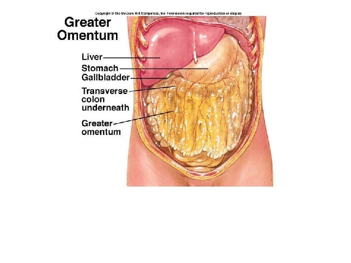
Figure 22. 30 c Mesenteries of the abdominal digestive organs. Greater omentum Transverse colon Transverse mesocolon Descending colon Jejunum Mesentery Sigmoid mesocolon Sigmoid colon Ileum © 2014 Pearson Education, Inc.