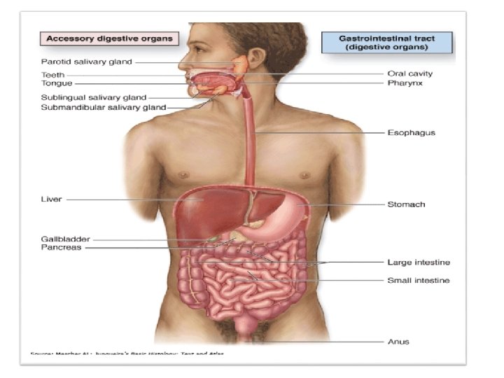
Digestive System General Structure of the Digestive Tract


- Slides: 32
Digestive System
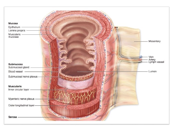
General Structure of the Digestive Tract • Mucosa: • Epithelium • Lamina propria • Muscularis mucosa • Submucosa: • Glands • Bl. Vs • Submucosal nerve plexus • Muscularis: • Inner circular • Outer longitudinal • Serosa: • C. T covered by mesothelium • If mesothelium is absent Adventitia
The Oral Cavity • The oral cavity is lined with stratified squamous epithelium, keratinized or nonkeratinized, depending on the region. • The keratin layer protects the oral mucosa from damage during masticatory function and is best developed on the dorsum side of the tongue and hard palate. The lamina propria in these regions has many papillae and rests directly on bony tissue. • Nonkeratinized squamous epithelium covers the soft palate, lips, cheeks, and the floor of the mouth.
Esophagus It is a muscular tube whose function is to transport food from the mouth to the stomach. • It is lined by nonkeratinized stratified squamous epithelium. • In the submucosa are groups of small mucus-secreting glands, the esophageal glands, secretions of which facilitate the transport of foodstuffs and protect the mucosa.
Esophagus • In the proximal third of the esophagus the muscularis is e skeletal muscle. • The middle third contains a combination of skeletal and smooth muscle. • In the distal third the muscularis contains only smooth muscle. • Also, only the most distal portion of the esophagus is covered by serosa. • The rest is enclosed by a layer of loose connective tissue, the adventitia, which blends into the surrounding tissue
Esophagus
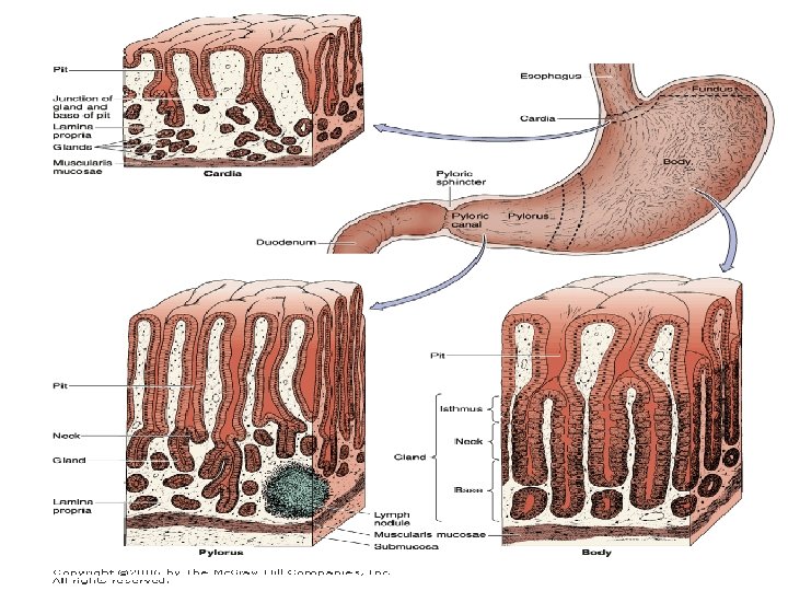
Stomach • It is a dilated segment of the digestive tract whose main functions are to continue the digestion of carbohydrates, add an acidic fluid to the ingested food, promote the initial digestion of proteins with the enzyme pepsin. And gastric lipase that digests triglycerides. • The food is transformed by it’s muscular activity into a viscous mass (chyme). • Gross inspection reveals four regions: cardia, fundus, body, and pylorus.
Stomach • The mucosa and submucosa of the empty stomach have longitudinally directed folds known as rugae, which flatten when the stomach is filled with food. The wall in all regions of the stomach is made up of all four major layers. • the mucosa of the stomach consists of a simple columnar surface epithelium that invaginates into the lamina propria, forming gastric pits. • Emptying into the gastric pits are branched, tubular glands. • The vascularized lamina propria that surrounds and supports these pits and glands contains smooth muscle fibers and lymphoid cells. • Separating the mucosa from the underlying submucosa is a layer of smooth muscle, the muscularis mucosae.
cells of the gastric glands The cells of the gastric glands provide key stomach functions. Important properties of each are as follows: • Parietal cells are present mainly in the upper half of gastric glands central spherical nucleus and cytoplasm that is intensely eosinophilic due to the high density of mitochondria. Parietal cells secrete both hydrochloric acid (HCl) and intrinsic factor, a glycoprotein required for uptake of vitamin B 12 in the small intestine. • Chief cells : near the muscularis mucosae seen as clusters of cells with condensed, basal nuclei and basophilic cytoplasm. Their apical ends chief cells secrete pepsinogen (the zymogen precursor for pepsin, a major protease).
Stomach h
A. Representation of the structure of the gastric mucosa showing a section through the wall of the stomach. B. Detail of the structure of gastric glands and cell types in the mucosa
Stomach h
Other Layers of the Stomach • The submucosa is composed of connective tissue containing blood and lymph vessels; it is infiltrated by lymphoid cells, macrophages, and mast cells. • The muscularis is composed of smooth muscle fibers oriented in three main directions. The external layer is longitudinal, the middle layer is circular, and the internal layer is oblique. • The stomach is covered by a thin serosa.
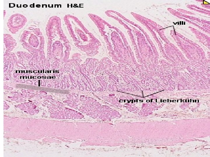
Small Intestine • The small intestine is the site of terminal food digestion, nutrient absorption, and endocrine secretion. • The processes of digestion are completed in the small intestine, where the nutrients are absorbed by cells of the epithelial lining. • The small intestine is relatively long (approximately 5 m) and consists of three segments: duodenum, jejunum, and ileum.
Small Intestine • Mucous Membrane: with the naked eye, the lining of the small intestine shows a series of permanent circular or semilunar folds (plicae circulares), consisting of mucosa and submucosa. • Intestinal villi are mucosal outgrowths (epithelium plus lamina propria) and project into the lumen. • Villi are covered by a simple columnar epithelium of absorptive cells and goblet cells. • Enterocytes are the absorptive cells, are tall columnar cells, each with an oval nucleus in the basal half of the cell. With densely packed microvilli.
Small Intestine
Small Intestine
Lamina Propria through Serosa • The lamina propria of the small intestine is composed of loose connective tissue with blood and lymph vessels, nerve fibers, and smooth muscle cells. • Smooth muscle fibers inside the villi are responsible for their rhythmic movements, which are important for efficient absorption. The muscularis mucosae also produces local movements of the villi and plicae circulares.
Lamina Propria through Serosa • The proximal part of the duodenum has primarily in its submucosa but extending into the mucosa, large clusters of branched tubular mucous glands, the duodenal (or Brunner) glands. The product of the glands is distinctly alkaline which neutralizes chyme entering the duodenum from the pylorus, protecting the mucous membrane. • In the ileum both the lamina propria and submucosa contain the lymphoid nodule aggregates known as Peyer patches. • The muscularis is well developed in the small intestine, composed of an internal circular layer and an external longitudinal layer, and is covered by a thin serosa with mesothelium
• Submucosal glands – Brunner’s gland
Jejunum
Ileum
Ileum
Large Intestine • The large intestine or bowel consists of a mucosal membrane with No villi. • The mucosa is penetrated throughout its area by tubular intestinal glands lined by goblet and absorptive cells, with a small number of enteroendocrine cells. • The absorptive cells or colonocytes are columnar and have short, irregular microvilli. • The large intestine is well suited to its main functions: absorption of water, formation of the fecal mass from undigestible material, and production of mucus that lubricates the intestinal surface.
Large Intestine
Large Intestine • The lamina propria is rich in lymphoid cells and in lymphoid nodules that frequently extend into the submucosa. • The muscularis comprises longitudinal and circular strands, but differs from that of the small intestine, with fibers of the outer layer gathered in three longitudinal bands called taeniae coli. • The colon are covered by serosa, which is characterized by small, pendulous protuberances of adipose tissue.
Large Intestine
THANK YOU