Gastrointestinal tract activities GI tract activities include ingestion
 digestion propulsion,")
")
 A lateral view, showing the relative positions of the salivary")

 This sequence based on a series of X rays, shows")
 A surface view of the gastric mucosa of the full")
 The organization of the stomach wall, (d) A gastric gland")

")
")
")
 A section of the intestine (LM X 2. 5), (b)")
 Internal structures in a single villus, showing the capillary and")
 The gross anatomy of the pancreas. The head of the pancreas")
 A single liver lobule and its cellular components, (b) A diagrammatic")
 A section through liver lobules from a pig liver (LM X")
 A view of the inferior surface of the liver, showing the")


")


- Slides: 31
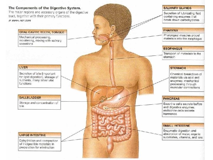
Gastrointestinal tract activities GI tract activities include ingestion, mechanical digestion, chemical (enzymatic) digestion propulsion, absorption, and defecation. Sites of chemical digestion are also sites that produce enzymes or that receive enzymes or other secretions made by acessory organs outside the alimentary canal. The mucosa of the GI tract secretes mucus, which protects and lubricates. (E. Marieb, HA&P, 2004)
The regulation of digestive activities The major factors responsible for regulating digestive activities: 1) neural mechanisms 2) hormonal mechanisms 3) local mechanisms (F. Martini, A&P, 2004)
The salivary glands (a) A lateral view, showing the relative positions of the salivary glands and ducts on the left side of the head (b) The submandibular gland secretes a mixture of mucins, produced by mucous cells, and enzymes, produced by serous cells (LM X 303) (c) (F. Martini, A&P, 2004)
The swallowing process This sequence based on a series of X rays, shows the stages of swallowing and the movement of materials from the mouth to the stomach. (F. Martini, A&P, 2004)
The swallowing process (continue) This sequence based on a series of X rays, shows the stages of swallowing and the movement of materials from the mouth to the stomach. (F. Martini, A&P, 2004)
The stomach lining (a) A surface view of the gastric mucosa of the full stomach, showing the entrances to the gastric pits (SEM X 35) (b) A section through gastric pits and gastric glands (LM X 300) (c) (F. Martini, A&P, 2004)
The stomach lining (c) The organization of the stomach wall, (d) A gastric gland (F. Martini, A&P, 2004)
The secretion of hydrochloric acid An active parietal cell generates H+ by the dissociation of carbonic acid within the cell. The bicarbonate is exchanged for Cl- in the interstitial fluid; the chloride ions diffuse into the lumen of the gastric gland as the hydrogen ions are transported out of the cell. (F. Martini, A&P, 2004)
The phase of the gastric secretion (F. Martini, A&P, 2004)
The phase of the gastric secretion (F. Martini, A&P, 2004)
The phase of the gastric secretion (F. Martini, A&P, 2004)
The intestinal wall. (a) A section of the intestine (LM X 2. 5), (b) A single plica and multiple villi, (c) The organization of the intestinal wall (F. Martini, A&P, 2004)
The intestinal wall (d) Internal structures in a single villus, showing the capillary and lymphatic supplies (e) A villus in sectional view (LM X 252) (F. Martini, A&P, 2004)
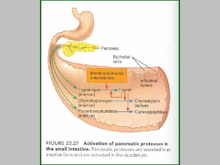
The pancreas. (a) The gross anatomy of the pancreas. The head of the pancreas is tucked into a C-shaped curve of the duodenum that begins at the pylorus of the stomach. The cellular organization of the pancreas is shown (b) diagrammatically and (c) in a micrograph (LM X 86) (F. Martini, A&P, 2004).
Liver histology. (a) A single liver lobule and its cellular components, (b) A diagrammatic view of liver structure, showing relationships among lobules (F. Martini, A&P, 2004).
Liver histology (c) A section through liver lobules from a pig liver (LM X 38) (d) A portal area (LM X 31) (F. Martini, A&P, 2004)
The gallbladder. (a) A view of the inferior surface of the liver, showing the position of the gallbladder and ducts that transport bile from the liver to the gallbladder and duodenum. (b) An interior view of the duodenum, showing the duodenum ampulla and related structures (F. Martini, A&P, 2004).
The activities of major digestive tract hormones The primary actions of gastrin, secretin, CCK, GIP and VIP (F. Martini, A&P, 2004)
Digestive secretion and absorption of water The green arrows indicate secretion and the white arrows indicate reabsorption. (F. Martini, A&P, 2004)
Ion and vitamin absorption by the digestive track (F. Martini, A&P, 2004)
Protein digestion and absorption in the small intestine Proteins and protein fragments are digested to amino acids by the action of pancreatic proteases (trypsin, chymotrypsin, and carboxypeptidase), and by brush border enzymes of the intestinal mucosal cells. The amino acids are then absorbed by active transport into the capillary blood and villi. (E. Marieb, HA&P, 2004)
Role of bile salts in fat emulsification As large aggregates of fats enter the small intestine, bile salts cling via their nonpolar parts to the fat molecules (triglycerides). Their polar parts, facing the aqueous phase, interact with water and repel each other, causing the fatty globule to be physically broken up into small fat droplets. (E. Marieb, HA&P, 2004)
Fatty acid absorption Digestion products of fat breakdown, lecithin, and cholesterol associate with bile salts to form micelles, which serve to “ferry” them to the intestinal mucosa. They then dissociate and enter the mucosa cells by diffusion. In the mucosal epithelial cells, they are recombined to lipids and packaged with other lipoid substances and protein to form chylomicrons. The chylomicrons are extruded from the epithelial cells by exocytosis and enter the lacteal for distribution in the lymph. Free fatty acids and monoglycerides enter the capillary bed. (E. Marieb, HA&P, 2004)