Anatomy Physiology Ch 8 Special Senses The somatic


![Types of Receptors: • Chemoreceptors: respond to changes in [chemicals] • Pain receptors: respond](https://slidetodoc.com/presentation_image_h2/53631a3ca1fdf38ef9578136a5913bb5/image-3.jpg "Types of Receptors: • Chemoreceptors: respond to changes in [chemicals] • Pain receptors: respond")


: – The cornea (a")
: – Choroid coat which nourishes the tissues of the")
: – The retina which contains the photoreceptors")
 meaning it can change its")


- Slides: 29
Anatomy & Physiology Ch. 8: Special Senses
• The somatic senses are receptors associated with touch, pressure, temperature & pain • The special senses are receptors associated with the senses (touch, smell, hearing, taste, vision & equilibrium)
Types of Receptors: • Chemoreceptors: respond to changes in [chemicals] • Pain receptors: respond to tissue damage • Thermoreceptors: respond to changes in temperature • Mechanoreceptors: respond to changes in movement or pressure • Photoreceptors: respond to changes in light energy
The Eye & Vision: • The organs of sight are the eyes, the eyelids, & the lacrimal apparatus • The eye orbit contains the above organs & fat, nerves, muscles, & blood vessels • The eyelids protect the eye (open & close) • The conjunctiva is within the eyelids that provides mucous (is a mucous membrane) to wash the eye.
• The lacrimal apparatus contains the lacrimal gland & a series of ducts that connect the eye to the nose & throat. This secretes tears. • This has 2 ducts which collect tears: – Lacrimal sac flows into the: – Nasolacrimal duct (empties into nasal cavity) • Tears have lysozymes (enzymes that aid in eye infection prevention) • There are 6 extrinisic muscles of the eyes, which allow for movements in all directions.
http: //members. aol. com/dcaronejr/ezmed/lacrimal. jpg
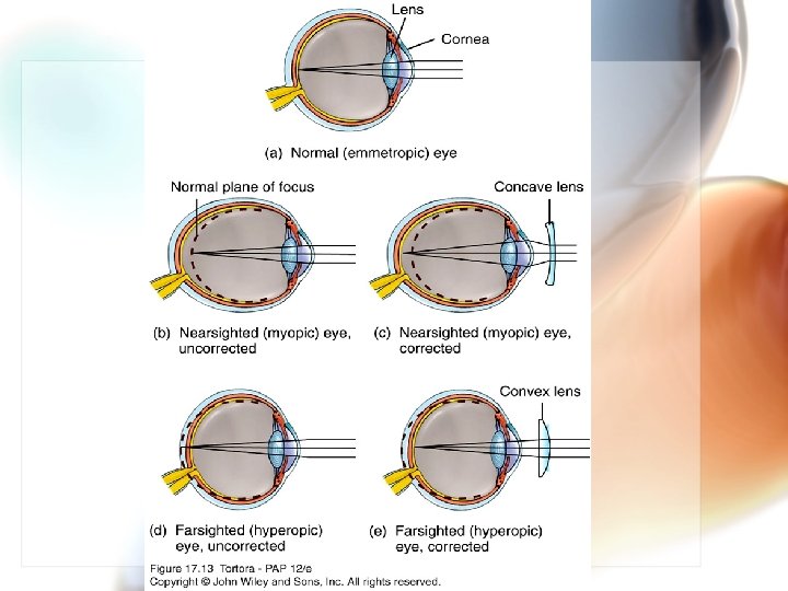
The Structure of the Eye: The Fibrous Layer (outer layer): – The cornea (a transparent, thin layer of epithelium that allows for light transmission into the eye) – The sclera which is connected to the cornea (the white part of the eye) which protects the eyes & is the attachment for the extrinsic muscles – The optic nerve is in the back of the eye & blood vessels which attaches to the sclera.
The Vascular Layer (middle layer): – Choroid coat which nourishes the tissues of the eye & provides the pigment (melanocytes) – The ciliary body forms the ring around the front of the eye; these hold the lens (transparent) in place – The iris (a muscle) is the colored portion of the eye (the lens is directly behind it) – The pupil is the opening of the eye that responds to light.
• The Sensory Layer (innermost layer): – The retina which contains the photoreceptors (visual receptor cells). This is the inner lining of the wall. – The vitreous humor is the jellylike fluid that maintains the globular shape of the eyeball; this fills the posterior cavity of the eye.
• The lens is clear & elastic (flexible) meaning it can change its shape to focus. – This is called accommodation. • The iris separates the anterior chamber (between cornea & iris) & posterior chamber (between iris & vitreous body which contains the lens) of the eye. • Aqueous humor is the watery fluid in the eye. http: //www. google. com/imgres
http: //adam. about. com/b/a/eye. jpg
*Refraction is the means of light bending in the eye. This is the focusing of an image. *There are 2 types of visual receptors: rods and cones. *Rods: more sensitive to light, provide vision in dim light, produce colorless vision, & provide general outlines of vision (less precise images) http: //www. google. com/imgres *Cones: provide sharp images & detect color. http: //www. veriluxstore. com/images/aboutnaturalspectrum/theretina. jpg
• https: //www. google. com/search? q=rod+and+cone+diagram&newwindow=1&tbm=isch&imgil=7 lg. UCVq. U 7 X 3 TM%253 A%253 BDbr. J 6 H 29 eh. DC_M%253 Bhttp%25253 A%25252 Fwww. ib. bioninja. com. au%25252 Foptions%25252 Foption-e-neurobiology-and 2%25252 Fe 2 -perception-of-stimuli. html&source=iu&pf=m&fir=7 lg. UCVq. U 7 X 3 TM%253 A%252 CDbr. J 6 H 29 eh. DC_M%252 C_&usg=__D 3 Bj. Ue. Vd. Oo. OL 0 z. X_H 8 y. Va. KMjvx. I%3 D&biw=1280&bih=887&ved=0 CDMQyjc&ei=Bs. LPVOre. N 43 Ls ASmg 4 Kw. Dg#imgdii=_&imgrc=7 lg. UCVq. U 7 X 3 TM%253 A%3 BDbr. J 6 H 29 eh. DC_M%3 Bhttp%253 A%252 Fwww. ib. bioninja. com. au%252 F_Media%252 Fretina. jpeg%3 Bhttp%253 A%252 Fww w. ib. bioninja. com. au%252 Foptions%252 Foption-e-neurobiology-and-2%252 Fe 2 -perception-of-stimuli. html%3 B 600%3 B 454
The Ear: Hearing & Balance: • The ear is the hearing organ. • It contains 3 parts: the external, middle & internal parts. • The external ear: 2 parts: – the auricle (a. k. a. pinna) collects sounds & directs them through the external auditory meatus (a. k. a. external auditory canal).
The middle ear: – contains the tympanic cavity – the eardrum (a. k. a. tympanic membrane): pressure is changed by the entering sound waves & reproduces vibrations – the auditory ossicles (3 small bones: ) bridge the eardrum & the inner & transmit the impulses as they increase the force (amplify) the force of vibrations. • Malleus (hammer) • Incus (anvil) • Stapes (stirrup)
• There is a tube that connects the inner ear to the throat. This is the auditory tube (eustacian). • This maintains air pressure on both sides of the eardrum (enables proper hearing) • When there is a change in altitude, the pressure of the eardrum is off and hearing is impaired. • A popping sound in the ear is the result of pressure equalizing (enabling hearing)
The inner ear: • contains chambers & tubes referred to as a labyrinth. This includes: – 3 semicircular canals which enable equilibrium – Cochlea which enables hearing – The Organ of Corti contains the hearing receptors & also contains hair cells.
http: //www. google. com/imgres
Equilibrium: • Static equilibrium is located within the vestibular apparatus. This is the maintenance & stability of the head when the head & body are still. • Dynamic equilibrium is the balancing of the head & body during sudden movement. This is due to the semicircular canals of the ear.
Static vs. Dynamic Equilibrium: http: //www. google. com/imgres
Sense of Smell: • Olfactory receptors: chemoreceptors; only work when chemicals are dissolved in a liquid (for stimulation). Smell & taste work together. • Olfactory organs: – located in the nasal cavity – contain olfactory receptors – Contain bipolar neurons with cilia http: //www. google. com/imgres
• Gases enter the nasal cavity & are dissolved into watery fluids for the receptors to detect them. • Odorant molecules are substances that trigger the sense of smell. • Olfactory receptors adapt quickly. • Anosmia is the partial or complete loss of smell http: //www. google. com/imgres
Sense of Taste: • The taste organs are the taste buds. • These are located on the tongue, roof of the mouth & pharynx. • They have papillae, tiny elevations that contain the taste receptors; the cells that respond are gustatory cells (taste cells). • These are chemoreceptors & detect chemicals when dissolved in liquids. • This fluid is provided by the salivary glands
http: //www. google. com/imgres
• There are 4 types of taste cells: – Sweet, sour, salty, & bitter • Some scientists recognize 3 other types: – Alkaline, metallic & umami (tasting MSG) • These receptors adapt quickly. • Taste is the [stimulating chemicals] • Flavor is the taste, odor, texture (touch), & temperature. http: //www. usc. edu/dept/gero/Age. Works/core_courses/gero 500_core/biology_b_lect/images/Tongue 1. GIF http: //www. google. com/imgres
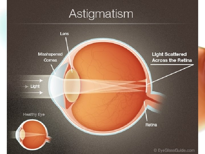
• Know the following: Conjunctivitis, night blindness, colorblindness, cataracts, glaucoma, myopia, hyperopia, astigmatism, deafness, otosclerosis, Meniere’s syndrome, vertigo, olfactory auras, strabismus, and presbyopia.
• • This slide show was developed by Dana Halloran, Cardinal Mooney High School, Sarasota, FL. • • • Used with her personal permission, adapted and amended by Rosa Whiting, Manatee School for the Arts, Palmetto, FL.