Human Responses 3 Senses SENSE ORGANS Animals have


![Ø The fovea is where our best vision is [mainly cones] Ø The front Ø The fovea is where our best vision is [mainly cones] Ø The front](https://slidetodoc.com/presentation_image_h/12353b799a6151a1ec5f8341f26ac546/image-7.jpg)


![Ø Ciliary body [muscle] — thickened edge of the choroid that controls the shape Ø Ciliary body [muscle] — thickened edge of the choroid that controls the shape](https://slidetodoc.com/presentation_image_h/12353b799a6151a1ec5f8341f26ac546/image-10.jpg)


![Ø Middle ear—air-filled cavity containing three small bones [ossicles] and the Eustachian tube Middle Ø Middle ear—air-filled cavity containing three small bones [ossicles] and the Eustachian tube Middle](https://slidetodoc.com/presentation_image_h/12353b799a6151a1ec5f8341f26ac546/image-25.jpg)


![LEARNING CHECK • What is the function of the [a] pinna, [b] 3 ossicles, LEARNING CHECK • What is the function of the [a] pinna, [b] 3 ossicles,](https://slidetodoc.com/presentation_image_h/12353b799a6151a1ec5f8341f26ac546/image-33.jpg)


- Slides: 37

Human Responses 3 Senses

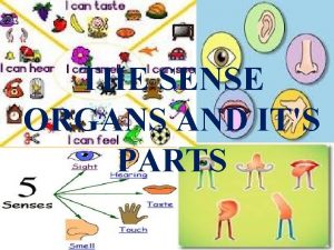
SENSE ORGANS Ø Animals have specialised senses to provide them with information about their environment. Ø The five senses are sight, hearing, touch, taste and smell. Ø A receptor is a cell that can detect a stimulus Ø A stimulus is any change in your environment, e. g. light, sound. 2

Sense Organ Sight Eye Hearing Ear Touch Skin Taste Tongue Smell Nose Stimulus detected light [by rods and cones in the retina] sound [receptors in cochlea] touch, pressure, temperature and pain [receptors spread throughout body] chemicals [taste buds detect sweet, sour, salt and bitter]. chemicals [receptors in the nasal cavity detect vapours] 3

4

The EYE Ø Eyelids can cover and protect the eyes. Eyelid Conjunctiva Cornea Ø Conjunctiva — thin transparent lining protecting the cornea. Ø Cornea—front transparent part of the sclera. It focuses light 5 rays on the retina.

Sclera Choroid Retina Ø Sclera—tough fibrous outer layer – the ‘white’ of the eye; it maintains the shape of the eyeball. Ø Choroid—contains blood vessels supplying food and oxygen to the cells of the eye. Ø Retina—the innermost layer that contains the receptor cells [rods and cones]. 6
![Ø The fovea is where our best vision is mainly cones Ø The front Ø The fovea is where our best vision is [mainly cones] Ø The front](https://slidetodoc.com/presentation_image_h/12353b799a6151a1ec5f8341f26ac546/image-7.jpg)
Ø The fovea is where our best vision is [mainly cones] Ø The front region of the choroid is specialised into the iris Fovea Iris Ø Iris—contains blood vessels and melanin [giving us our eye colour], and controls the amount of light entering the eye [through the pupil]. 7

Pupil 8

Ø In bright light, pupil constricts to protect the retina Ø due to circular muscles contracting in iris. Ø In dim light, the pupil dilates to allow more light in Ø Due to radial muscles contracting in the iris. 9
![Ø Ciliary body muscle thickened edge of the choroid that controls the shape Ø Ciliary body [muscle] — thickened edge of the choroid that controls the shape](https://slidetodoc.com/presentation_image_h/12353b799a6151a1ec5f8341f26ac546/image-10.jpg)
Ø Ciliary body [muscle] — thickened edge of the choroid that controls the shape of the lens Ø Suspensory ligaments — hold the lens in place. Ciliary muscle Suspensory ligaments Lens Ø Lens—like a magnifying glass, it focuses the light rays on the retina. 10

Ø Lens—focuses the light rays on the retina. Ø Accommodation is the ability of the lens to change its shape 11 (focal length) to form a clear image.

LEARNING CHECK • Name the 5 senses and the organs involved. • Name the 3 main layers of the eye and the function of each. • Which part of the eye is only an opening – a hole in another part? • What is the function of the [a] iris, • [b] lens, • [c] cornea, • [d] fovea • What is accommodation? 12

Close Vision Ø For close vision, the ciliary muscle contracts, the suspensory ligaments relax, the lens becomes thicker. 13

Distant Vision Ø When the eye is at rest, the lens is thin, has a long focal length and is adapted for seeing distant objects. 14

Ø Accommodation is the ability of the lens to change its shape (focal length) to form a clear image. 15

Seeing things at different distances For distant objects, the ciliary muscle relaxes and so the suspensory ligaments pull tight, pulling the lens thinner – the light doesn’t bend as much. For close objects the ciliary muscle contracts, allowing the lens to go fat, thus bending the light more. 16

Ø Aqueous humour—watery liquid that supplies the lens and cornea with nutrients and helps keep the shape of the cornea and lens. Aqueous humour Vitreous humour Ø Vitreous humour—gel that helps maintain the shape of the 17 eye.

Ø When light rays focus on the retina, receptor cells are stimulated and impulses are carried along the optic nerve to the brain. (optic nerve = communication between eye and brain) Optic nerve Blind Spot Ø Blind spot—where the optic nerve fibres pass through the 18 retina and there is no room for receptors.

Eye Defects Ø Long-sighted : You are long-sighted if you can clearly see objects a long way off, but you cannot see things close by. Ø Reading glasses [convex lenses] can correct the problem. 19

Eye Defects Ø Short-sighted You are short-sighted if you can clearly see objects close to you, but you cannot see things in the distance. Ø Glasses with concave lenses can correct the problem. 20

Eye Defects Ø NOTE Ø You have to learn either eye defect or Ø Ear defect – recommend this …. 21

LEARNING CHECK • Explain how the ciliary body and suspensory ligaments alter the lens. • What is the function of the [a] humours, [b] optic nerve? • If you are longsighted, what does it mean? • What could be a possible cause? • What type of lens can rectify it? 22

23

The EAR Ø Pinna—outer visible ear, funnels sound into the ear canal. Ø Ear canal —tube leading to the ear drum. It has hairs and wax glands to trap dirt and germs. Ø Eardrum—membrane of skin that vibrates when sound waves hit it. Eardrum Pinna Ear Canal 24
![Ø Middle earairfilled cavity containing three small bones ossicles and the Eustachian tube Middle Ø Middle ear—air-filled cavity containing three small bones [ossicles] and the Eustachian tube Middle](https://slidetodoc.com/presentation_image_h/12353b799a6151a1ec5f8341f26ac546/image-25.jpg)
Ø Middle ear—air-filled cavity containing three small bones [ossicles] and the Eustachian tube Middle Ear Ossicles Ø Ossicles— 3 small bones [hammer, anvil and stirrup], that amplify the sound. Ø Eustachian tube—keeps air pressure equal on each side of the eardrum. Ø It opens when we swallow, cough, etc. Eustachian tube 25

Ø Inner ear—contains a coiled, fluid-filled tube called the cochlea and the semi-circular canals. Inner Ear Semi-circular canals Ø Cochlea—contains nerves that convert sound vibrations into electrical impulses. Ø Semi-circular canals—help us keep our balance and posture. Cochlea 26

Ø The pinna (ear lobe) channels the sound (vibrations in the air) towards the eardrum, which then vibrates. Ø In turn, this vibrates the hammer, anvil and stirrup bones, 27 which amplify the sound.

Ø The stirrup pushes on the oval window of the cochlea, moving the liquid inside. Ø Special hairs on 30, 000 receptor cells detect the movement and send signals to the brain along the auditory nerve. Ø The brain interprets these as sounds, and we ‘hear’. 28

Ø Semi-circular canals—help us keep our balance and posture. Ø The three semicircular canals are curved tubes, each about 29 15 mm long and filled with fluid.

Ø Head movements are detected by nerves inside the canals. Ø The brain responds by sending messages through the cerebellum, which trigger reflex actions in our muscles. Ø This helps us keep our whole body balanced as we move. 30

Ear Defects Deafness ØDeafness can be caused by long exposure to a high level of noise, drugs, or ear infections. ØDamage to the eardrum, ossicles [bones], and cochlea, which can be caused by loud sounds, produces incurable deafness. ØWorkers exposed to prolonged sounds of over 90 decibels [d. B] are obliged by law to wear protection. ØAny exposure to 140 d. B causes immediate damage to hearing. 31

The SKIN as a Sense Organ 32
![LEARNING CHECK What is the function of the a pinna b 3 ossicles LEARNING CHECK • What is the function of the [a] pinna, [b] 3 ossicles,](https://slidetodoc.com/presentation_image_h/12353b799a6151a1ec5f8341f26ac546/image-33.jpg)
LEARNING CHECK • What is the function of the [a] pinna, [b] 3 ossicles, [c] cochlea, [d] semi-circular canals, [e] eustachian tube? • Outline how vibrations in the air are eventually “heard” by our brain. • Name a common ear defect. • Give some possible causes & treatments. • How might you reduce your risks of this defect? 33

3. 5. 3 Responses in the Human Nervous System Objectives – What you will need to know from this section Ø Outline the nervous system components: central nervous system (CNS) and the peripheral nervous system (PNS) Ø Receptor messages are carried through these systems by nerve cells or neurons. Ø Outline the structure & function of the neuron including: cell body, dendrites, axon, myelin sheath, schwann cell, and neurotransmitter vesicles & synaptic cleft Ø Outline impulse movement & synapse. Ø Explain activation & inactivation of neurotransmitter. 34

Ø The structure and function of a neuron: variation in size and shape. Ø Neuron -- Three part structure: > dendrite(s) receive information and carry it towards the cell body, > the axon conducts nerve impulses away from the cell body, > the cell body contains the nucleus and other organelles and produces neurotransmitter chemicals. Ø Explain the role & position of 3 types of neuron -- sensory/motor/inter Ø Movement of nerve impulse. (Detailed knowledge of electrochemistry not required. ) Ø Knowledge that the conduction of nerve impulses along a neuron involves movement of ions (details not required). 35

Ø Outline the senses with the brain as an interpreting centre. Ø Outline the CNS, brain & spinal cord. ØState location & function of cerebrum / hypothalamus / pituitary gland / cerebellum / medulla oblongata ØLabel &/or draw diagrams of spinal cord (cross section) indicating : white matter, grey matter, central canal, 3 layer protective tissue-meninges. Ø Spinal nerves containing dorsal and ventral roots that project from the spinal cord 36

Ø Outline disorders from NS disorders: paralysis or Parkinson's including: Cause/Prevention/Treatment Ø Outline PNS including the location nerve fibres & cell bodies. Ø State the role, structure & mechanism of the Reflex arc/action. Ø The sense organs contain receptors, with the brain as an interpreting centre for received information. Ø Knowledge of the five senses and related organs. Ø Study the eye and the ear – recognition and fuction of the main parts. • Corrective measures for long and short sight or for a hearing defect. 37
 Distinguish between general senses and special senses.
 General senses vs special senses
 Sense organs
 Images of sense organs with names
 Sense organs for class 1
 Amphibians sense organs
 Ten sense organs
 Take care of sense organs
 Dominant genetic variance
 Narrow sense heritability vs broad sense heritability
 The human inputs and outputs information through
 How many senses do humans have
 It is a bony cage enclosing vital human organs
 Internal organs human body
 Gametophytes have gamete-producing organs called _____.
 Kyle douglass
 Is a horse a producer consumer or decomposer
 Parasitic food chain example
 Animals that eat both plants and animals
 12 edges of cube
 Do owls have a sense of smell
 Personification for animals
 What was the first human language
 Animals and human language chapter 2
 Examples of commensalism relationships
 Chordata taxonomy
 Why do desert animals have longer loop of henle
 Prokaryotic cells vs eukaryotic cells
 Why do desert animals have longer loop of henle
 Do animals have consciousness
 Every living plants and animals must have
 Alpine biome
 Does animal have language
 Division of animal kingdom
 Animal whose ears we cannot see
 Phylum with spiny skin
 Invertebrates without legs
 Spiny skinned animals have an endoskeleton formed with

