ANATOMY PHYSIOLOGY THE DIGESTIVE SYSTEM Digestive System Ingestion


 § Mucosa – secrete mucus,")


 Serous")


 § Distension of")


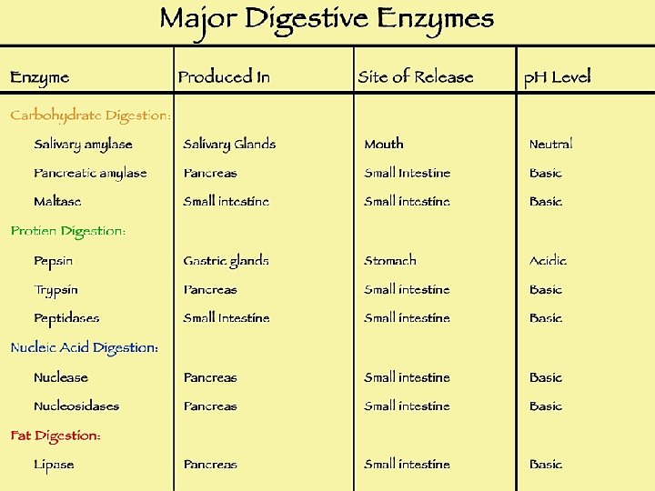
 • Digestive enzymes - break down carbs, proteins, and fats. •")


 that may affect any part")

 in")


- Slides: 32
ANATOMY & PHYSIOLOGY THE DIGESTIVE SYSTEM
Digestive System § Ingestion: receiving food § Digestion: breakdown of food particles small enough to pass through cell membranes § Absorption: transfer of food into circulation
A. Alimentary Canal – The Tube from beginning to end ~ 9 m or 30 ft. § § § § Oral Cavity Pharynx Esophagus Stomach Small Intestine Large Intestine Rectum/Anal Canal
Walls of Alimentary Canal – 4 layers (inside outside) § Mucosa – secrete mucus, protects surrounding tissue, absorbs nutrients § Submucosa – nourishes surrounding tissues, carry away absorbed nutrients § Muscular layer – move food to stomach (peristalsis) 1) Circular: changes diameter of tube 2) Longitudinal: changes length of tube § Serous layer/serosa – produce serous fluid to prevent friction
Tube Movements § Mixing – some sections of the tube contract to mix and breakdown food § Propelling (peristalsis) – wave-like movements push food through the canal
B. Mouth/ Oral Cavity § Cheeks – muscles for chewing § Lips – sensory and manipulation of food § Tongue – manipulated food; taste buds/receptors • Frenulum connects the tongue to the bottom of the mouth § Palate (roof of mouth) – hard anterior, soft posterior • Uvula is extension of soft palate hangs down to direct food
§ Teeth • 20 as a child, 32 as an adult • Incisors and canines: cutting and tearing • Premolars and molars: mastication
§ Salivary Glands • Saliva – mostly water; slightly acidic, some antibodies 1) Serous cells: secrete serous fluid with digestive enzymes (amylase enzyme of the mouth) to lubricate food and begin digestion 2) Mucous cells: produce mucous to clump food into a ball/bolus 3) Three Glands a. Parotid – below the ear b. Submandibular – under the mandible (jaw) c. Sublingual – under the tongue
Pharynx – cavity; posterior end of the mouth, leads to the esophagus § Step 1 – chew, breakdown, mix with saliva into bolus § Step 2 – swallowing reflex, once near the pharynx • Soft palate is raised to block the nasal passageway • Hyoid bone – larynx elevated to block trachea epiglottis then covers the opening • Muscle relax, lower pharynx and open esophagus • Peristaltic wave begins pushing food down esophagus
Stomach – located on left portion of body, j-shaped § Holds between 50 m. L – 4 L, folded when empty § Digest proteins § Absorbs very little – only few liquids, drugs, alcohol, and salts § Mechanical and chemical digestion
Regions of the Stomach § Cardia –small region at the esophageal-stomach junction § Fundus – formed by the upper curvature of the organ; temporary storage area for food § Body – main, central region; holds max 4 Liters, avg. 0. 9 L § Pylorus – small region at the stomach-duodenal junction; holds 30 m. L only releases 3 m. L
Cells of the Stomach/ Gastric Glands § Mucous cells – secrete mucous for protection § Chief cells – secrete digestive enzymes; pepsinogen § Parietal cells – secrete HCL (hydrochloric acid); p. H~ 2 § Pepsinogen (an inactive enzyme) is converted to Pepsin when it reacts with HCl/acidic environment § Pepsin breaks down proteins into amino acids
3 Layers of the Stomach Wall § Serosal: outermost; thick connective tissue binds to other organs § Muscularis: thick-middle; three layers of smooth muscle tissue. § Mucosal: innermost; relatively thick and contains numerous tubular glands called gastric pits • Little to no digestion occurs here; alkaline (high p. H) to combat HCl • Ulcers- holes in stomach, esophagus, duodenum brought on by stress
Control of Gastric Juices § Sight, smell, taste stimulates secretions (parasympathetic) § Distension of the stomach and production of chemicals due to breakdown of food • Gastrin: hormone released with secretion also stimulates increase of secretion • Decrease juices if lots of fat/protein in the duodenum or if the intestine is extended.
Digestion and Absorption in Stomach § HCl + churning action breaks down food § Smaller particles does what to surface area? What does this have to do with enzyme digestion? – Enzymes able to work better because they can attack a higher surface area § Chyme: mixture of small food particles + acid + mucous § Little absorption occurs through the walls of the stomach; foods high in fat stay in stomach the longest
Exiting the Stomach § Chyme: Ingested substances combine with the secretions of the glands of the stomach, producing a viscous, highly acidic soupy mixture of partially digested food
Accessory Organs § Liver § Gallbladder § Pancreas
§ Liver: Largest gland/organ of the body; produces bile that is • • stored in the gallbladder supplies quick energy metabolizes alcohol makes proteins stores vitamins and minerals regulates blood clotting regulates cholesterol production detoxifies poisons § 3 lbs. § Requires 25% of cardiac output
§ Gallbladder: stores and concentrates bile that is not needed immediately for digestion § Bile: yellow-green, alkaline solution containing bile salts, bile pigments (primarily bilirubin), cholesterol, neutral fats, phospholipids, and a variety of electrolytes • Fat emulsification via bile salts • Bile does not usually enter the small intestine until the gallbladder contracts
§ Pancreas (exocrine) • Digestive enzymes - break down carbs, proteins, and fats. • Base, bicarbonate ion (alkaline). - Neutralizes chyme from the stomach, permitting digestive enzymes to function. • Secretin stimulates production of bicarbonate ions • Cholecystokinin (CCK) stimulates production of enzymes Enzymes include: proteases, pancreatic lipase, and pancreatic amylase
Small Intestine § Receives chyme, pancreatic and liver secretions to complete digestion of nutrients • Duodenum: 25 cm long x 5 cm wide; c-shaped, most absorption of nutrients occurs here • Jejunum: middle; thicker more vascular walls, greater in diameter • Ileum: last section; suspended from abdominal wall by mesentery
Duodenum § Pancreatic enzymes & bile enter, continuing digestion. § Villi: microscopic hair-like structures; absorption of nutrients • Active transport of sugars, amino acids, and fatty acids across membranes. • Pass through the epithelial wall into the bloodstream. § Enzymes: break down carbs, proteins, and fats into sugars, amino acids, & fatty acids § This is where most nutrients are absorbed in the digestive system
Large Intestine § Large Intestine: serves to compact the solids remaining after digestion. • Lacks villi: little to no absorption of nutrients • Water and a few ions (e. g. sodium) are absorbed through the walls of intestine; only sig. secretion is mucous • Undigested material then passes into the rectum and is finally eliminated through the anus.
Sections of the Large Intestine § Cecum: beginning of the large intestine; receives chyme from the ileum, and connects to the ascending colon; appendix attached § Ascending Colon: begin the process of extracting water and electrolytes; peristalsis required to move up to transverse colon § Transverse Colon: attached to the stomach by a wide band of tissue called the greater omentum; mobile section
§ Descending Colon: store undigested wastes before moving into the rectum; IBS and colon cancer most common here § Sigmoid Colon: s-shaped; walls are muscular and contract to increase pressure inside the colon to move wastes into the rectum § Rectum: final straight portion of the large intestine; acts as a temporary storage site for undigested wastes; stretch receptors located in the walls
Ailments & Diseases of the Digestive System § Dysphagia: difficulty swallowing; common in old age or after a stroke/nervous system malfunction § Gastroesophageal Reflux Disease (GERD): stomach contents leak backwards from the stomach into the esophagus irritating the esophagus, causing heartburn
§ Crohn’s Disease: type of inflammatory bowel disease (IBD) that may affect any part of the gastrointestinal tract; primarily causes abdominal pain, diarrhea, vomiting, or weight loss; caused by interactions between environmental, immunological and bacterial factors in genetically susceptible individuals
§ Ulcers: Peptic ulcers are holes/breaks in the lining of the duodenum or the stomach, areas that come into contact with stomach acids and enzymes. Duodenal ulcers are more common than stomach ulcers; recent theory holds that bacterial infection is the primary cause is the bacterium Helicobacter pylori (H. pylori) § Celiac Disease: a disease that damages the small intestine and interferes with absorption of nutrients from food. Cannot tolerate gluten, a protein in wheat, rye, and barley. Gluten is found mainly in foods but may also be found in everyday products such as medicines, vitamins, and lip balms.
§ Ulcerative colitis: a chronic, long-lasting disease that causes inflammation and sores (ulcers) in the lining of the large intestine, especially the colon and rectum § Irritable Bowel Syndrome (IBS): a common disorder that affects the large intestine (colon); commonly causes cramping, abdominal pain, bloating gas, diarrhea and constipation § Diverticulosis: small pouches may form along the walls of the large intestine called diverticuli; may collect and not be able to empty fecal material which can lead to inflammation
§ Jaundice: “yellow” in french; backup of bile by-products from the blood into body tissues. May result from blockage of the ducts draining bile into the intestines § Gallstones: hard, pebble-like deposits that form inside the gallbladder; may lead to inflammatory condition characterized by retention of bile in the gallbladder § Cirrhosis: is scarring of the liver and poor liver function. It is the final phase of chronic liver disease; most commonly caused by alcoholism, hepatitis B and hepatitis C
§ Hepatitis: inflammation of the liver; viruses cause most cases of hepatitis. Drug or alcohol use can also cause hepatitis. In other cases, your body mistakenly attacks healthy cells in the liver. • A - hepatitis A virus (HAV); usually spread by eating or drinking food or water contaminated with infected feces, undercook shellfish, or close contact with an infectious person. After a single infection a person is immune for the rest of their life. • B - hepatitis B virus (HBV); transmitted by exposure to infectious blood or body fluids; causes liver inflammation, vomiting, jaundice, and, rarely, death. Chronic hepatitis B may eventually cause cirrhosis and liver cancer. • C - hepatitis C virus (HCV); spread primarily by blood-to-blood contact associated with intravenous drug use, poorly sterilized medical equipment, and transfusions; often asymptomatic, but chronic infection can lead to scarring of the liver and ultimately to cirrhosis, liver failure, or liver cancer. § Vaccines are readily available in developed countries