The SKIN SELECTED INTEGUMENTARY SYSTEM DISEASES CONDITIONS Wound


:")
 – continuous low-level negative pressure")


 may protrude through a wound or")


 Stevens-Johnson Syndrome")

 Note however: Staphlococcus skin infections may look the")


 skin")

 Transverse and longitudinal over -curvature of")


 Hyperbaric oxygen therapy (HBO) involves the breathing of pure oxygen while")


; nevi (plural) 1. Any birthmark. 2. A benign pigmented spot on")


 Under negative pressure, V. A. C. Therapy with proprietary")


 An edematous, transitory papule or plaque having a ring of erythema known")


- Slides: 138
The SKIN… SELECTED INTEGUMENTARY SYSTEM DISEASES & CONDITIONS Wound Infections Wound Dehiscence & Evisceration Pressure Ulcers Arterial and Venous Ulcers Skin Cancer Skin Diseases Lesions/Wounds Rashes Herpes Zoster/ Shingles Part I
Trivia Question How many square feet of skin does the average adult have?
Trivia Question How many square feet of skin does the average adult have? Answer: ~ 18 -20 square feet…… 5’ x 4’
Integumentary Diseases Learning Outcomes: 1. Use proper terminology to describe different skin lesions and problems 2. Identify signs of wound infections 3. Describe the ABCD(E) method of assessing skin cancer 4. Identify clients at risk for pressure ulcer development 5. Differentiate between the clinical manifestations for Stage I through Stage IV pressure ulcers, and their treatments. 6. Identify appropriate treatment for prevention of pressure ulcers. 7. Compare the clinical manifestations and modes of transmission for bacterial, viral, and fungal skin infections. 8. Identify the difference between arterial ulcers and insufficiency versus venous stasis ulcers.
Key Terminology angioma carcinoma cellulitis dermatitis atopic dermatitis contact dermatitis eczema edema ecchymosis erythema eschar folliculitis furuncles granulation hemangioma hirsutism hyperbaric oxygen (HBO) keloid lichenification macule melanoma nevus nodule papule paronychia petechiae pressure ulcer pruritis psoriasis purpura pustule vacuum assisted closure (Wound Vac) turgor urticaria vesicle wheal xerosis
Wound Infections Wound Dehiscence & Evisceration Pressure Ulcers Arterial and Venous Ulcers Skin Cancer Skin Diseases Skin Lesions / Wounds Rashes Herpes Zoster/ Shingles
Raised Depressed Flat /Macular Surface Change Fluid Filled Purpura / Vascular Configurations papule plaque nodule cyst comedo keloid horn wheal atrophy erosion ulcer sinus striae burrow poikolderma macule patch erythema erythroderma scale crust excoriation eschar lichenification exfoliation ichthyosiform vesicle bulla pustule abscess furuncle petechiae purpura ecchymosis telangiectasia linear grouped scattered polycyclic reticular serpinginous targetoid whorled arcuate annular “Beauty is Only Skin Deep…”
Raised Depressed Flat /Macular Surface Change Fluid Filled Purpura / Vascular Configurations papule plaque nodule cyst comedo keloid horn wheal atrophy erosion ulcer sinus striae burrow poikolderma macule patch erythema erythroderma scale crust excoriation eschar lichenification exfoliation ichthyosiform vesicle bulla pustule abscess furuncle petechiae purpura ecchymosis telangiectasia linear grouped scattered polycyclic reticular serpinginous targetoid whorled arcuate annular “Beauty is Only Skin Deep…” “But UGLY goes down to the bone!”
Raised Depressed Flat /Macular Surface Change Fluid Filled Purpura / Vascular Configurations papule plaque nodule cyst comedo keloid horn wheal atrophy erosion ulcer sinus striae burrow poikolderma macule patch erythema erythroderma scale crust excoriation eschar lichenification exfoliation ichthyosiform vesicle bulla pustule abscess furuncle petechiae purpura ecchymosis telangiectasia linear grouped scattered polycyclic reticular serpinginous targetoid whorled arcuate annular “Skin Changes” Associated Terminology
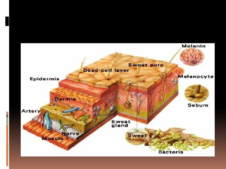
Integumentary Diseases Review of anatomy & physiology
Integumentary diseases Skin Function: �Skin, hair, and nails are external structures �Serve a variety of specialized functions �Sebaceous and sweat glands cool and lubricate the body and get rid of wastes �The skin, hair, and nails have many vital functions, but the most important is protection. �Once the skin has been pierced or damaged the body is much more susceptible to infection, even from organisms which would not normally be pathogenic, e. g. , staphylococci.
Integumentary diseases Wound Infections & Risks for infection include any pathophysiological states which cause diminished circulation, poor immune response, or delayed repair of tissues These include: �Radiation or trauma �Poor circulation �Immunosuppression, drug induced or disease induced �Impaired oxygenation �Nutritional deficiencies �Coagulation disorders �Immobility �Diabetes, lupus, and other immunodeficiencies
Integumentary diseases Etiology: THINK – WHAT IS CAUSING THIS? And you might learn how to prevent it For example – If the patient is immobile and starting to break down because they cannot move, how do we prevent the skin from breaking down? If the patient has an immunodeficiency, how do we prevent skin breakdown? If the patient has a nutritional deficiency, how do we promote wound healing?
Diagnostic Procedures and Nursing Interventions Wound Culture and Sensitivity (swab cultures and/or wound biopsies): Definitively identify and quantify wound bacteria. Complete Blood Count (CBC) with Differential: Assess immune response. Blood Cultures: Rule out sepsis. Serum Albumin (normal > 3. 5 g/d. L) and Prealbumin (normal 17 to 40 mg/d. L): Assess nutritional status (low levels indicate malnutrition).
Therapeutic Procedures and Nursing Interventions Vacuum-assisted Wound Closure (Wound-Vac) – continuous low-level negative pressure is applied to a sponge-covered suction tube for several hours. Hyperbaric Oxygen Therapy (HBO / HBOT)– administration of high-pressurized 100% oxygen directly over the wound for 60 to 90 min. Surgical Debridement and / or Wound Grafting – surgical excision of nonviable tissue to promote wound healing and/or grafting of skin from donor sites to clean granulating or freshly excised wound bed.
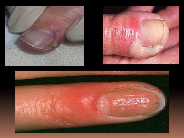
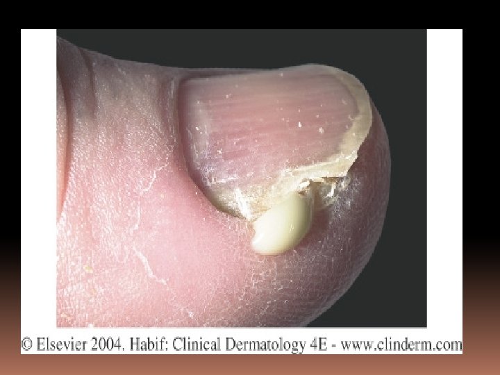
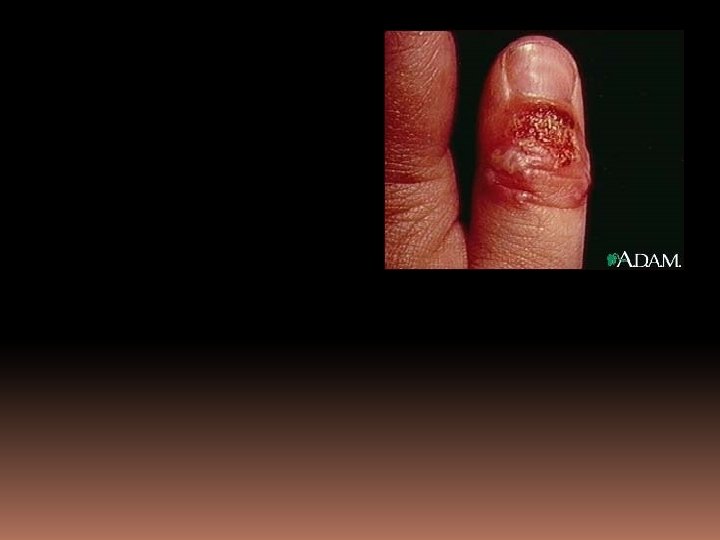
Integumentary diseases Lesions & Infections Signs and symptoms of infection include: Drainage Odor Abscess Fever Discoloration Heat / warmth at site Edema Erythema Poor healing
Integumentary diseases Lesions & Infections
Integumentary diseases Lesions & Infections
Integumentary diseases Lesions & Infections
Integumentary diseases Lesions & Infections
Integumentary diseases Rashes – is it macular or papular?
Integumentary diseases Rashes – is it macular or papular? This rash is actually vesicular, and is herpes zoster, or viral in origin
Integumentary diseases Rashes – is it macular or papular? This rash is actually vesicular, and is herpes zoster, or viral in origin “chicken pox”
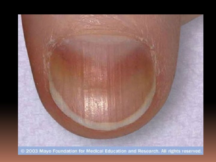
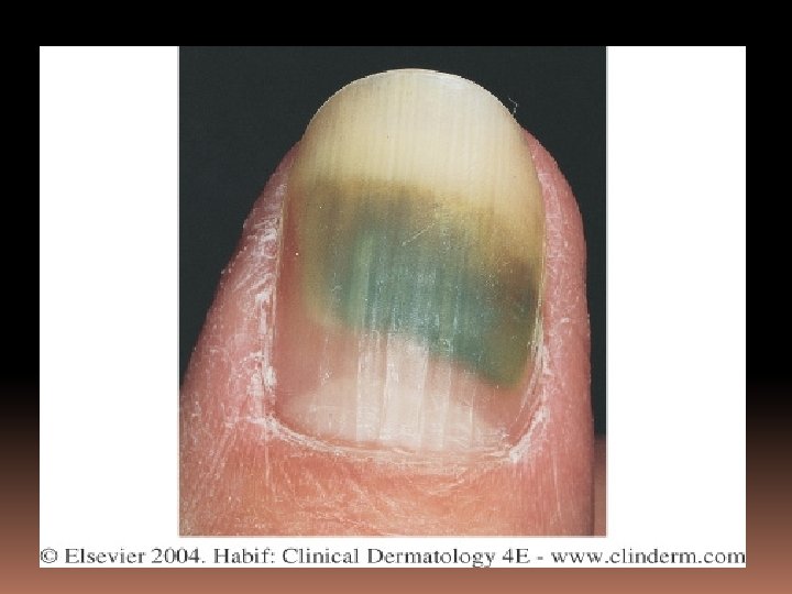
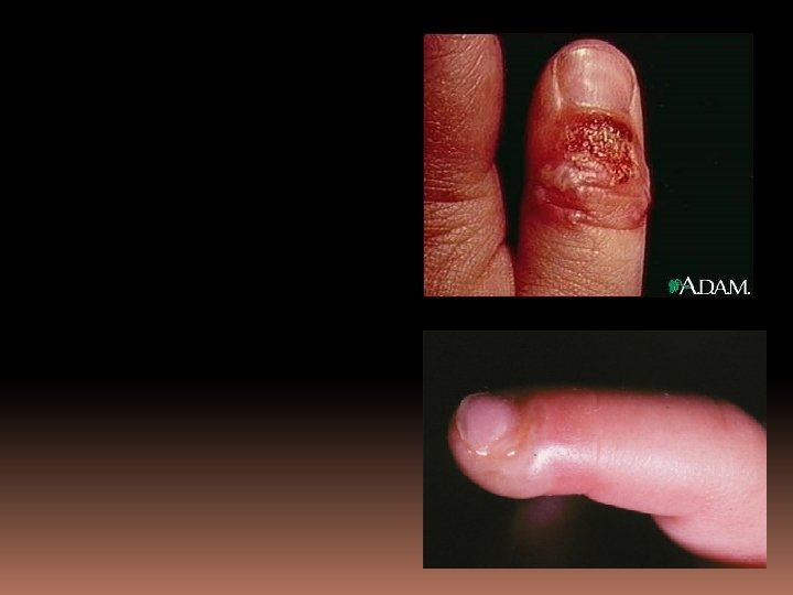
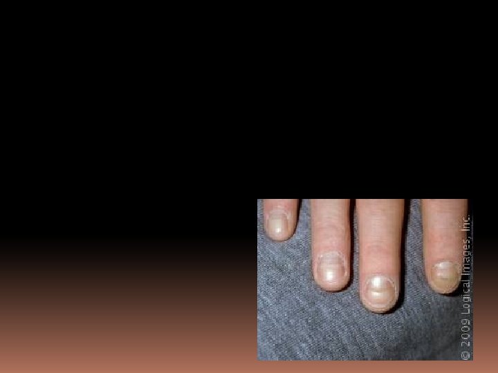
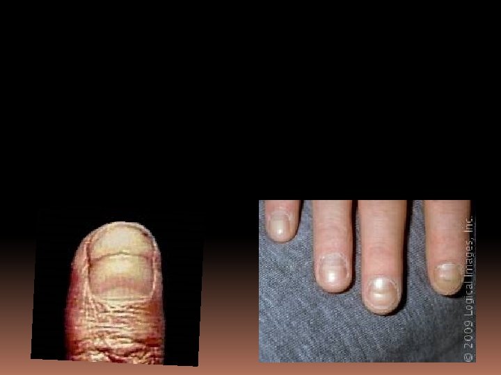
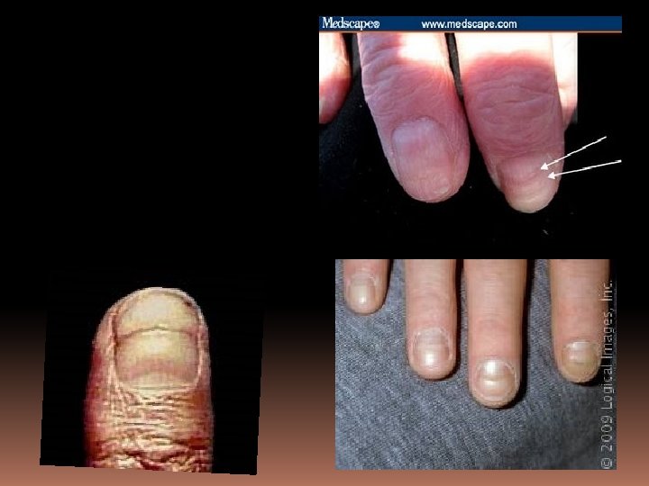
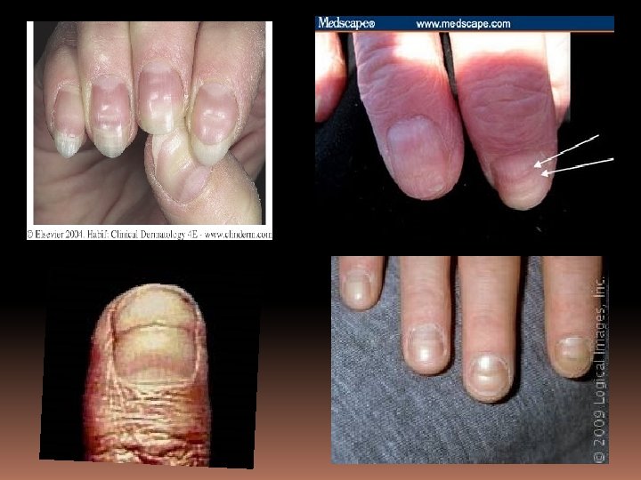
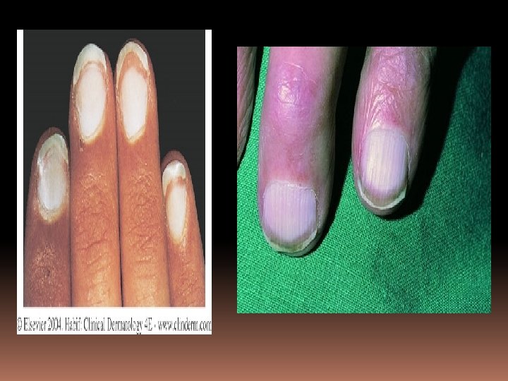
Integumentary diseases Infected hair &/or nails also count These are Beau’s lines
Integumentary diseases
Integumentary diseases Dehiscence – opening of a wound or surgical site
Integumentary diseases Evisceration – internal body parts (viscera) may protrude through a wound or opening. See below. Eviscerated bowel through a clean stab wound
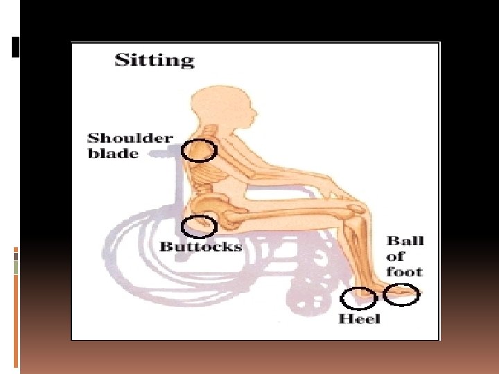
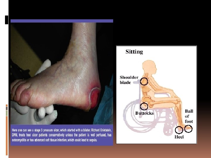
Integumentary diseases Pressure sites – cause pressure ulcers, the number one risk is immobility and boney prominences
Integumentary diseases Stage I ulcer Defined as non-blanching erythema of intact skin, discoloration of skins with warmth or hardness
Integumentary diseases Stage II Ulcer Defined – partial thickness skin loss involving epidermis and/or dermis layer; the ulcer is superficial and presents clinically as an abrasion, blister or shallow crater. Coccyx
Integumentary diseases Stage III Defined as a full thickness skin loss involving damage or necrosis of subcutaneous tissue which may extend down to , but not through underlying fascia; the ulcer presents clinically as a deep crater with or without undermining or adjacent tissue.
Integumentary diseases Stage IV Defined as full-thickness skin loss with extensive destruction, tissue necrosis, or damage to muscle, bone, or supporting structures Bone visible under damage
Integumentary diseases Venous Stasis Ulcers – no stages Caused by poor circulation and poor venous return (PVD) Peripheral vascular disease
Integumentary diseases Venous Stasis Ulcers – no stages Caused by poor circulation and poor venous return (PVD) Peripheral vascular disease Harder to heal, May need a bypass to restore circulation, in addition to dressings. etc. Edema and discoloration are primary symptoms prior to skin breakdown.
Integumentary diseases Treatments of ulcers #1 = prevention and turning, ROM Dressings and packings, from simple to complex colloids
Integumentary diseases Drug therapy Debridement Diet therapy with vitamins and supplements wound – vacuum (Wound Vac) Hyperbaric oxygenation (HBO) Surgical repair
Integumentary diseases Documentation of ulcers It is necessary to identify any skin damage or breakdown on admission, as if these are missed, the hospital or facility can be liable If it was not documented, it was not done
Integumentary diseases Documentation of ulcers Documentation should include a photograph, measuring the ulcer or wound with a sterile paper tape or circle; the depth, the width, the breadth and the total area involved, and any breaking of the skin, any color and any drainage (including color and odor). Then “stage” it.
Integumentary diseases The ABCDE’s of Skin Cancer: Asymmetry Border Irregularity Color - changes Diameter – increasing (Elevation--variable)
Integumentary diseases A-B-C-D----What is it?
Integumentary diseases Malignant Melanoma - Stage I
Integumentary diseases Malignant Melanoma – an aggressive form of cancer, that is mostly preventable, has a genetic component, and may spread into other systems before it is noticed.
Integumentary diseases Rashes and Lesions – Dermatology, the great mimic- is it bacterial, auto-immune, infected, viral, or parasitic?
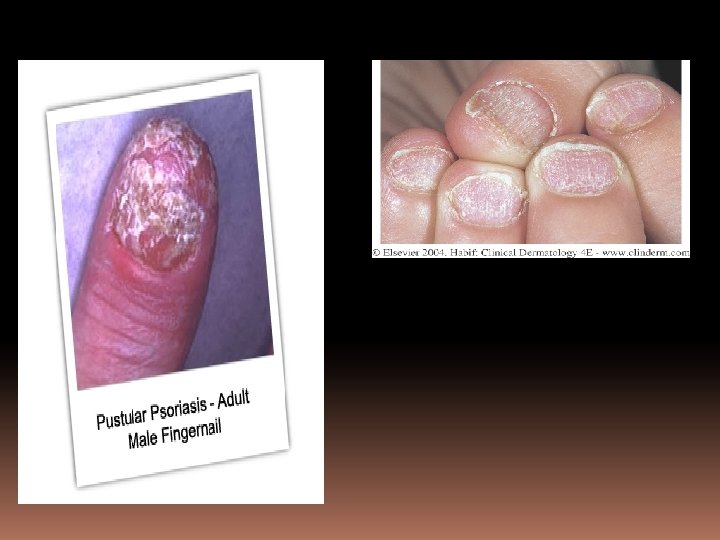
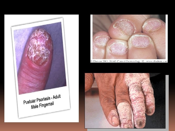
Integumentary diseases Psoriasis – an autoimmune disease which causes overstimulation of the keratinocytes in the skin. This can be caused initially by skin trauma, irritation, or stress.
Integumentary diseases Psoriasis – may appear like eczema at first, but the plaques become thicker and widespread. Eczema tends to be lighter and scalier
eczema
psoriasis
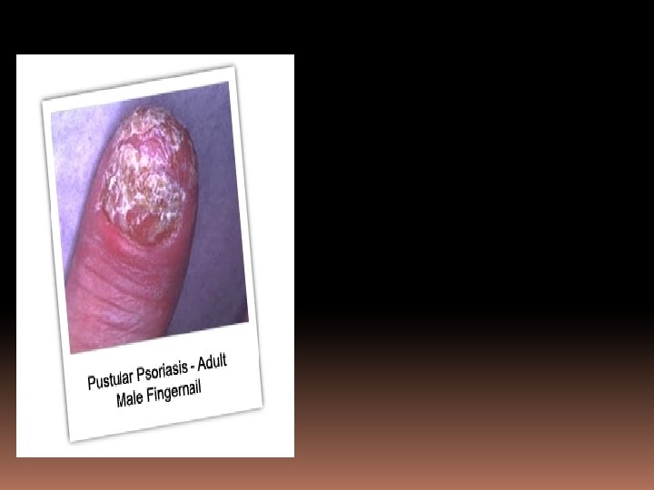
Integumentary diseases Psoriasis – can erode and disfigure fingers and toes that are affected
Integumentary diseases Psoriasis Treatments Steroids Topical zinc ointments Topical steroids Tar and oil preparations Ultraviolet light therapy – sunlight often helps
Integumentary diseases Psoriasis Treatments ‘Older’ drugs include methotrexate & cyclosporine to suppress the immune system, but this requires monitoring of liver enzymes. New treatments include monoclonal antibodies (A form of chemotherapy—the ‘-mabs’ i. e. efalizumab (Raptiva) for severe forms
Integumentary diseases Lesions – see handouts of definitions and terms, be able to use these. A rash is a rash…. or is it? Common causes of rashes include Allergic dermatitis, usually localized to the areas of contact, hives Drug reactions, usually all over the body Bacterial infections Viral infections
Integumentary diseases What kind of rash is this? If you can’t tell, describe it. Describe the area as well.
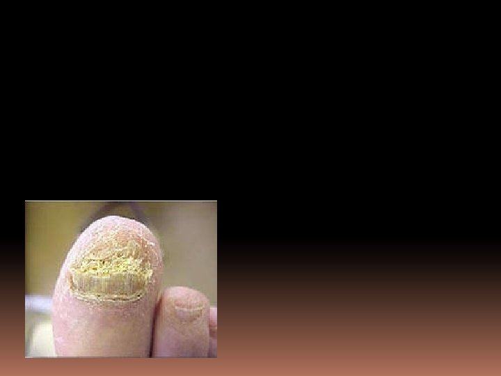
Integumentary diseases ‘Ringworm’ – tinea capitus – a fungal infection
Integumentary diseases Etiology: Systemic or local?
toxic epidermal necrolysis (T. E. N. ) Stevens-Johnson Syndrome
Integumentary diseases ‘Viral rashes’
Integumentary diseases Chicken POX (Herpes Zoster) Note however: Staphlococcus skin infections may look the same
Integumentary diseases
Terminology Appendix Terminology Associated With the Integumentary System
The “ABCDE’s of Skin Cancer” Asymmetry Border Irregularity Color Diameter: ¼ inch <or> 6 mm Elevation
angioma Angiomas : benign tumors that are made up of small blood vessels or lymph vessels. Cherry angiomas Spider angiomas
asymmetric 1. Pertaining to an individual lesion: Unequal shape from side to side. 2. Pertaining to a body distribution: Unequal distribution of lesions on both sides of body.
Beau’s lines Beau's lines Transverse depressions or grooves in the nail plate typically occurring at corresponding positions within each nail plate. Often a sign of a prior severe illness such as malnutrition, a systemic disease, or trauma.
carcinoma Carcinoma refers to an invasive malignant tumor consisting of transformed epithelial cells. Alternatively, it refers to a malignant tumor composed of transformed cells of unknown histogenesis, but which possess specific molecular or histological characteristics that are associated with epithelial cells, such as the production of cytokeratins or intercellular bridges. Carcinoma in situ (CIS) refers to a small, localized focus of carcinoma that has not yet invaded Basal Cell Carcinoma
dermatitis Atopic dermatitis~ atopic dermatitis is a very common, often chronic (long-lasting) skin disease that affects a large percentage of the world's population. It is also called eczema, dermatitis, or atopy. Most commonly, it may be thought of as a type of skin allergy or sensitivity. The atopic dermatitis triad includes asthma, allergies (hay fever), and eczema. There is a known hereditary component of the disease, and it is seen more in some families. The hallmarks of the disease include skin rashes and itching. Contact dermatitis~ Contact dermatitis is an inflammation of the skin caused by direct contact with an irritating substance. Symptoms: Itching (pruritus) of the skin in exposed areas ; Skin redness or inflammation in the exposed area. Treatment: remove irritating exposure; topical corticosteroids.
café’ au lait patch café au lait patch A well circumscribed macule or patch varying from pale brown in lighter skin to dark brown in darker skin patients. The macule or patch may have a serrated or irregular margin.
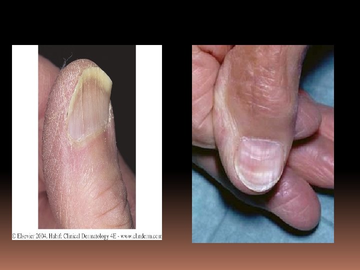
Clubbing clubbing (Hippocratic fingers, watch-glass nails, drumstick fingers) Transverse and longitudinal over -curvature of the nail plate. The distal digit may have associated enlargement. Lovibond's angle is greater than 180 degrees. Typically associated with chronic hypoxia, e. g in those with congenital heart disease, or COPD. .
Dermal dermal Relating to the layer of skin below the epidermis, but above the panniculus, consisting of papillary and reticular layers and containing blood and lymphatic vessels.
eczema : a general term for many types of skin inflammation, also known as dermatitis. The most common form of eczema is atopic dermatitis (some people use these two terms interchangeably).
edema Edema is swelling that is caused by fluid trapped in your body’s tissues. Edema happens most often in the feet, ankles, and legs. Other parts of the body, such as the face and hands, can also be affected
ecchymosis Extravasation of blood into the skin or mucous membranes forming large macules or patches; flat color changes over time may go from blue-black, to brown-yellow, or green.
excoriation A focal erosion usually due to scratching.
erythema Localized, blanchable redness of the skin or mucous membranes.
eschar An adherent, thick, dry black crust.
folliculitis Folliculitis is inflammation of one or more hair follicles. It can occur anywhere on the skin. Common symptoms include a rash, itching, and pimples or pustules near a hair follicle in the neck, groin , or other areas. . . “Hot Tub Folliculitis”:
furuncle A follicle-centered nodule caused by a suppurative infection characterized by pain, redness, and potentially visible pus. Usually greater than 1 cm in diameter.
hematoma A collection of extravasated blood that is relatively or completely confined within a space. The blood is usually clotted (or partly clotted), and depending on time may manifest various degrees of organization and color.
hirsutism Unwanted, excess hair growth; generally in women.
Hyperbaric oxygen (HBO) Hyperbaric oxygen therapy (HBO) involves the breathing of pure oxygen while in a sealed chamber that has been pressurized at 1 1/2 to 3 times normal atmospheric pressure. used in conventional treatment for decompression sickness (the bends); severe carbon monoxide poisoning; certain kinds of wounds, injuries, and skin infections; delayed radiation injury; and certain bone or brain infections.
keloid A firm, usually elevated, proliferation of scar tissue exceeding the area of the preceding skin injury or wound.
lichenification Thickened skin with accentuated markings usually due to repeated rubbing and scratching of skin.
maceration Softened or broken down skin resulting from prolonged exposure to wetness causing whitening and thickening of the keratin sometimes with redness, oozing, and/or scaling.
macule A flat, generally less than. 5 cm area of skin or mucous membrane with different color from surrounding tissue. Macules may have non-palpable, fine scale.
melanoma Melanoma is a malignant tumor that originates in melanocytes, the cells which produce the pigment melanin that colors our skin, hair, and eyes. The majority of melanomas are black or brown. However, some melanomas are skin-colored, pink, red, purple, blue or white. Melanoma is the most serious form of skin cancer.
nevus Nevus (singular); nevi (plural) 1. Any birthmark. 2. A benign pigmented spot on the skin such as a mole (a cluster of melanocytes and supportive tissue that appears as a tan, brown, or flesh-colored spot on the skin). 3. A benign blood vessel tumor on the skin such as a vascular nevus, a local collection of capillaries of the skin (a strawberry birthmark, stork mark, or port wine stain). From the Latin naevus meaning a body mole, especially a birthmark. The plural of nevus is nevi.
nodule A dermal or subcutaneous firm, well-defined lesion usually greater than. 5 cm in diameter.
papule A discrete, solid, elevated body usually less than. 5 cm in diameter. Papules are further classified by shape, size, color, and surface change.
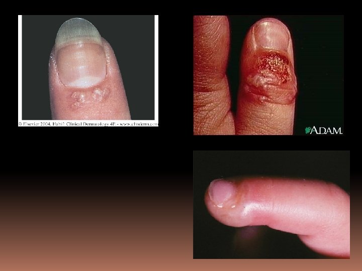
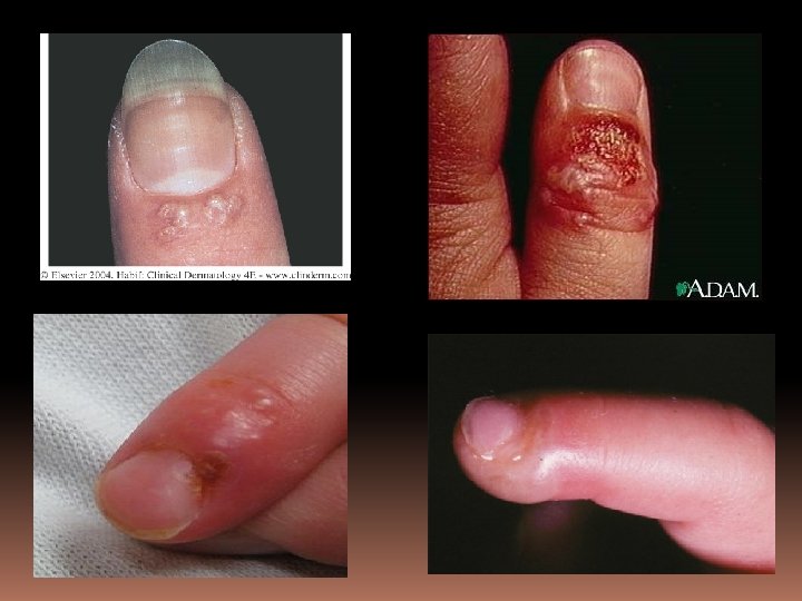
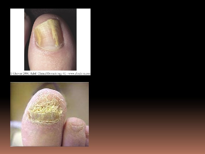
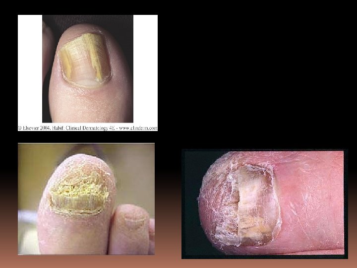
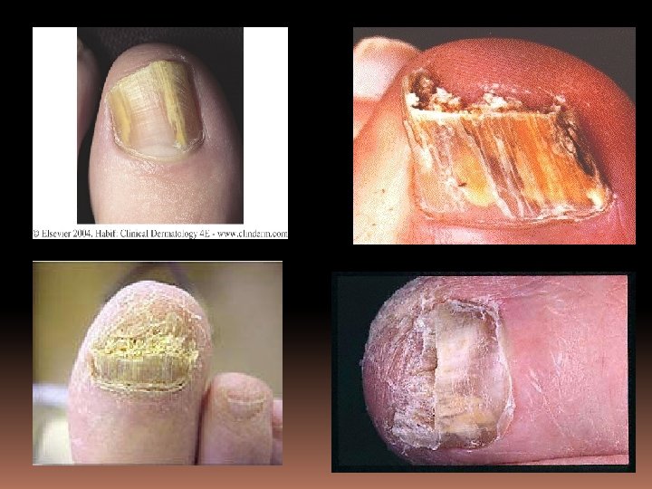
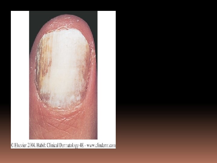
paronychia Paronychia: Inflammation of the folds of tissue surrounding the nail due to infection. The infection may be bacterial (most commonly, staph or strep species) or fungal. The term "paronychia" is compounded from "para-", next to + the Greek "onyx", nail = next to the nail. Paronychia is synonymous with perionychia.
petechiae Tiny, 1 -2 mm nonblanchable purpuric macules resulting from tiny hemorrhages.
plaque A discrete, solid, elevated body usually broader than it is thick measuring more than. 5 cm in diameter. Plaques may be further classified by shape, size, color, and surface change.
Pressure ulcer …an area of skin that breaks down when you stay in one position for too long without shifting your weight. Pressure ulcers are categorized by severity, from Stage I (earliest signs) to Stage IV (worst): Stage I: A reddened area on the skin that, when pressed, is "nonblanchable" (does not turn white). This indicates that a pressure ulcer is starting to develop. Stage II: The skin blisters or forms an open sore. The area around the sore may be red and irritated. Stage III: The skin breakdown now looks like a crater where there is damage to the tissue below the skin. Stage IV: The pressure ulcer has become so deep that there is damage to the muscle and bone, and sometimes tendons and joints.
pruritis Pruritus is an itch or a sensation that makes a person want to scratch. Pruritus can cause discomfort and be frustrating. If it is severe, it can lead to sleeplessness, anxiety, and depression. The exact cause of an itch is unknown. It is a complex process involving nerves that respond to certain chemicals like histamine that are released in the skin, and the processing of nerve signals in the brain. Pruritus can be a part of skin diseases, internal disorders, or due to faulty processing of the itch sensation within the nervous system.
psoriasis A common skin condition that causes skin redness & irritation. Most persons with psoriasis have thick, red skin with flaky, silver-whit patches called scales. There are five main types of psoriasis. Erythrodermic -- The skin redness is very intense and covers a large area. Guttate -- Small, pink-red spots appear on the skin. Inverse -- Skin redness and irritation occurs in the armpits, groin, and in between overlapping skin. Plaque -- Thick, red patches of skin are covered by flaky, silver-white scales. This is the most common type of psoriasis. Pustular -- White blisters are surrounded by red, irritated skin.
purpura Hemorrhage into skin or mucous membranes which varies in size and ranges in color related to duration. Types of purpura include palpable purpura, ecchymosis, and petechiae.
pustule A circumscribed elevation that contains pus. Pustules are usually less than. 5 cm in diameter.
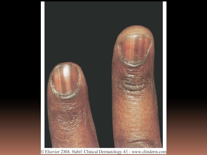
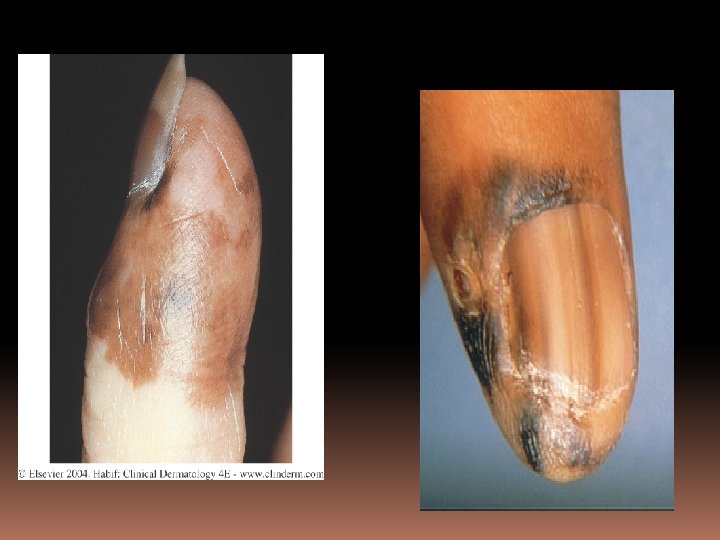
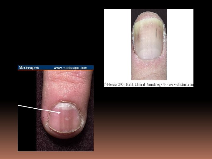
splinter hemorrhages Dark-red, sometimes black thin longitudinal lines appearing to be within the nail plate or nail bed. The shape of the hemorrhages is due to the longitudinal orientation of nail bed capillaries. Splinter hemorrhages are caused by injury to the nail or by certain drugs and diseases. However, trauma is the most common cause. Splinter hemorrhages resolve spontaneously
symmetric 1. Pertaining to an individual lesion: Equal shape from side to side. 2. Pertaining to a body distribution: Equal distribution of lesions on both sides of body.
striae A flat or atrophic, usually linear, area of skin that may vary in color from pink to red that eventually becomes hypopigmented.
telangiectasia Visible, persistent, dilation of small, superficial cutaneous blood vessels.
ulcer A circumscribed loss of the epidermis and at least upper dermis. Ulcers are further classified by their depth, border, shape, edge, and tissue at its base.
Vacuum assisted closure (Wound VAC) Under negative pressure, V. A. C. Therapy with proprietary V. A. C. Granu. Foam Dressings applies mechanical forces to the wound to create an environment that promotes wound healing. These forces are known as macrostrain and microstrain. Macrostrain: Draws wound edges together Provides direct and complete wound bed contact Evenly distributes negative pressure Removes exudate and infectious materials Microstrain Reduces edema Promotes perfusion Promotes granulation tissue formation by facilitating cell migration and proliferation
vesicle A fluid filled cavity or elevation less than. 5 cm in diameter. Fluid may be clear, serous, hemorrhagic or pusfilled.
turgor Skin turgor test a fold of skin is picked up and then quickly let go. The amount that it will stretch is an indication of its extensibility. The speed with which it returns to a normal position is determined by the degree of hydration of the skin and subcutaneous tissue and the amount of fat in the subcutaneous tissue, e. g. in an animal that is 10 to 12% dehydrated the skin fold will not disappear until 20 to 45 seconds have elapsed. The best place to assess skin turgor on a patient who has been laying in the bed for prolonged amounts of time is: forehead or sternum.
urticaria (“hives”) An edematous, transitory papule or plaque having a ring of erythema known as a flare and surrounded often by a narrow peripheral zone of pallor or vasoconstriction. Also known as a wheal.
wheal Wheal An edematous, transitory papule or plaque having a ring of erythema known as a flare and surrounded often by a narrow peripheral zone of pallor or vasoconstriction. Also known as urticaria.
xerosis Dry flaking skin