
Chapter 48 Skin Integrity and Wound Care Copyright






- Slides: 57
Chapter 48 Skin Integrity and Wound Care Copyright © 2013, 2009, 2005 by Mosby, an imprint of Elsevier Inc.
Future Nursing student practicing how to best view skill demonstration. Copyright © 2013, 2009, 2005 by Mosby, an imprint of Elsevier Inc. 2
Prevention Economic consequences The Centers for Medicare and Medicaid Services (CMS) implemented a policy effective October 1, 2008, whereby hospitals no longer receive additional reimbursement for care related to eight conditions, including stage III and IV pressure ulcers that occur during the hospitalization. Copyright © 2013, 2009, 2005 by Mosby, an imprint of Elsevier Inc. 3
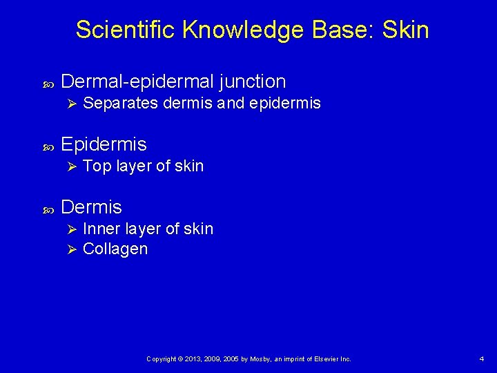
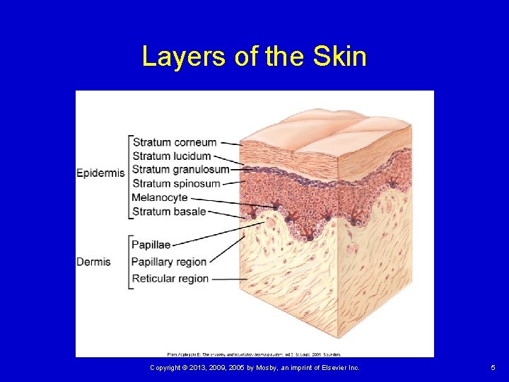
Scientific Knowledge Base: Skin Dermal-epidermal junction Ø Epidermis Ø Separates dermis and epidermis Top layer of skin Dermis Ø Ø Inner layer of skin Collagen Copyright © 2013, 2009, 2005 by Mosby, an imprint of Elsevier Inc. 4
Layers of the Skin Copyright © 2013, 2009, 2005 by Mosby, an imprint of Elsevier Inc. 5
Pressure Ulcers Pressure ulcer Ø Pressure sore, decubitus ulcer, or bed sore Pathogenesis Pressure intensity • Tissue ischemia • Blanching Ø Pressure duration Ø Tissue tolerance Ø Copyright © 2013, 2009, 2005 by Mosby, an imprint of Elsevier Inc. 6
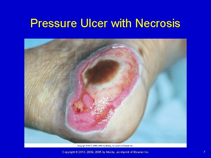
Pressure Ulcer with Necrosis Copyright © 2013, 2009, 2005 by Mosby, an imprint of Elsevier Inc. 7
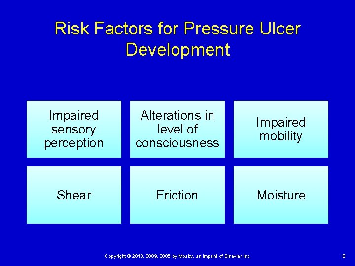
Risk Factors for Pressure Ulcer Development Impaired sensory perception Alterations in level of consciousness Impaired mobility Shear Friction Moisture Copyright © 2013, 2009, 2005 by Mosby, an imprint of Elsevier Inc. 8
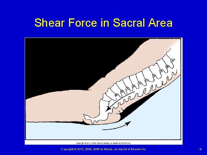
Shear Force in Sacral Area Copyright © 2013, 2009, 2005 by Mosby, an imprint of Elsevier Inc. 9
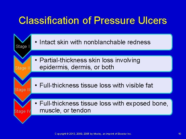
Classification of Pressure Ulcers Stage III Stage IV • Intact skin with nonblanchable redness • Partial-thickness skin loss involving epidermis, or both • Full-thickness tissue loss with visible fat • Full-thickness tissue loss with exposed bone, muscle, or tendon Copyright © 2013, 2009, 2005 by Mosby, an imprint of Elsevier Inc. 10
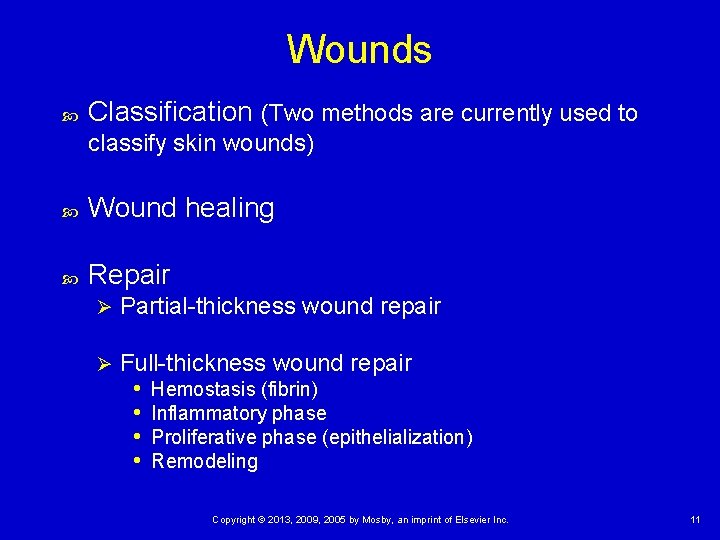
Wounds Classification (Two methods are currently used to classify skin wounds) Wound healing Repair Ø Partial-thickness wound repair Ø Full-thickness wound repair • Hemostasis (fibrin) • Inflammatory phase • Proliferative phase (epithelialization) • Remodeling Copyright © 2013, 2009, 2005 by Mosby, an imprint of Elsevier Inc. 11
Wounds Classification (Two methods are currently used to classify skin wounds) 1. Describe the status of skin integrity, the cause of the wound, the severity or extent of injury or damage, and the cleanliness of the wound (see Table 48 -1 on p. 1181). 2. Or the descriptive qualities of the wound tissue such as color (see Table 48 -2 on p. 1183). Copyright © 2013, 2009, 2005 by Mosby, an imprint of Elsevier Inc. 12
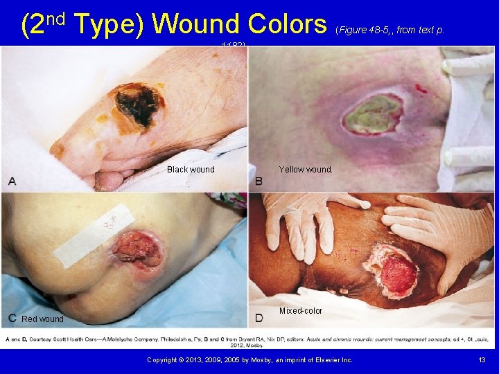
(2 nd Type) Wound Colors (Figure 48 -5, , from text p. 1182) Black wound Red wound Yellow wound. Mixed-color Copyright © 2013, 2009, 2005 by Mosby, an imprint of Elsevier Inc. 13
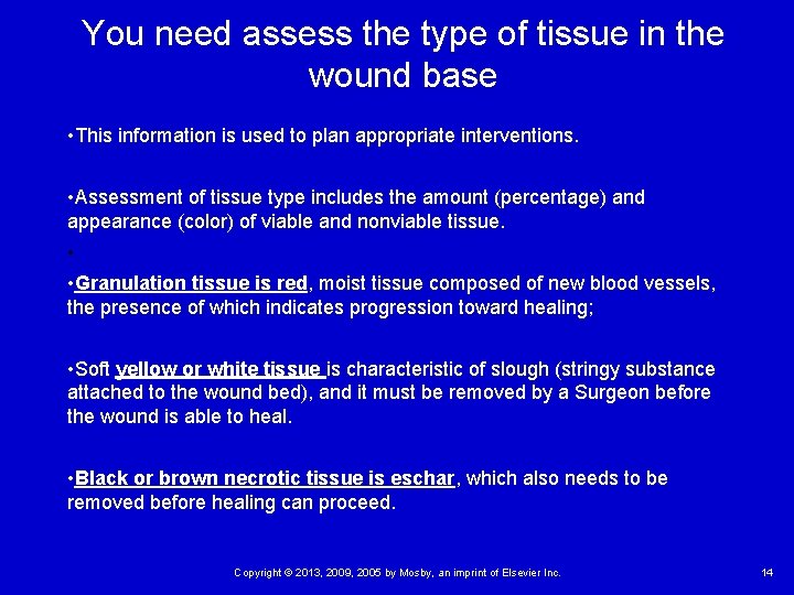
You need assess the type of tissue in the wound base • This information is used to plan appropriate interventions. • Assessment of tissue type includes the amount (percentage) and appearance (color) of viable and nonviable tissue. • • Granulation tissue is red, moist tissue composed of new blood vessels, the presence of which indicates progression toward healing; • Soft yellow or white tissue is characteristic of slough (stringy substance attached to the wound bed), and it must be removed by a Surgeon before the wound is able to heal. • Black or brown necrotic tissue is eschar, which also needs to be removed before healing can proceed. Copyright © 2013, 2009, 2005 by Mosby, an imprint of Elsevier Inc. 14
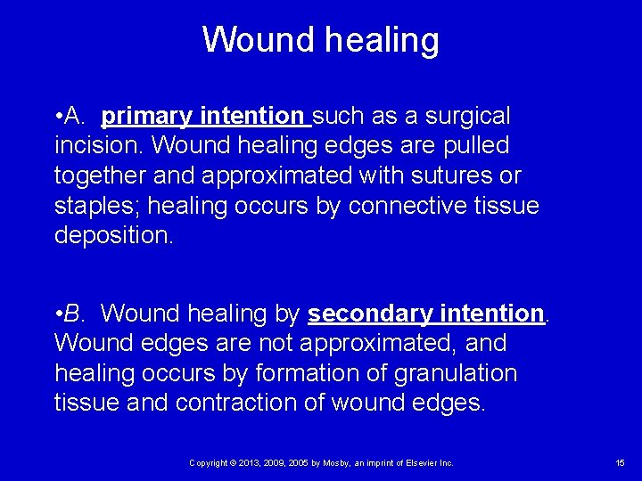
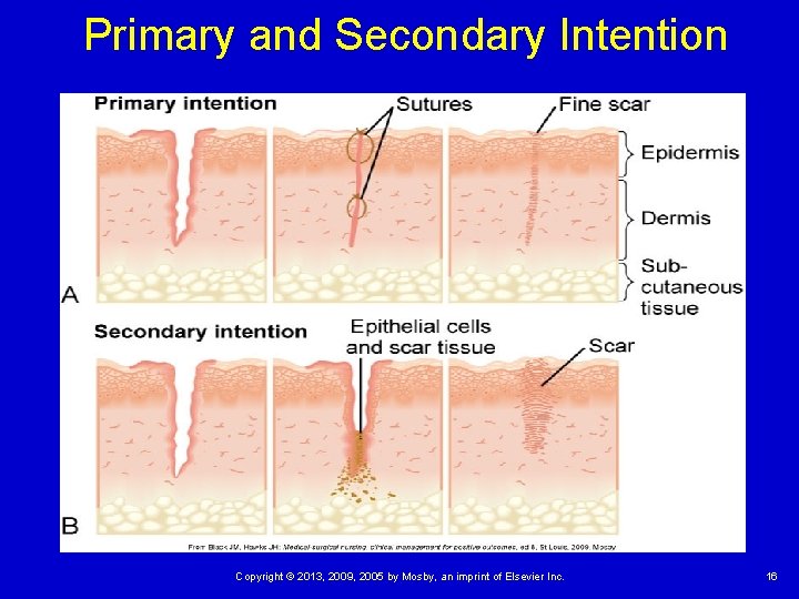
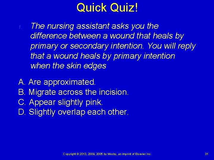
Wound healing • A. primary intention such as a surgical incision. Wound healing edges are pulled together and approximated with sutures or staples; healing occurs by connective tissue deposition. • B. Wound healing by secondary intention. Wound edges are not approximated, and healing occurs by formation of granulation tissue and contraction of wound edges. Copyright © 2013, 2009, 2005 by Mosby, an imprint of Elsevier Inc. 15
Primary and Secondary Intention Copyright © 2013, 2009, 2005 by Mosby, an imprint of Elsevier Inc. 16
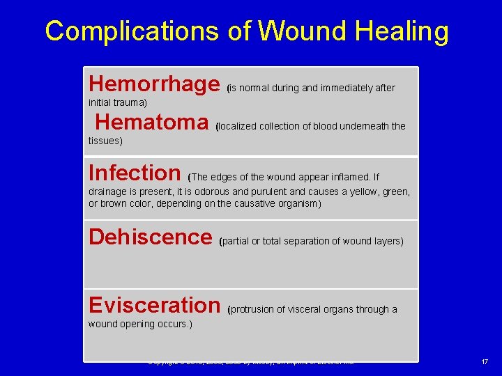
Complications of Wound Healing Hemorrhage (is normal during and immediately after initial trauma) Hematoma (localized collection of blood underneath the tissues) Infection (The edges of the wound appear inflamed. If drainage is present, it is odorous and purulent and causes a yellow, green, or brown color, depending on the causative organism) Dehiscence (partial or total separation of wound layers) Evisceration (protrusion of visceral organs through a wound opening occurs. ) Copyright © 2013, 2009, 2005 by Mosby, an imprint of Elsevier Inc. 17
Nursing Knowledge Base Prediction and prevention of pressure ulcers Ø Risk assessment • Braden scale [Table 48 -3 on p. 1185. ] Ø Sensory perception, moisture, activity, mobility, nutrition, and friction and shear Ø Prevention • Economic consequences Copyright © 2013, 2009, 2005 by Mosby, an imprint of Elsevier Inc. 18
Prevention Economic consequences When a patient develops a pressure ulcer, the length of stay is extended and the overall cost of care increases. Prevention includes special beds and mattresses, good hygiene, good nutrition, adequate hydration, and impeccable nursing care. Copyright © 2013, 2009, 2005 by Mosby, an imprint of Elsevier Inc. 19
Factors Influencing Pressure Ulcer Formation and Wound Healing Nutrition Tissue perfusion Infection Age Psychosocial impact of wounds Copyright © 2013, 2009, 2005 by Mosby, an imprint of Elsevier Inc. 20
Assessment Skin Pressure ulcers Ø Ø Ø Predictive measures Mobility Nutritional status Body fluids Pain Copyright © 2013, 2009, 2005 by Mosby, an imprint of Elsevier Inc. 21
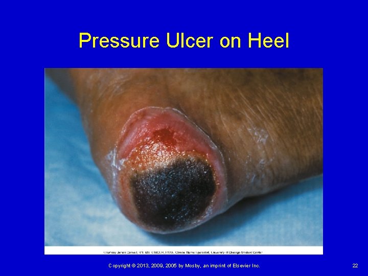
Pressure Ulcer on Heel Copyright © 2013, 2009, 2005 by Mosby, an imprint of Elsevier Inc. 22
Assessment Wounds Ø Ø Ø Ø Emergency setting Stable setting Wound appearance Character of wound drainage Drains Wound closures Palpation of wound Wound cultures Copyright © 2013, 2009, 2005 by Mosby, an imprint of Elsevier Inc. 23
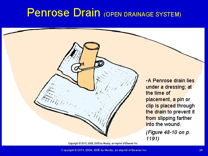
Penrose Drain (OPEN DRAINAGE SYSTEM) • A Penrose drain lies under a dressing; at the time of placement, a pin or clip is placed through the drain to prevent it from slipping farther into the wound. (Figure 48 -10 on p. 1191) Copyright © 2013, 2009, 2005 by Mosby, an imprint of Elsevier Inc. 24
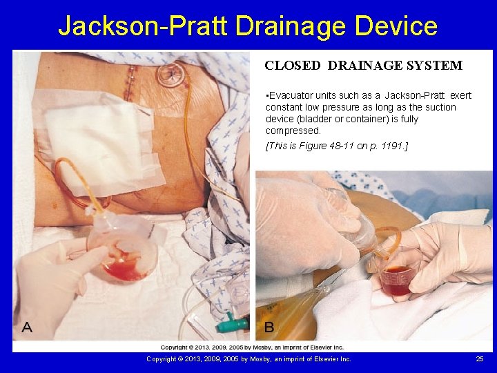
Jackson-Pratt Drainage Device CLOSED DRAINAGE SYSTEM • Evacuator units such as a Jackson-Pratt exert constant low pressure as long as the suction device (bladder or container) is fully compressed. [This is Figure 48 -11 on p. 1191. ] Copyright © 2013, 2009, 2005 by Mosby, an imprint of Elsevier Inc. 25
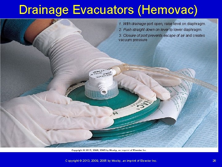
Drainage Evacuators (Hemovac) 1. With drainage port open, raise level on diaphragm. 2. Push straight down on lever to lower diaphragm. 3. Closure of port prevents escape of air and creates vacuum pressure Copyright © 2013, 2009, 2005 by Mosby, an imprint of Elsevier Inc. 26
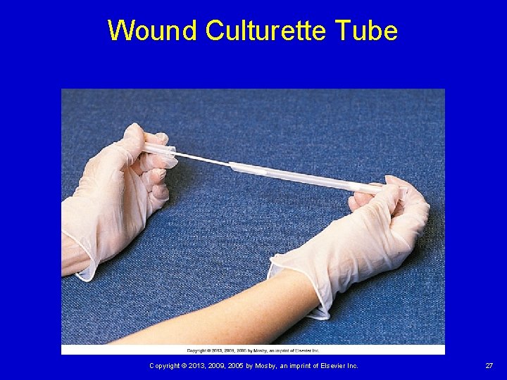
Wound Culturette Tube Copyright © 2013, 2009, 2005 by Mosby, an imprint of Elsevier Inc. 27
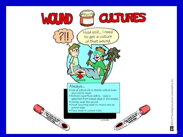
Copyright © 2013, 2009, 2005 by Mosby, an imprint of Elsevier Inc. 28
Copyright © 2013, 2009, 2005 by Mosby, an imprint of Elsevier Inc. 29
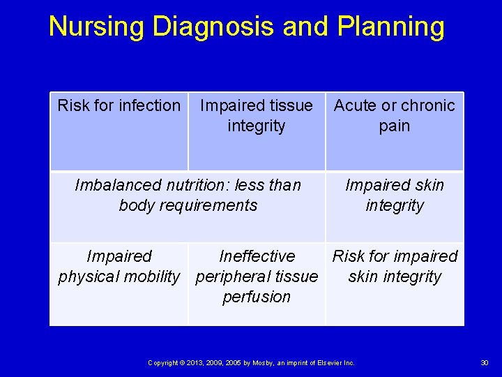
Nursing Diagnosis and Planning Risk for infection Impaired tissue integrity Imbalanced nutrition: less than body requirements Acute or chronic pain Impaired skin integrity Impaired Ineffective Risk for impaired physical mobility peripheral tissue skin integrity perfusion Copyright © 2013, 2009, 2005 by Mosby, an imprint of Elsevier Inc. 30
Quick Quiz! 1. The nursing assistant asks you the difference between a wound that heals by primary or secondary intention. You will reply that a wound heals by primary intention when the skin edges A. Are approximated. B. Migrate across the incision. C. Appear slightly pink. D. Slightly overlap each other. Copyright © 2013, 2009, 2005 by Mosby, an imprint of Elsevier Inc. 31
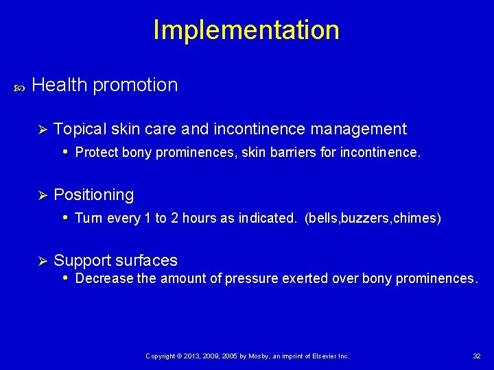
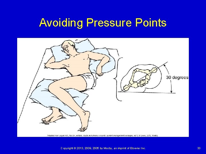
Implementation Health promotion Ø Topical skin care and incontinence management • Protect bony prominences, skin barriers for incontinence. Ø Positioning • Turn every 1 to 2 hours as indicated. (bells, buzzers, chimes) Ø Support surfaces • Decrease the amount of pressure exerted over bony prominences. Copyright © 2013, 2009, 2005 by Mosby, an imprint of Elsevier Inc. 32
Avoiding Pressure Points Copyright © 2013, 2009, 2005 by Mosby, an imprint of Elsevier Inc. 33
Acute Care Management of pressure ulcers Wound management Debridement (removal of nonviable, necrotic tissue) • Mechanical, autolytic, chemical, or sharp/surgical Ø Education Ø Nutritional status Ø Protein status Ø Hemoglobin Ø Copyright © 2013, 2009, 2005 by Mosby, an imprint of Elsevier Inc. 34
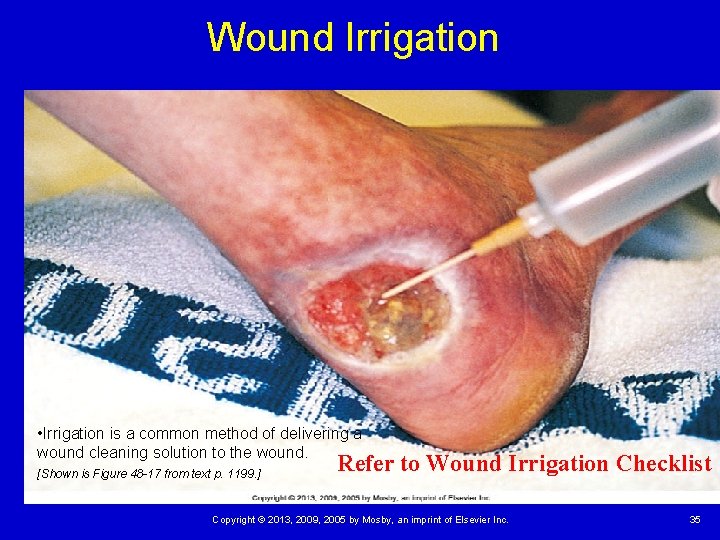
Wound Irrigation • Irrigation is a common method of delivering a wound cleaning solution to the wound. [Shown is Figure 48 -17 from text p. 1199. ] Refer to Wound Irrigation Checklist Copyright © 2013, 2009, 2005 by Mosby, an imprint of Elsevier Inc. 35
First Aid for Wounds Hemostasis Control bleeding. • Allow puncture wounds to bleed. • Do not remove a penetrating object. Ø Bandage Ø Cleaning Ø Ø Gentle Normal saline Protection Copyright © 2013, 2009, 2005 by Mosby, an imprint of Elsevier Inc. 36
Purposes of Dressings Protect a wound from microorganism contamination Aid in hemostasis Promote healing by absorbing drainage and debriding a wound Support or splint the wound site Protect patients from seeing the wound (if perceived as unpleasant) Copyright © 2013, 2009, 2005 by Mosby, an imprint of Elsevier Inc. 37
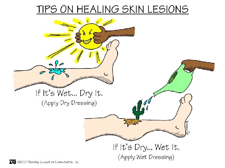
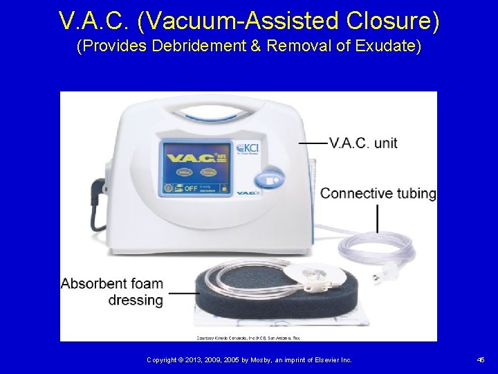
Dressings Dry or moist (wet) Ø Gauze Film dressing Hydrocolloid—protects the wound from surface contamination Hydrogel—maintains a moist surface to support healing Wound vacuum assisted closure (V. A. C. )—uses negative pressure to support healing Copyright © 2013, 2009, 2005 by Mosby, an imprint of Elsevier Inc. 38
Copyright © 2013, 2009, 2005 by Mosby, an imprint of Elsevier Inc. 39
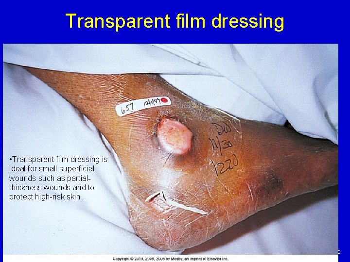
Transparent film dressing • Transparent film dressing is ideal for small superficial wounds such as partialthickness wounds and to protect high-risk skin. Copyright © 2013, 2009, 2005 by Mosby, an imprint of Elsevier Inc. 40
Dressings (cont’d) Changing Ø Know type of dressing, placement of drains, and equipment needed. Prepare the patient for a dressing change Ø Ø Ø Evaluate pain. Describe procedure steps. Gather supplies. Recognize normal signs of healing. Answer questions about the procedure or wound. Copyright © 2013, 2009, 2005 by Mosby, an imprint of Elsevier Inc. 41
During a Dressing Change Assess the skin beneath the tape. Perform thorough hand hygiene before and after wound care. Wear sterile gloves before directly touching an open or fresh wound. Remove or change dressings over closed wounds when they become wet or if the patient has signs or symptoms of infection, and as ordered. Copyright © 2013, 2009, 2005 by Mosby, an imprint of Elsevier Inc. 42
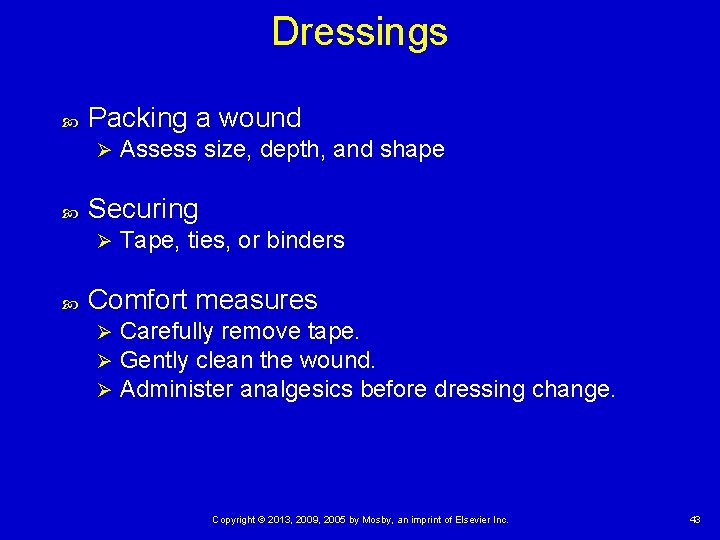
Dressings Packing a wound Ø Securing Ø Assess size, depth, and shape Tape, ties, or binders Comfort measures Ø Ø Ø Carefully remove tape. Gently clean the wound. Administer analgesics before dressing change. Copyright © 2013, 2009, 2005 by Mosby, an imprint of Elsevier Inc. 43
Copyright © 2013, 2009, 2005 by Mosby, an imprint of Elsevier Inc. 44
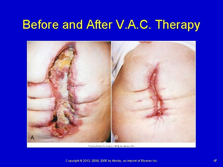
V. A. C. (Vacuum-Assisted Closure) (Provides Debridement & Removal of Exudate) Copyright © 2013, 2009, 2005 by Mosby, an imprint of Elsevier Inc. 45
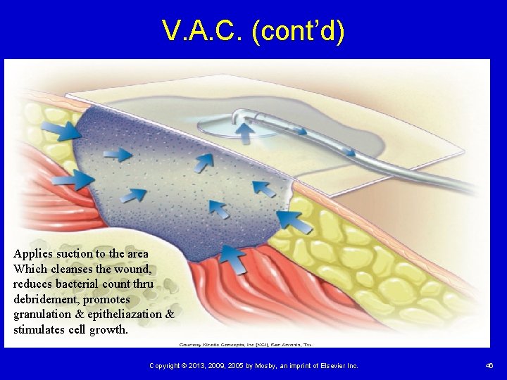
V. A. C. (cont’d) Applies suction to the area Which cleanses the wound, reduces bacterial count thru debridement, promotes granulation & epitheliazation & stimulates cell growth. Copyright © 2013, 2009, 2005 by Mosby, an imprint of Elsevier Inc. 46
Before and After V. A. C. Therapy Copyright © 2013, 2009, 2005 by Mosby, an imprint of Elsevier Inc. 47
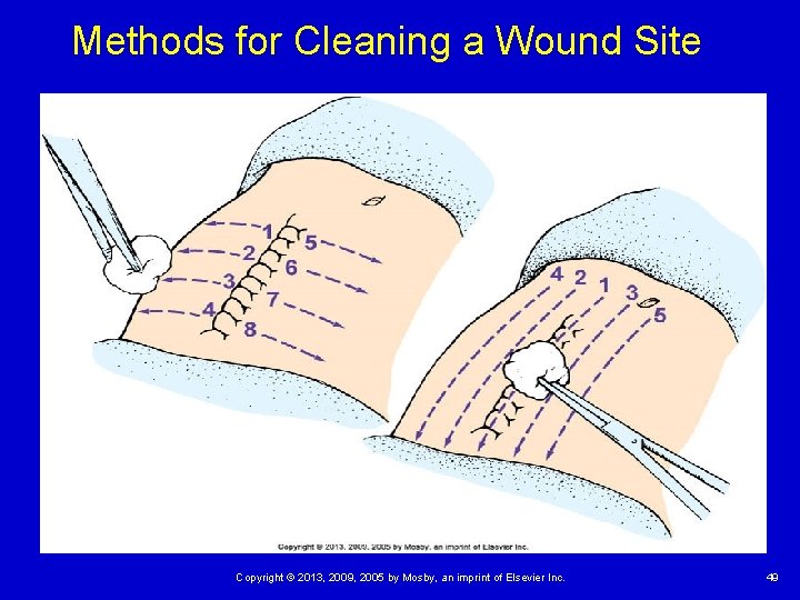
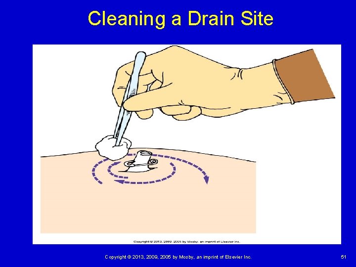
Cleaning Skin 1. Clean in a direction from the least contaminated area such as from the wound or incision to the surrounding skin or from an isolated drain site to the surrounding skin. 2. Use gentle friction when applying solutions locally to the skin. 3. When irrigating, allow the solution to flow from the least to the most contaminated area. Copyright © 2013, 2009, 2005 by Mosby, an imprint of Elsevier Inc. 48
Methods for Cleaning a Wound Site Copyright © 2013, 2009, 2005 by Mosby, an imprint of Elsevier Inc. 49
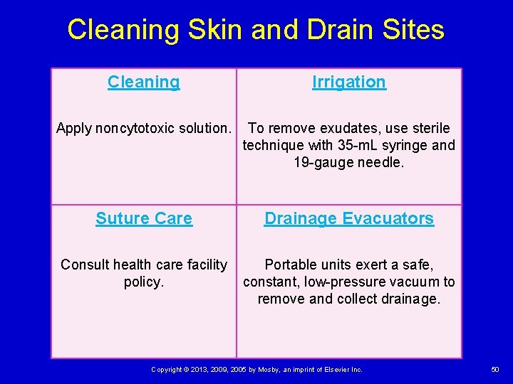
Cleaning Skin and Drain Sites Cleaning Irrigation Apply noncytotoxic solution. To remove exudates, use sterile technique with 35 -m. L syringe and 19 -gauge needle. Suture Care Drainage Evacuators Consult health care facility policy. Portable units exert a safe, constant, low-pressure vacuum to remove and collect drainage. Copyright © 2013, 2009, 2005 by Mosby, an imprint of Elsevier Inc. 50
Cleaning a Drain Site Copyright © 2013, 2009, 2005 by Mosby, an imprint of Elsevier Inc. 51
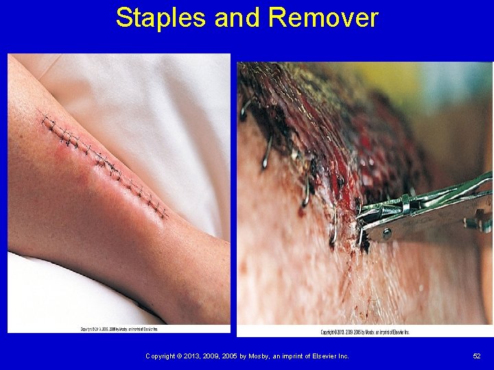
Staples and Remover Copyright © 2013, 2009, 2005 by Mosby, an imprint of Elsevier Inc. 52
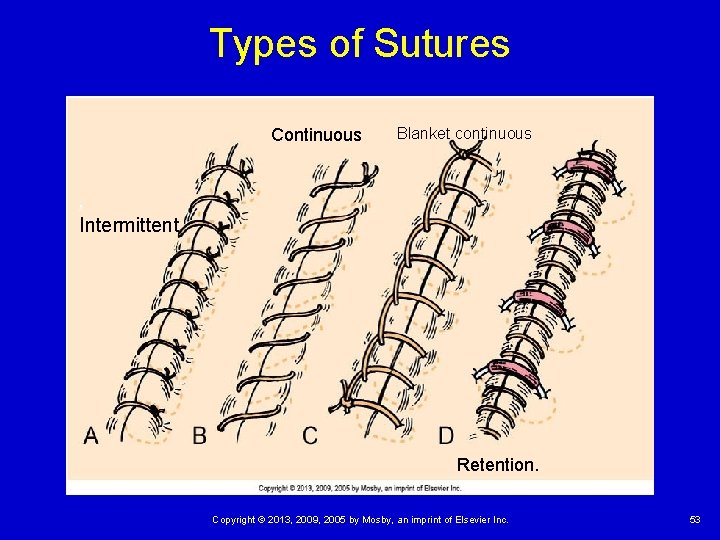
Types of Sutures Continuous Blanket continuous , Intermittent Retention. Copyright © 2013, 2009, 2005 by Mosby, an imprint of Elsevier Inc. 53
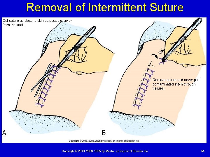
Removal of Intermittent Suture Cut suture as close to skin as possible, away from the knot. Remove suture and never pull contaminated stitch through tissues. Copyright © 2013, 2009, 2005 by Mosby, an imprint of Elsevier Inc. 54
Quick Quiz! 2. A postoperative patient arrives at an ambulatory care center and states, “I am not feeling good. ” Upon assessment, you note an elevated temperature. An indication that the wound is infected would be A. It has no odor. B. A culture is negative. C. The edges reveal the presence of fluid. D. It shows purulent drainage coming from the incision site. Copyright © 2013, 2009, 2005 by Mosby, an imprint of Elsevier Inc. 55
Case Study (cont’d) In preparation for her husband’s discharge, Mrs. Ahmed is interested in learning how to change Mr. Ahmed’s pressure ulcer dressing. Lynda develops a teaching plan to include Mrs. Ahmed, with the outcome goal that “At the end of the teaching session, Mrs. Ahmed will perform an acceptable return demonstration of dressing application. ” What teaching and evaluation strategies would be appropriate? Copyright © 2013, 2009, 2005 by Mosby, an imprint of Elsevier Inc. 56
Quick Quiz! 3. A surgical wound requires a Hydrogel dressing. The primary advantage of this type of dressing is that it provides A. An absorbent surface to collect wound drainage. B. Decreased incidence of skin maceration. C. Protection from the external environment. D. Moisture needed for wound healing. Copyright © 2013, 2009, 2005 by Mosby, an imprint of Elsevier Inc. 57