Integumentary System Part 1 The Integumentary system skin

 is made up of several layers: • The hypodermis •")


 • These cells originate in the bone marrow and")
 cells • These cells are found at the dermal/epidermal junction (stratum basale)")

 • This is the deepest epidermal layer.")
 • This is the deepest epidermal layer. • Cells in")
 • This is the deepest epidermal layer. • Cells in")
 • This layer is several cells thick.")
 • This layer is several cells thick. • The cells")
 • This layer is several cells thick. • The cells")


 are found in this")


 • This is the outermost layer and is between 20")
 • This is the outermost layer and is between 20")
 • This is the outermost layer and is between 20")


- Slides: 77
Integumentary System Part 1
The Integumentary system (skin) is made up of several layers: • The hypodermis • The epidermis • The dermis
The Hypodermis This layer is under the dermis and is not considered part of the skin. It serves to separate the skin from the underlying muscle.
The Hypodermis This layer contains fat tissue and is loosely anchored to the skin. This allows the skin to slide while the fat serves as an insulator and a cushion
The Hypodermis
The Hypodermis • This layer is the major site of fat accumulation • Distribution differs among the sexes. Its in the thighs and breasts of females and the belly of males
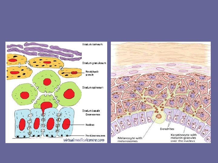
Epidermis • This is the outer most layer and consists of a stratified squamous epithelium. • Four cell types populate it:
Epidermis • This is the outer most layer and consists of a stratified squamous epithelium. • Four cell types populate it: – Keratinocytes – Melanocytes – Epidermal dendritic cells (Langerhan’s cells) – Tactile cells (Merkel cells)
Keratinocytes • These cells produce keratin, a protein made of intermediate fibers.
Keratinocytes • These cells produce keratin, a protein made of intermediate fibers. • These cells attach to each other by desmosomes.
Keratinocytes
Keratinocytes • These cells are found in the lowest level of the epidermis, the stratum basale (basal cell layer). • These cells divide and give rise to the major epithelial cell type in the epidermis.
Keratinocytes • These cells divide and are pushed towards the upper layers of the epidermis.
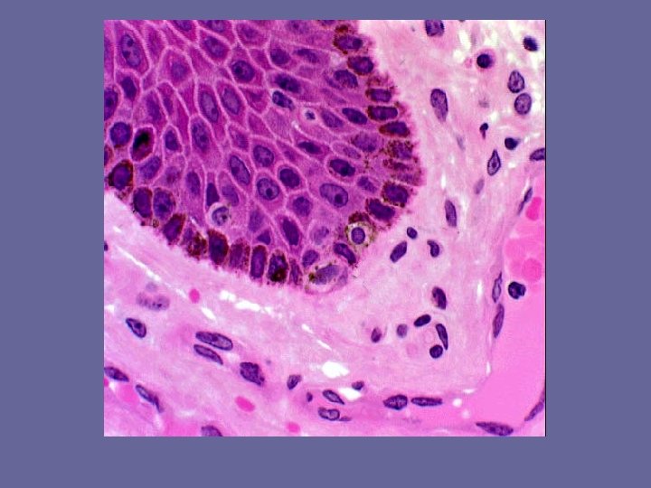
Melanocytes • These cells produce melanin and are found occupying the stratum basale.
Melanocytes • These cells produce melanin and are found occupying the stratum basale. • Melanin is stored in intracellular vesicles called melanosomes.
Melanocytes
Melanocytes • The melanocytes are “spider” shaped. • The melanosomes migrate to the distal portions of the melanocyte where it is secreted. • The melanin is then taken up by the keratinocytes.
Epidermal dendritic cells (Langerhan’s cells) • These cells originate in the bone marrow and migrate to the epidermis. • They play an important role in the skin’s immune response.
Merkel (tactile) cells • These cells are found at the dermal/epidermal junction (stratum basale) and serve as a touch receptor
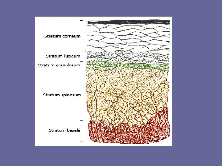
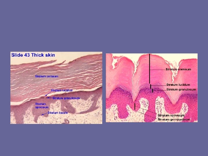
Five layers which make up the epidermis. • These are: – Stratum basale – Stratum spinosum – Stratum granulosum – Stratum lucidum – Stratum corneum
Stratum basale (basal layer) • This is the deepest epidermal layer.
Stratum basale (basal layer) • This is the deepest epidermal layer. • Cells in the lowest level undergo constant mitosis in an effort to replace cells lost in the upper layers.
Stratum basale (basal layer) • This is the deepest epidermal layer. • Cells in the lowest level undergo constant mitosis in an effort to replace cells lost in the upper layers. • 10 to 25% of the cells in this layer are melanocytes.
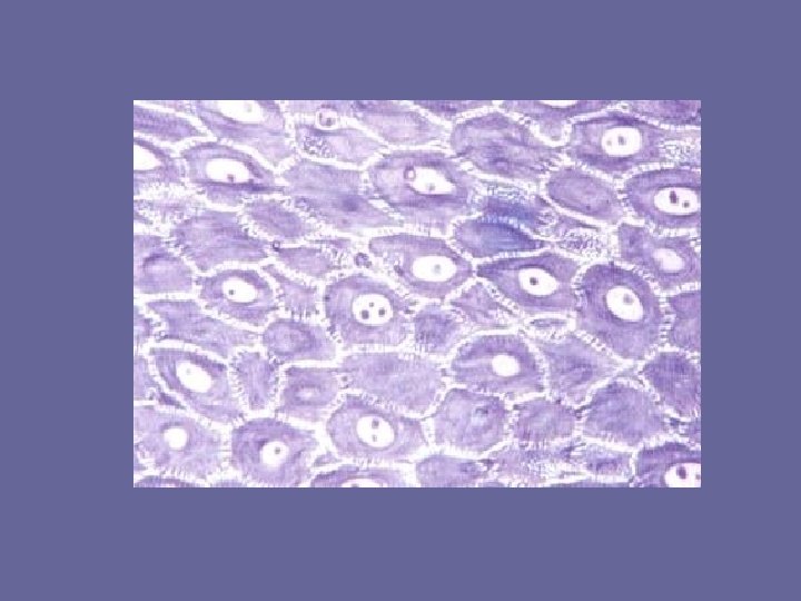
Stratum spinosum (prickly layer) • This layer is several cells thick.
Stratum spinosum (prickly layer) • This layer is several cells thick. • The cells are filled with intermediate fibers which attach to the desmosomes.
Stratum spinosum (prickly layer) • This layer is several cells thick. • The cells are filled with intermediate fibers which attach to the desmosomes. • The desmosomes appear on the cells surface like spikes giving rise to the name prickly.
Stratum granulosum • This layer is 3 to 5 layers thick and is where keratinization occurs
Stratum granulosum • This layer is 3 to 5 layers thick and is where keratinization occurs • Here the cells begin to flatten and the organelles begin to break down.
Stratum granulosum Two types of granules (non membrane bound inclusions) are found in this cell type: – Keratohyaline granules which contain keratin – Lamellated granules contain glycolipids and serve as a water proofing
Stratum lucidum • Seen only where the epidermis is very thick, typically the feet and palms of the hand.
Stratum lucidum • Seen only where the epidermis is very thick, typically the feet and palms of the hand. • It is a histological term for a clear area of cells seen between the stratum granulosum and the stratum corneum.
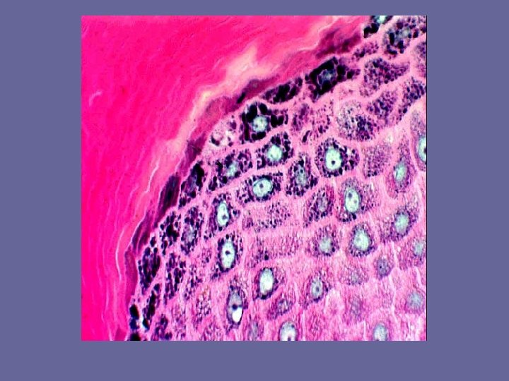
Stratum corneum (horny layer) • This is the outermost layer and is between 20 to 30 cells thick.
Stratum corneum (horny layer) • This is the outermost layer and is between 20 to 30 cells thick. • It protects against abrasions and is made up of dead cells.
Stratum corneum (horny layer) • This is the outermost layer and is between 20 to 30 cells thick. • It protects against abrasions and is made up of dead cells. • The glycolipids from the lamellated granules waterproof this layer
The Dermis • This layer consists of connective tissue and resident cell types of macrophages, fibroblasts and masts cells.
The Dermis • This layer consists of connective tissue and resident cell types of macrophages, fibroblasts and masts cells. Interspersed in this layer are blood vessels, nerve tissue, hair follicles, oil and sweat glands.
The Dermis The dermis consists of two layers: • Papillary layer • Reticular layer
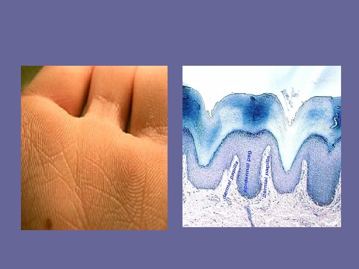
The Papillary Layer • This consists of areolar connective tissue and lies just below the stratum basale. • The loose nature of this layer allows the cells of the immune system to act quickly on any breach of the epidermal cells
The Papillary Layer • This layer has dermal papillae which project into the epidermis.
The Papillary Layer • This consists of areolar connective tissue and lies just below the stratum basale.
The Papillary Layer • This consists of areolar connective tissue and lies just below the stratum basale. • These carry nerve and capillary networks and touch receptors known as Meissner’s corpuscles to the epidermis through dermal papillae.
The Papillary Layer
• The dermal papillae make up the dermal ridges and by extension help form the epidermal ridges. • These are prominent on the hands and feet and form friction ridges and improve gripping. • On the fingers these are known as finger prints.
The Reticular Layer • This layer accounts for about 80% of the thickness of the dermis
The Reticular Layer • This layer accounts for about 80% of the thickness of the dermis • It is made up of dense irregular connective tissue with scattered fat deposits
The Reticular Layer • This layer accounts for about 80% of the thickness of the dermis • It is made up of dense irregular connective tissue with scattered fat deposits • The collagen fibers run is planes and are responsible for the furrow in the brow and neck.
The Reticular Layer • If this area is stretched as occurs in pregnancy or obesity, the dermis becomes torn and gives rise to the so called “stretch marks or cellulite”
Cellulite
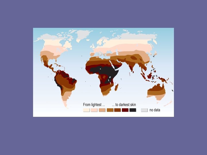
Skin Color There are 3 pigments that contribute to your skin color: • Melanin • Carotene • Hemoglobin
Skin Color • Melanin is the only pigment produced in the skin.
Skin Color • Melanin is the only pigment produced in the skin. • Melanin is a polymer of the amino acid tyrosine.
Skin Color • The color ranges from yellow to tan to reddish brown to black.
Skin Color • The color ranges from yellow to tan to reddish brown to black. • Tyrosinase is the enzyme responsible for making the melanin. This is formed in the melanocytes.
Skin Color
Skin Color
Skin Color • All humans have the same number of melanocytes; racial differences are due to the type of melanin formed
Skin Color • All humans have the same number of melanocytes; racial differences are due to the type of melanin formed • Those groups with dark brown to black skin have the darker forms and retain it longer.
Skin Color • Freckles are locale accumulations of melanin. • They are most often seen in the fair skinned • Often located on the face and become darker with sun exposure
Skin Color • Moles occur when cells in the skin grow in a cluster instead of being spread throughout the skin. These cells are called melanocytes. • As the years pass, moles usually change slowly, becoming raised and/or changing color. Often, hairs develop on the mole. Some moles may not change at all, while others may slowly disappear over time.
Skin Color
Skin Color • Carotene is a yellow to orange pigment and can accumulate in the stratum corneum. Is often seen in the palms and soles of the feet where this epidermal layer is thickest. Is usually associated with diet.
Skin Color • Hemoglobin reflects through the skin of lightly pigmented individuals. • More prominent in lighter pigmented individuals, albinos represent the extreme.
Skin Conditions • Skin conditions can be a sign of underlying health issues. These include: – Redness or erythema - blushing fever – Pallor – pale – Jaundice- liver issues – Bronzing- Addison’s disease, deep tan – Bruises – blood accumulating in the dermis
Redness or erythema • It occurs with any skin injury, infection, or inflammation • This is caused by the capillaries leaking
Pallor • Pallor is a reduced amount of blood flow to the skin or mucous membranes. • It is a pale color which can be caused by illness, emotional shock or stress. • It can develop suddenly or gradually, depending on the cause.
Jaundice • Typically seen with patients having problems with their liver and gall bladder.
Bronzing • Seen with certain types of endocrine orders which result in over productions of MSH (Melanin Stimulating Hormone) • Addison’s disease, a type of adrenal insufficiency, is the most common example
Bruises • Bruises are caused by blood accumulating under the skin. This is usually caused by trauma.