Integumentary System Skin and accessory structures hair nails
 Integument means “covering”")


 are produced by Golgi apparatus of the melanocytes")


- Slides: 24
Integumentary System Skin and accessory structures (hair, nails) Integument means “covering”
Functions of the Integumentary System Protection Against UV light, entry of pathogens, abrasions, water loss Sensation Sensory receptors detect heat, cold, touch, pressure, pain Temperature regulation By controlling blood flow and sweat glands Vitamin D production In UV light, skin produces a molecule that can be transformed into Vitamin D
Vitamin D Necessary for normal absorption of calcium from intestine Rickets in children is a disease from reduced mineralization of bone matrix
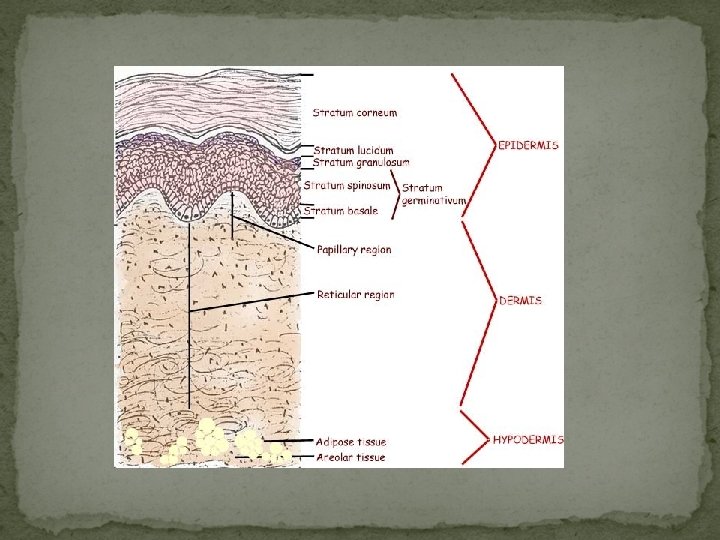
Skin in composed of two layers
The Skin Consists of TWO layers The Epidermis and the Dermis 1. Epidermis Outer layer Stratified squamous Contains no blood vessels Has four types of cells Most cells are Keratinocytes Produce keratin Malanocytes Skin color, produced from melanin Langerhans’ cells Part of the immune system Merkel’s cells Detect light and pressure
Epidermis Cells are produced by mitosis in the deepest layer New cells push older, dead cells to surface to slough off or desquamate Keratinization is the process of moving older cells to surface because they fill with keratin. These cells will resist abrasion and form a permeability barrier
5 layers of epidermal cells Strata are regions of the epidermis Stratum Corneum Stratum Lucidum Stratum Granulosum Stratum Spinosum Stratum Basale
An easy trick to remember these 5 epidermal layers in order from the most superficial to the deepest: "Can Little Girls Speak British" Can Corneum Little Lucidum Gi. Rl GRanulosum Speak SPinosum British Basale
Stratum Corneum 25 + layers of dead skin Cornified cells (dead cells with a hard protein envelope) Hard keratin (nails, hair) Soft keratin (skin)
Stratum Lucidum Thin, clear region 3 -5 layers Dead cells Only found in thick skin (palms, soles of feet, fingertips)
Strata Granulosum 2 -5 layers of diamond shaped cells Most cells are dead Keratohyalin granules Protein envelope that forms below plasma membrane
Strata Spinosum 8 -10 layers of flatten cells Some mitosis occurs Lamellar bodies form These structures move to the cell membrane and release their lipid (adipose) content into the intercellular space The lipids are responsible for the permeability of the epidermis. Abundant Langerhans (used for immunity) cells
Strata Basale Deepest layer Single layer of cuboidal/columnar cells Mitosis occurs every 19 days One daughter cell pushes to surface to become keratinized (40 -56 days)
Thick and Thin Skin Thick All 5 strata Areas subject to pressure and friction (soles of feet, palms of hands, fingertips) Thin 4 layers Hair present Callus is increased layer of strata corneum Corn is a cone-shaped structure that develops in thin or thick skin over a bony prominence.
Dermis Deeper layer Exchanges gases, nutrients, wastes from epidermis Comprised of 2 layers (papillary and reticular) Responsible for strength and flexibility
Structures of the dermis Sebaceous glands Sweat glands Sensory receptors Blood vessels Collagen and Elastin Hair follicle
2 Layers of the Dermis 1. Papillary region Loose connective Dermal Papillae projections are the bumps that are visible These make your fingerprints 2. Reticular region Dense, irregular connective Collagen and elastin fibers Cleavage/tension lines seen on the surface of your skin Stretch marks (overstretched, ruptured dermis) http: //www. youtube. com/watch? v=y. KAz. VC 0 Wcm. I&NR= 1
Background Information about Acne Sebum is produced by the sebaceous glands in the skin. The Latin meaning of sebum is fat. It comes out of the glands in the skin and coats the hair follicles. It reaches the skin’s surface through the follicle. There it mixes with other lipids and sweat and forms a coating on the skin. This coating is called acid mantle. Acid mantle protects the skin against bacteria and keeps the skin hydrated So……. what is acne?
Acne Sebum is one of the main causes of acne. When sebum gets blocked in the hair follicle pores, it forms acne. Acne tends to run in families and can be triggered by: Hormonal changes related to menstrual periods, pregnancy, birth control pills, or stress Greasy or oily cosmetic and hair products Certain drugs (such as steroids, testosterone, estrogen, and phenytoin) High levels of humidity and sweating
Melanosomes (vesicle for carrying melanin) are produced by Golgi apparatus of the melanocytes Melanin is a term used to describe a group of pigments responsible for color of hair, skin, eyes. (UV protection) Melanosomes move into cell processes of the melanocyte Epithelial cells engulf melanosomes Production due to genes, hormones, sun exposure Skin Color
Skin Color Freckles = large amounts of melanin Albinism = inability to produce melanin Tanning= UV light darkens melanin and stimulates production Carotene = yellow pigment in carrot/ corn that accumulates in stratum corneum and tints skin yellow Cyanosis = bluish color from shock due to decrease in blood oxygen
Freckles Albinism Tanning Carotene Cyanosis
Hypodermis Subcutaneous tissue Not a part of skin Functions as the Foundation for the dermis Supplies blood vessels and nerves Adipose abundant Accounts for half of the body’s fat!