Integumentary System Unit 12 Introduction to Medical Terminology


, water,")
 that lubricates the skin and")


 are tiny, coiled glands found on")


, itchy wheals caused by")


 fungus on")


, surgical reduction")
, replacement of")
- Slides: 55
Integumentary System Unit 12 Introduction to Medical Terminology, Ehrlich
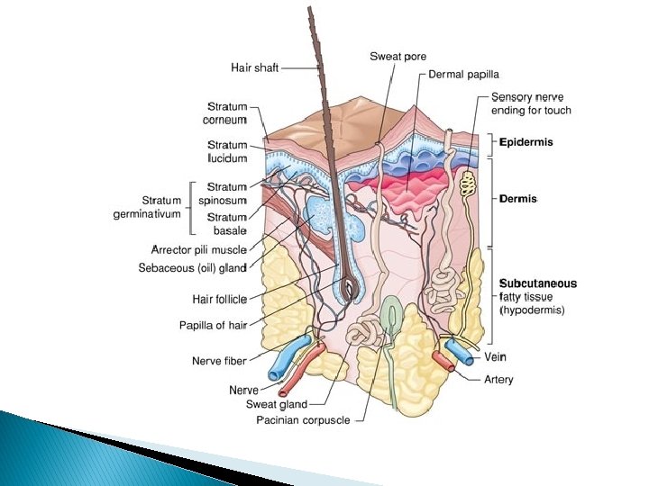
Components of the Skin
Functions of Skin � Protection – Barrier to sun & invasion of pathogens, holds moisture in & prevents body tissues from drying out � Sensory Perception – Nerves in the skin help body respond to pain, pressure, temperature & touch sensations � Body Temperature – Blood vessels in skin help body retain or lose heat. Sudoriferous glands help cool body through evaporation of perspiration
Functions of Skin � Storage - Tissues for temporary storage of fat, glucose(sugar), water, vitamins, & salts � Absorption – Substances can be absorbed through skin, ex. Medications (creams, patches) called transdermal medications � Excretion – Eliminate salt, a minute amount of waste, and excess water and heat through perspiration � Production – Helps in the production of vitamin d by using ultraviolet rays from sun to form an initial molecule of vitamin D that matures in the liver
Related Structures � Sebaceous Glands – secrete sebum (oil) that lubricates the skin and discourages the growth of bacteria on skin � Sudoriferous Glands – (Sweat), help regulate body temperature and water content by secreting sweat. Small amount of metabolic waste also secreted. � Hair – Helps control loss of body heat � Nails – Protect the dorsal surface of the last bone of each toe & finger
Layers of Skin � Covers the external surfaces of the body. Average adult has two square yards of skin, making it the largest bodily organ � Cutaneous means relating to skin � Skin is a complex system of specialized tissues & is made up of three basic layers: ◦ Epidermis ◦ Dermis ◦ Subcutaneous layer
The Epidermis � Epidermis - outermost layer of skin, made up of several specialized epithelial tissues ◦ Epithelial Tissues – form protective covering for all internal & external surfaces of body ◦ Squamous Epithelial Tissues – Forms upper layer of epidermis, consists of flat, scaly cells that are continuously shed ◦ Does not contain blood vessels or connective tissue, is dependent on lower layers for nourishment
The Epidermis � Epidermis ◦ Basal Layer – Lowest layer of epidermis. Here cells are produced and then pushed upward. When these cells reach the surface, they die & become filled with keratin. ◦ Keratin – fibrous, water-repellent protein. Soft keratin is primary component of the epidermis. Hard keratin found in hair & nails
The Epidermis � Epidermis ◦ Melanocytes – Special cells found in the basal layer. Produce & contain a dark brown to black pigment called melanin. Type & amount of melanin pigment determines color of skin. ◦ Melanin – has important function of protecting the skin against some of the harmful ultraviolet rays of the sun.
The Dermis � Known as the corium & the true skin, thick layer of living tissue directly below the epidermis. � Contains connective tissue, blood & lymph vessels, and nerve fibers. � Contains the associated structures of the skin, which are the hair follicles plus the sebaceous & sudoriferous glands � Sensory nerve endings in dermis are the sensory receptors stimuli such as touch, temperature, pain and pressure
Tissues Within the Dermis � Collagen – Means glue, is a tough flexible, fibrous protein material found in the skin and in the bones, Cartilage, tendons & ligament � Mast Cells – Found in the connective tissues of the dermis, respond to injury, infection, or allergy by producing and releasing substances, including heparin & histamine
Tissues Within the Dermis � Heparin – is released in response to an injury, is an anticoagulant. An anticoagulant prevents blood clotting � Histamine – Which is released in response to allergens, causes the signs of an allergic response, including itching and increased mucus secretion
The Subcutaneous Layer � Subcutaneous Layer – Located just below the skin, connects the skin to the surface muscles � Layer is made up of loose connective tissue and adipose tissue. Adipose means fat � Cellulite – Term sometimes used to describe deposits of dimpled fat, is really just simply ordinary fat. � Lipocytes – (fat cells) are predominant in the subcutaneous layer where they manufacture and store large quantities of fat
The Sebaceous Glands � Sebaceous Glands – Located in the dermis layer of the skin and are closely associated with hair follicles ◦ Secretes sebum which is released through ducts opening into the hair follicles. From here, the sebum moves onto the surface & lubricates the skin ◦ Sebum is slightly acidic, discourages the growth of bacteria on the skin ◦ Milk-producing mammary glands, which are modified sebaceous glands, are sometimes classified with the integumentary system. They are also part of the reproductive system
Sudoriferous Glands � Sudoriferous Glands – (Sweat Glands) are tiny, coiled glands found on almost all body surfaces. Most numerous in palms or hands, soles of feet, forehead, & armpits ◦ Pores – are openings on the surface of the skin for the ducts of the sweat glands ◦ Perspiration – (Sweat) is secreted by sweat glands and is made up of 99% water plus some salt & metabolic waste products ◦ Perspiring – (Sweating) one way the body excretes excess. Body odor associated with sweat comes from interaction of perspiration with bacteria on the skin’s surface ◦ Hidrosis – Production & excretion of sweat
The Hair � Hair - rod-like structures composed of tightly fused dead protein cells filled with hard keratin. Darkness & color of the hair is determined by the amount & type of melanin produced by the melanocytes that surround the core of the hair shaft ◦ Hair Follicles – sacs that hold the root of the hair fibers. Shape of the follicle determines whether the hair is straight or curly ◦ Dead Hair Tissue – appears to grow because cells at the base of the follicle divide rapidly & push old cells upward. As they are pushed upward they harden and undergo pigmentation
The Hair � Hair ◦ Arrector pili – tiny muscles fibers attached to the hair follicles that cause the hair to stand erect. ◦ In response to cold or fright, these muscles contract, causing raised areas of skin known as goose bumps. This action reduces heat loss through the skin.
The Nails � Unguis – Commonly know as a fingernail or toenail, is the keratin plate protecting the dorsal surface of the last bone of each finger and toe. Each nail consists of these parts: ◦ Nail Body – translucent, closely molded to the surface of the underlying tissues. Made up of hard, keratinized plates of epidermal cells ◦ Nail Bed – joins the nail body to the underlying connective tissue, nourished the nail. The blood vessels here give he nail its characteristic pink color
The Nails � Unguis ◦ Free Edge – Portion of the nail not attached to the nail bed, extends beyond the tip of the finger or toe ◦ Lunula – Pale half moon-shaped region at every nail root that is generally most easily seen in thumb nail. This is the active area of the nail, where new keratin cells form. ◦ Cuticle – Narrow band of epidermis attached to the surface of the nail in front of the root protecting the new keratin cells as they form. ◦ Nail Root – Fastens the nail to the finger or toe by fitting into a groove in the skin
Related Medical Specialties � Dermatologist – A physician, who specializes in diagnosing & treating disorders of the skin � Cosmetic Surgeon – (plastic surgeon) a physician who specializes in the surgical restoration & reconstruction of body structures
Pathology of Integumentary: Sebaceous Glands � Acne Vulgaris – Commonly known as acne. Chronic inflammatory disease characterized by pustular eruptions of the skin caused by an overproduction of sebum. Often triggered by a hormones in puberty & adolescence
Pathology of Integumentary: Sebaceous Glands � Seborrhea – over activity of sebaceous glands, results in the production of an excessive amount of sebum � Seborrheic Dermatitis – Inflammation that causes scaling & itching of the upper layers of skin or scalp.
Pathology of Integumentary Surface Lesions Lesion – Pathologic change of the tissues due to disease or injury. Skin lesions are described by their appearance, location, color, and size as measured in centimeters
Pathology of Integumentary: Surface Lesions � Crust – scab, a collection of dried serum and cellular debris � Macule – freckle, discolored, flat spot usually <1 cm in diameter
Pathology of Integumentary: Surface Lesions � Nodule – solid, raised skin lesion > 0. 5 cm in diameter & deeper than a papule � Papule – small, raised red lesion < 0. 5 in diameter & does not contain pus, ex. small pimples & insect bites
Pathology of Integumentary: Surface Lesions � Plaque – scaly, solid raised area of closely spaced papules, ex. psoriasis lesions � Scales – flakes or dry patches made up of excess dead epidermal cells, ex. Psoriasis scales
Pathology of Integumentary: Surface Lesions � Verrucae – warts, small, hard skin lesions caused by the human papilloma virus � Wheal – welt, small bump that itches, can appear as a symptom of an allergic reaction
Pathology of Integumentary: Fluid-Filled Lesions � Abscess – closed pocket containing pus, caused by a bacterial infection � Cyst – abnormal sac containing gas, fluid, or semisolid material, most common is sebaceous cyst
Pathology of Integumentary: Fluid-Filled Lesions � Pustule – pimple, small circumscribed lesion containing pus � Vesicle – small blister <0. 5 cm, contains watery fluid � Bulla – Large blister >0. 5 cm, contains watery fluid
Pathology of Integumentary: Lesions Through the Skin � Abrasion – injury, superficial layers of skin are scraped or rubbed away � Pressure Sore, decubitus ulcer or bed sore. Ulcerated area caused by prolonged pressure that caused tissue death
Pathology of Integumentary: Lesions Through the Skin � Fissure – groove or crack-like break in skin � Laceration – torn or jagged wound, or an accidental cut wound
Pathology of Integumentary: Lesions Through the Skin � Puncture wound – deep hole made by sharp object such as a nail. Increased risk of infection � Ulcer – Open lesion of skin or mucous membrane resulting in tissue loss around the edges
Pathology of Integumentary: Dermatitis – inflammation of the skin � Contact Dermatitis. Localized allergic response caused by contact with an irritant, ex diaper rash, jewelry rash
Pathology of Integumentary: Dermatitis � Eczema – form of persistent or recurring dermatitis characterized by redness, itching, & dryness � Pruitus – itching that is associated with most forms of dermatitis
Pathology of Integumentary: Erythema – Redness of the skin due to capillary dilation � Erythema multiform. Results from generalized allergic reaction to illness, infection or medication. Characterized by rash (nodules, papules, vesicles or bullae)
Pathology of Integumentary: Erythema � Erythema infectiosum – fifth disease, mildly contagious viral infection common in childhood. Red lace-like rash on face
Pathology of Integumentary: Erythema � Exfoliative Dermatitis – Widespread scaling of the skin, often with pruritus, erythroderma & hair loss. May occur in severe cases of many common skin conditions, ex. Eczema, psoriasis, & allergic reactions
Pathology of Integumentary: General Skin Conditions � Psoriasis – Chronic noncontagious, inherited. Has flareups with red plaque covered with silvery scales occur on elbows knees, scalp back or buttocks.
Pathology of Integumentary: General Skin Conditions � Urticaria – (hives), itchy wheals caused by an allergic reaction � Xeroderma – (xerosis) excessively dry skin
Pathology of Integumentary: Bacterial Skin Infections � Cellulitis – acute, rapidly spreading infection within tissues with malaise, swelling, warmth & red streaks
Pathology of Integumentary: Bacterial Skin Infections � Gangrene – tissue necrosis, caused by a loss of circulation to tissues. Tissue death is followed by bacterial invasion that causes putrefaction & if this infection enters bloodstream it can be fatal
Pathology of Integumentary: Bacterial Skin Infections � Impetigo – highly contagious bacterial skin infection that commonly occurs in children. Isolated pustules become crusted & ruptures
Pathology of Integumentary: Bacterial Skin Infections � Necrotizing Fasciitis – Severe infection caused by group A strep bacteria. (flesh eating) If bacteria enter the body serious infection can result. If untreated body tissue is destroyed & can be fatal
Pathology of Integumentary: Fungal Skin Infections Tinea – Fungal infection that can grow on skin, hair, or nails. (ringworm) � Tinea Capitis – on the scalps of children
Pathology of Integumentary: Fungal Skin Infections � Tinea Corporis – fungal infection on the body � Tinea Cruis – (jock itch) found on genital area
Pathology of Integumentary: Fungal Skin Infections � Tinea pedis – (athlete’s foot) fungus on foot & between toes � Tinea Versicolor – fungal infection that causes painless, discolored areas on skin
Pathology of Integumentary: Skin Cancer � Basal Cell Carcinoma – malignant tumor of basal cell layer of epidermis. Slow growing & rarely spreads to other parts of the body.
Pathology of Integumentary: Skin Cancer � Squamous Cell Carcinoma – malignant tumor of the scaly squamous cells of the epithelium. Can quickly spread to other body systems
Pathology of Integumentary: Skin Cancer � Malignant Melanoma – skin cancer that occurs in the melanocytes. Most serious type of skin cancer
Pathology of Integumentary: Burns � Injury to the body tissues caused by heat, flame, electricity, sun, chemicals, or radiation. Degree of burn is determined by the layer of skin involved
Pathology of Integumentary: Diagnostic Procedures Biopsy – Removal of a small piece of living tissue for examination to confirm or establish a diagnosis � Incisional Biopsy – a piece but not all, of the tumor or lesion is removed
Pathology of Integumentary: Diagnostic Procedures � Excisional Biopsy – entire tumor or lesion & a margin of surrounding tissue are removed � Needle Biopsy – a hollow needle is used to remove a core of tissue for examination
Pathology of Integumentary Treatment Procedures: Cosmetic Procedures � Belpharoplasty – (lid lift), surgical reduction of the upper & lower eyelids � Botox – formulation of botulinum toxin type A, temporarily blocks the nerve signals to injected muscle to reduce frown lines
Pathology of Integumentary Treatment Procedures: Cosmetic Procedures � Dermatoplasty – (skin graft), replacement of damaged skin with healthy tissue taken from a donor site on patient’s body