INTEGUMENTARY SYSTEM Introduction to the SKIN Integumentary System

 �Nails �Hair Follicles �Skin")


 �Associated with")


- Slides: 21
INTEGUMENTARY SYSTEM
Introduction to the SKIN! �Integumentary System �Skin (aka- cutaneous membrane) �Nails �Hair Follicles �Skin Glands �The skin is the largest organ by weight �Functions: �Protective covering �Slows water loss �Regulates body temperature �Houses sensory receptors �Excretes small amounts of waste �Helps in vitamin D formation
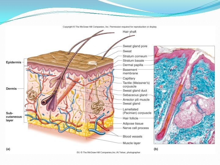
Tissues of the Skin- Into to Layers � 2 distinct layers �Epidermis � Outer layer � Stratified squamous epithelium �Dermis � Inner layer � Connective tissue � Smooth muscle tissue � Nervous tissue � Blood �Layers are separated by a basement membrane
Tissues of the Skin- Hypodermis �Subcutaneous layer �Aka- hypodermis �Not part of the skin �Areolar and adipose tissues �Binds skin to underlying organs �Helps insulate �Contains blood vessels that supply the skin �The dermis and hypodermis lack a sharp boundary
Tissues of the Skin- Injections �Intradermal injections �Injected into the skin �Subcutaneous injections �Injected into the hypodermis �Administered through a hollow needle �Also called hypodermic injections �Transdermal patches �Drug passes into the epidermis through a permeable membrane �Motion sickness, chest pain, blood pressure, smoking cessation
The Epidermis- Characteristics and Functions �Lacks blood vessels �Thickest on palms and soles (0. 0 -1. 4 mm) �Other body regions average 0. 07 -0. 12 mm �Production of new epidermal cells balances loss of dead cells in healthy skin �Skin does not completely wear away �Cell division increases where skin is rubbed/pressed regularly � Leads to calluses and corns �Protective Functions �Shields against water loss �Prevents injury �Protects against harmful chemicals �Keeps out pathogens
The Epidermis-Layers � 4 main layers: �Stratum corneum � Outermost layer � Keratinized, dead epithelial cells � Keratinization- hardening of cells, waterproof keratin proteins made and stored in the cells �Stratum granulosum �Strantum spinosum �Stratum basale � Deepest layer � Able to divide and grown � Receive nutrients from dermis � Contains melanocytes
The Epidermis- Pigmentation �The epidermis contains melanocytes �Produce the pigment melanin �Found in the stratum basale �Melanin �Provides skin color �Absorbs UV radiation
�Effect of Environmental Factors �Sunlight, UV light, X rays � Rapidly darken melanin � Stimulate melanocytes to produce more pigment � Pigment is transferred to nearby cells �Effect of Blood �Well oxygenated blood is bright red � Light-complexioned people may appear pink �Dilated vessels redden the skin � Overheated, embarrassed, under the influence of alcohol �Constriction of vesssel cause loss of color � Low body temperature, frightened/anxious person
�Human Skin Color �Determined by heredity and environmental & physiological factors �All people have ~same number of melanocytes �Differences in skin color result from different amounts of melanin produced � Controlled by several genes � More melanin = darker skin
The Dermis- General Characteristics �The boundary between the epidermis and dermis is uneven �Dermal papillae extend from dermis into ridges of the epidermis �Increases surface area �Most abundant in hands and feet �Form fingerprints � Genes determine general patterns � Fetal movement forms distinct characteristics �The dermis binds epidermis to underlying tissues �Thickness ranges from 0. 5 mm to 3. 0 mm
The Dermis- Layers �Papillary Layer �Upper layer �Areolar connective tissue �Reticular Layer �Lower layer �Dense irregular connective tissue �Give skin toughness and elasticity
The Dermis- Additional Components �Smooth muscle fibers �Can wrinkle the skin (testes) �Associated with hair follicles and glands �Skeletal muscle fibers �Voluntary movements (facial expressions)
�Nerve cells �Carry impulses to dermal muscles and glands �Carry sensory impulses away from sensory receptors � Lamellated corpuscles � In deep dermis � Respond to heavy pressure � Tactile corpuscles � In upper dermis � Sense light touch and texture �Accessory Structures (blood, hair follicles, glands)
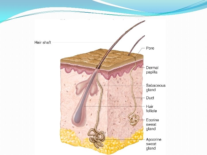
Accessory Structures of the Skin. Nails �Protective coverings �Components: �Nail plate �Nail bed � Skin surface �Lunula � Most actively growing region � Produces keratinized, dead cells �Wears away with normal use �Analogous to hoofs and claws of other animals
Accessory Structures of the Skin. Hair Follicles �Hair is present on all surfaces except: palms, soles, lips, nipples and parts of external reproductive organs �Not well developed on other surfaces (forehead) �Originates in epidermis �Nourished by dermis �Grow, divide and push older cells toward surface �Cells become keratinized and die �Create the hair shaft
�Average hair loss is 20 -100 hairs a day �A single hair grows 2 -6 years and is then replaced by a completely new hair �Genes determine hair color �direct the type and amount of pigment produced by melanocytes �Arrector pili muscles attaches to each hair follicle �Contraction causes hair to stand up �Causes goose bumps
Accessory Structures of the Skin. Glands �Sebaceous glands �Associated with hair follicles �Oil glands �Produce sebum (oil and cell fragments) �Ducts usually empty into hair follicles �Activated by hormones �Keep hairs and skin soft, pliable and waterproof �Not on palms or soles
�Sweat glands � ~2 million person � Widespread � Originates in deep dermis � Eccrine glands � Most numerous � Abundant on forehead, neck, back � Palms and soles � Respond to elevated body temperature � Also respond to emotional stress � Apocrine glands � Develop a scent as they mix with skin bacteria � Activated at puberty � React to emotional upset, fright, pain, sexual arousal � Most unmberous in axillary regions and groin �Specialized sweat glands � Ceruminous glands- ear wax � Female mammary glands- milk